Laminins are heterotrimeric, non-collagenous glycoproteins composed of alpha, beta, and gamma chains. Through interactions with integrins, dystroglycan, and other receptors, Laminins contribute to cell differentation, cell shape and migration, maintenance of tissue phenotypes, and survival. Laminin-1 is comprised of alpha 1, beta 1, and gamma 1 subunits.
Mouse Laminin alpha 1/beta 1 Antibody (AL-4)
R&D Systems | Catalog # MAB4656

Key Product Details
Species Reactivity
Validated:
Mouse
Cited:
Mouse, Fish - Danio rerio (Zebrafish), Transgenic Mouse
Applications
Validated:
Immunohistochemistry, Western Blot
Cited:
Immunohistochemistry, Western Blot, Immunocytochemistry
Label
Unconjugated
Antibody Source
Monoclonal Rat IgG2A Clone # AL-4
Loading...
Product Specifications
Immunogen
Purified fragment of chymotrypsin-digested mouse EHS tumor-derived Laminin-1
Specificity
Detects the Hep-1 heparin binding domain of mouse Laminin A/alpha 1 subunit (1).
Clonality
Monoclonal
Host
Rat
Isotype
IgG2A
Scientific Data Images for Mouse Laminin alpha 1/beta 1 Antibody (AL-4)
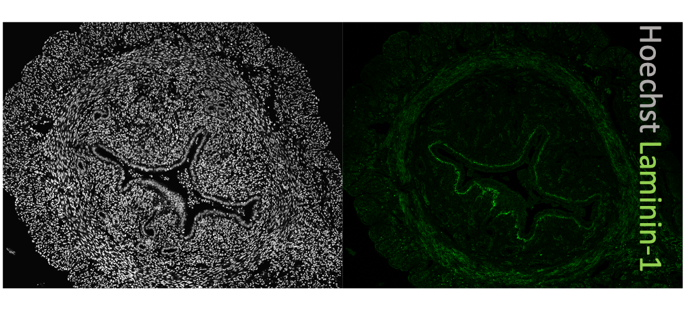
Laminin alpha 1/ beta 1 in Embryonic Mouse Decidua.
Laminin alpha 1/ beta 1 was detected in immersion fixed frozen sections of embryonic mouse decidua (E10.5) using 10 µg/mL Rat Anti-Mouse Laminin alpha 1/ beta 1 Monoclonal Antibody (Catalog # MAB4656) overnight at 4 °C. Tissue was stained with the NorthernLights™ 557-conjugated Anti-Rat IgG Secondary Antibody (red; Catalog # NL013) and counterstained with DAPI (blue). View our protocol for Fluorescent IHC Staining of Frozen Tissue Sections.
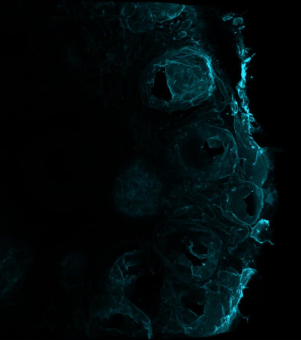
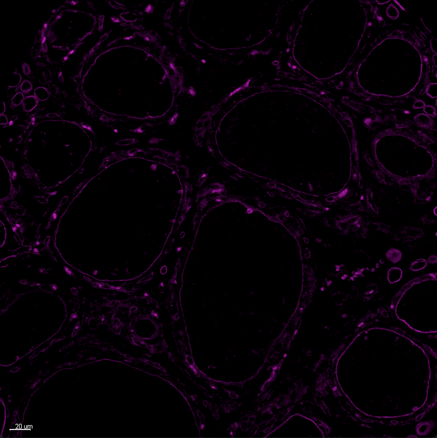
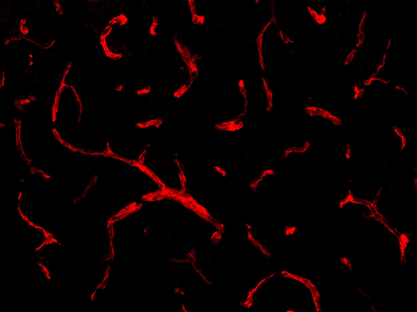
Laminin alpha 1/ beta 1 in Mouse Kidney, Skin, Intestine, and Skeletal Muscle.
Laminin alpha 1/ beta 1 was detected in perfusion fixed frozen sections of mouse kidney glomeruli (panel A), skin (panel B), intestine (panel C), and skeletal muscle (panel D) using Rat Anti-Mouse Laminin alpha 1/ beta 1 Monoclonal Antibody (Catalog # MAB4656) at 10 µg/mL overnight at 4 °C. Tissue was stained using the NorthernLights™ 557-conjugated Anti-Rat IgG Secondary Antibody (red; Catalog # NL013) and counterstained with DAPI (blue). Specific staining was localized to cell surfaces. View our protocol for Fluorescent IHC Staining of Frozen Tissue Sections.Applications for Mouse Laminin alpha 1/beta 1 Antibody (AL-4)
Application
Recommended Usage
Immunohistochemistry
8-25 µg/mL
Sample: Immersion fixed frozen sections of embryonic mouse decidua (E10.5), and perfusion fixed frozen sections of mouse kidney glomeruli, skin, intestine, and skeletal muscle
Sample: Immersion fixed frozen sections of embryonic mouse decidua (E10.5), and perfusion fixed frozen sections of mouse kidney glomeruli, skin, intestine, and skeletal muscle
Western Blot
Skubitz, A.P. et al. (1987) Exp. Cell Res. 173:349. This application was not tested by R&D Systems.
Reviewed Applications
Read 4 reviews rated 4.5 using MAB4656 in the following applications:
Formulation, Preparation, and Storage
Purification
Protein A or G purified from hybridoma culture supernatant
Reconstitution
Reconstitute at 0.5 mg/mL in sterile PBS. For liquid material, refer to CoA for concentration.
Loading...
Formulation
Lyophilized from a 0.2 μm filtered solution in PBS with Trehalose. *Small pack size (SP) is supplied either lyophilized or as a 0.2 µm filtered solution in PBS.
Shipping
Lyophilized product is shipped at ambient temperature. Liquid small pack size (-SP) is shipped with polar packs. Upon receipt, store immediately at the temperature recommended below.
Stability & Storage
Use a manual defrost freezer and avoid repeated freeze-thaw cycles.
- 12 months from date of receipt, -20 to -70 °C as supplied.
- 1 month, 2 to 8 °C under sterile conditions after reconstitution.
- 6 months, -20 to -70 °C under sterile conditions after reconstitution.
Calculators
Background: Laminin alpha 1/beta 1
References
- Skubitz, A.P. et al. (1988) J. Biol. Chem. 263:4861.
- Skubitz, A.P. et al. (1987) Exp. Cell Res. 173:349.
Additional Laminin alpha 1/beta 1 Products
Product Documents for Mouse Laminin alpha 1/beta 1 Antibody (AL-4)
Certificate of Analysis
To download a Certificate of Analysis, please enter a lot or batch number in the search box below.
Note: Certificate of Analysis not available for kit components.
Product Specific Notices for Mouse Laminin alpha 1/beta 1 Antibody (AL-4)
For research use only
Citations for Mouse Laminin alpha 1/beta 1 Antibody (AL-4)
 Powered by Bioz
Customer Reviews for Mouse Laminin alpha 1/beta 1 Antibody (AL-4) (4)
4.5 out of 5
4 Customer Ratings
Have you used Mouse Laminin alpha 1/beta 1 Antibody (AL-4)?
Submit a review and receive an Amazon gift card!
$25/€18/£15/$25CAN/¥2500 Yen for a review with an image
$10/€7/£6/$10CAN/¥1110 Yen for a review without an image
Submit a review
Customer Images
Showing
1
-
4 of
4 reviews
Showing All
Filter By:
-
Application: ImmunohistochemistrySample Tested: Ovary tissue and Ovary (developing oocytes)Species: MouseVerified Customer | Posted 11/15/2024Worked on mouse reconstituted ovary. IHC-P sample working concentration 1:100 and 1:200.

-
Application: Immunocytochemistry/ImmunofluorescenceSample Tested: Ovary tissueSpecies: MouseVerified Customer | Posted 11/08/2024The antibody was tested on a mouse P14 ovary paraffin section at a 1:200 dilution, yielding clear, specific staining. This concentration appears effective for immunohistochemistry in ovarian tissues. Further tests could confirm its optimal range for related applications.
-
Application: Immunocytochemistry/ImmunofluorescenceSample Tested: Uterus tissueSpecies: MouseVerified Customer | Posted 12/15/2022
-
Application: Immunocytochemistry/ImmunofluorescenceSample Tested: Adult brainSpecies: MouseVerified Customer | Posted 09/18/20191:200
There are no reviews that match your criteria.
Protocols
Find general support by application which include: protocols, troubleshooting, illustrated assays, videos and webinars.
- Antigen Retrieval Protocol (PIER)
- Antigen Retrieval for Frozen Sections Protocol
- Appropriate Fixation of IHC/ICC Samples
- Cellular Response to Hypoxia Protocols
- Chromogenic IHC Staining of Formalin-Fixed Paraffin-Embedded (FFPE) Tissue Protocol
- Chromogenic Immunohistochemistry Staining of Frozen Tissue
- ClariTSA™ Fluorophore Kits
- Detection & Visualization of Antibody Binding
- Fluorescent IHC Staining of Frozen Tissue Protocol
- Graphic Protocol for Heat-induced Epitope Retrieval
- Graphic Protocol for the Preparation and Fluorescent IHC Staining of Frozen Tissue Sections
- Graphic Protocol for the Preparation and Fluorescent IHC Staining of Paraffin-embedded Tissue Sections
- Graphic Protocol for the Preparation of Gelatin-coated Slides for Histological Tissue Sections
- IHC Sample Preparation (Frozen sections vs Paraffin)
- Immunofluorescent IHC Staining of Formalin-Fixed Paraffin-Embedded (FFPE) Tissue Protocol
- Immunohistochemistry (IHC) and Immunocytochemistry (ICC) Protocols
- Immunohistochemistry Frozen Troubleshooting
- Immunohistochemistry Paraffin Troubleshooting
- Preparing Samples for IHC/ICC Experiments
- Preventing Non-Specific Staining (Non-Specific Binding)
- Primary Antibody Selection & Optimization
- Protocol for Heat-Induced Epitope Retrieval (HIER)
- Protocol for Making a 4% Formaldehyde Solution in PBS
- Protocol for VisUCyte™ HRP Polymer Detection Reagent
- Protocol for the Preparation & Fixation of Cells on Coverslips
- Protocol for the Preparation and Chromogenic IHC Staining of Frozen Tissue Sections
- Protocol for the Preparation and Chromogenic IHC Staining of Frozen Tissue Sections - Graphic
- Protocol for the Preparation and Chromogenic IHC Staining of Paraffin-embedded Tissue Sections
- Protocol for the Preparation and Chromogenic IHC Staining of Paraffin-embedded Tissue Sections - Graphic
- Protocol for the Preparation and Fluorescent IHC Staining of Frozen Tissue Sections
- Protocol for the Preparation and Fluorescent IHC Staining of Paraffin-embedded Tissue Sections
- Protocol for the Preparation of Gelatin-coated Slides for Histological Tissue Sections
- R&D Systems Quality Control Western Blot Protocol
- TUNEL and Active Caspase-3 Detection by IHC/ICC Protocol
- The Importance of IHC/ICC Controls
- Troubleshooting Guide: Immunohistochemistry
- Troubleshooting Guide: Western Blot Figures
- Western Blot Conditions
- Western Blot Protocol
- Western Blot Protocol for Cell Lysates
- Western Blot Troubleshooting
- Western Blot Troubleshooting Guide
- View all Protocols, Troubleshooting, Illustrated assays and Webinars
Loading...