Mouse LIFR alpha PE-conjugated Antibody Summary
Leu44-Ser828
Accession # P42703
Applications
Please Note: Optimal dilutions should be determined by each laboratory for each application. General Protocols are available in the Technical Information section on our website.
Scientific Data
 View Larger
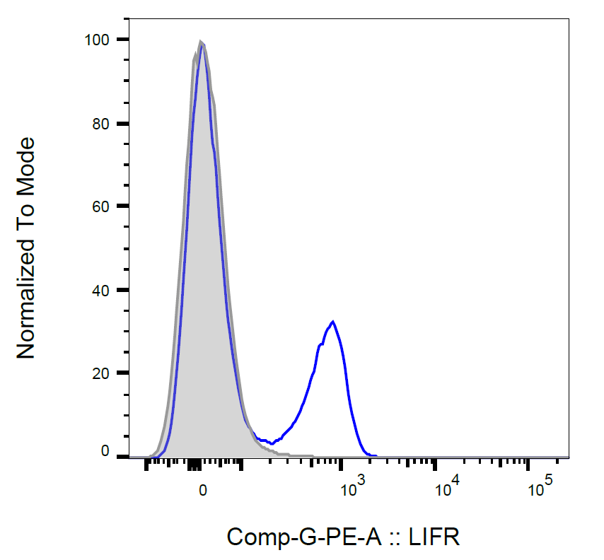
Detection of LIF R alpha in D3 Mouse Cell Line by Flow Cytometry. D3 mouse embryonic stem cell line was stained with Rat Anti-Mouse LIF Ra PE-conjugated Monoclonal Antibody (Catalog # FAB5990P, filled histogram) or isotype control antibody (Catalog # IC005P, open histogram). View our protocol for Staining Membrane-associated Proteins.
 View Larger
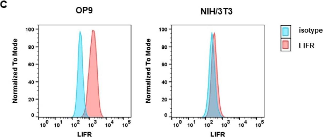
Detection of Mouse LIFR alpha by Flow Cytometry OP9 and NIH/3T3 cells show differential levels of OSMR and LIFR. (A) OP9 and NIH/3T3 cells were examined for LIFR and OSMR expression. BaF3 cells were used as a negative control. Protein molecular weight is labeled in kDa. Relative protein quantities are marked in red. (B) Microarray analysis of OP9 and NIH/3T3 cells showing Osmr and Lifr expression. The p-values were calculated by Transcriptome Analysis Console software. *** p < 0.001. ns = not significant. (C) OP9 and NIH/3T3 cells were analyzed for LIFR surface expression using FACS. (D) OP9 and NIH/3T3 cells were examined for expression of LIFR and OSMR in relation to different incubation periods with 10 ng/mL mOSM. Protein molecular weight is labeled in kDa. (E) OP9 and NIH/3T3 cells were examined for LIFR surface expression in relation to different incubation periods with 10 ng/mL mOSM. p-Values were calculated using one-way ANOVA and Bonferroni post-test. * p <0.05 and *** p < 0.001. ns = not significant. Image collected and cropped by CiteAb from the following open publication (https://pubmed.ncbi.nlm.nih.gov/34769079), licensed under a CC-BY license. Not internally tested by R&D Systems.
 View Larger
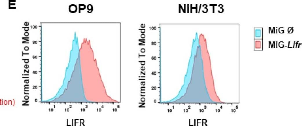
Detection of Mouse LIFR alpha by Flow Cytometry OSMR downregulation attenuates OSM effects on proliferation. (A,B) Immunoblot quantifying the shRNA-induced knockdown of Osmr (n = 4) and Lifr (n = 5) in (A) OP9 and (B) NIH/3T3 cells. The shRNAs Osmr 5 and Lifr 1 were used in subsequent experiments. Protein molecular weight is labeled in kDa. Relative protein quantities are indicated in red. (C) Relative quantification of S phase of OP9 and NIH/3T3 cells carrying a knockdown for either Osmr, Lifr, or a Renilla control in presence or absence of 10 ng/mL mOSM. Cell cycle analysis was performed as described before. One-way ANOVA and Bonferroni post-test. *** p < 0.001. ns = not significant. (D) Immunoblot quantifying the LIFR overexpression in OP9 and NIH/3T3 cells. Protein molecular weight is labeled in kDa. Relative protein quantities are indicated in red. (E) Lifr overexpressing OP9 and NIH/3T3 cells were examined for LIFR surface expression compared to cells carrying the mock control. (F) Relative quantification of S phase of OP9 and NIH/3T3 cells transduced with Lifr or empty vector (MiG). Cell cycle analysis was performed as described before. One-way ANOVA and Bonferroni post-test. * p < 0.05, ** p < 0.01, and *** p < 0.001. Image collected and cropped by CiteAb from the following open publication (https://pubmed.ncbi.nlm.nih.gov/34769079), licensed under a CC-BY license. Not internally tested by R&D Systems.
Reconstitution Calculator
Preparation and Storage
Background: LIFR alpha
Leukemia Inhibitory Factor Receptor alpha (LIF R alpha ), also known as LIFR beta and CD118, is a 185-190 kDa type I transmembrane protein that belongs to the Interleukin-6 receptor family. Members of this family mediate the biological effects of Cardiotrophin-1, CLC, CNTF, IL-6, IL-11, IL-27, and Oncostatin M. Mature mouse LIF R alpha consists of a 785 amino acid (aa) extracellular domain (ECD) with two cytokine receptor homology domains, one WSxWS motif, and three fibronectin type III repeats, followed by a 25 aa transmembrane segment and a 239 aa cytoplasmic domain. Within the ECD, mouse LIF R alpha shares 73% and 90% aa sequence identity with human and rat LIF R alpha, respectively. Alternative splicing generates a 90 kDa soluble form of the mouse LIF R alpha ECD. LIF R alpha binds the pleiotropic cytokine LIF with low affinity, and the soluble isoform retains LIF-binding activity. Binding affinity is increased by the ligand-induced association of LIF R alpha with the signal transducing subunit gp130. The LIF R alpha /gp130 receptor complex also transduces Oncostatin M signals, although LIF R alpha alone does not interact with Oncostatin M. gp130 and LIF R alpha also associate with different ligand-specific receptors to form signaling receptor complexes for other IL-6 family ligands such as CNTF, CT-1 and CLC. The CNTF receptor is a ternary complex that contains CNTF R alpha and gp130 as well as LIF R alpha. LIF R alpha is widely expressed, and cells reported to contain LIF R alpha include hepatic sinusoidal endothelium, steroidal adrenal cortical cells, uterine luminal columnar epithelium, cardiac muscle cells, embryonic stem cells, odontoblasts, forebrain neural precursors, fibroblasts (3T3), osteoblasts, megakaryocytes, osteocytes, activated macrophages and sympathetic neurons. LIF induces the proliferation, differentiation, and activation of cells in many tissues. In particular, LIF R alpha plays an important role in several aspects of early pregnancy such as blastocyst implantation in the uterus.
Product Datasheets
Citations for Mouse LIFR alpha PE-conjugated Antibody
R&D Systems personnel manually curate a database that contains references using R&D Systems products. The data collected includes not only links to publications in PubMed, but also provides information about sample types, species, and experimental conditions.
2
Citations: Showing 1 - 2
Filter your results:
Filter by:
-
ILC2-derived LIF licences progress from tissue to systemic immunity
Authors: Gogoi, M;Clark, PA;Ferreira, ACF;Rodriguez Rodriguez, N;Heycock, M;Ko, M;Murphy, JE;Chen, V;Luan, SL;Jolin, HE;McKenzie, ANJ;
Nature
Species: Transgenic Mouse
Sample Types: Whole Cells
Applications: Flow Cytometry -
Murine Oncostatin M Has Opposing Effects on the Proliferation of OP9 Bone Marrow Stromal Cells and NIH/3T3 Fibroblasts Signaling through the OSMR
Authors: Lena Jakob, Tony Andreas Müller, Michael Rassner, Helen Kleinfelder, Pia Veratti, Jan Mitschke et al.
International Journal of Molecular Sciences
Species: Mouse
Sample Types: Whole Cells
Applications: Flow Cytometry
FAQs
No product specific FAQs exist for this product, however you may
View all Antibody FAQsReviews for Mouse LIFR alpha PE-conjugated Antibody
Average Rating: 5 (Based on 1 Review)
Have you used Mouse LIFR alpha PE-conjugated Antibody?
Submit a review and receive an Amazon gift card.
$25/€18/£15/$25CAN/¥75 Yuan/¥2500 Yen for a review with an image
$10/€7/£6/$10 CAD/¥70 Yuan/¥1110 Yen for a review without an image
Filter by:

