Key Product Details

Species Reactivity

Validated:

Mouse

Cited:

Human, Mouse, Transgenic Mouse

Applications

Validated:

Immunohistochemistry, Western Blot, Flow Cytometry, Immunocytochemistry, CyTOF-ready

Cited:

Immunohistochemistry, Immunohistochemistry-Paraffin, Immunohistochemistry-Frozen, Neutralization, Flow Cytometry, Immunofluorescence, Immunocytochemistry, ELISA Detection, Intravital Microscopy, Labeling, CyTOF-reported

Label

Unconjugated

Antibody Source

Monoclonal Rat IgG2A Clone # 223322
Loading...

Product Specifications

Immunogen

BaF/3 mouse pro-B cell line transfected with mouse LYVE-1
Ala24-Thr234
Accession # Q8BHC0

Specificity

Detects mouse LYVE-1 in direct ELISAs and Western blots. In direct ELISAs and Western blots, no cross-reactivity with recombinant mouse CD44 or recombinant human LYVE-1 is observed.

Clonality

Monoclonal

Host

Rat

Isotype

IgG2A

Endotoxin Level

<0.10 EU per 1 μg of the antibody by the LAL method.

Scientific Data Images for Mouse LYVE‑1 Antibody

Detection of Mouse LYVE-1 antibody by Western Blot.

Detection of Mouse LYVE‑1 by Western Blot.

Western blot shows lysate of bEnd.3 mouse endothelioma cell line. PVDF membrane was probed with 2 µg/mL of Rat Anti-Mouse LYVE-1 Monoclonal Antibody (Catalog # MAB2125) followed by HRP-conjugated Anti-Rat IgG Secondary Antibody (HAF005). A specific band was detected for LYVE-1 at approximately 65 kDa (as indicated). This experiment was conducted under non-reducing conditions and using Immunoblot Buffer Group 1.
Detection of LYVE-1 antibody in bEnd.3 Mouse Cell Line antibody by Flow Cytometry.

Detection of LYVE‑1 in bEnd.3 Mouse Cell Line by Flow Cytometry.

bEnd.3 mouse endothelioma cell line was stained with Rat Anti-Mouse LYVE-1 Monoclonal Antibody (Catalog # MAB2125, filled histogram) or isotype control antibody (MAB006, open histogram), followed by Phycoerythrin-conjugated Anti-Rat IgG Secondary Antibody (F0105B).
LYVE-1 antibody in NS0 mouse myeloma cell line transfected with mouse LYVE-1 by Immunocytochemistry (ICC).

LYVE-1 in NS0 mouse myeloma cell line transfected with mouse LYVE-1.

LYVE-1 was detected in immersion fixed NS0 mouse myeloma cell line transfected with mouse LYVE-1 (positive control) and wild type NS0 mouse myeloma cell line (negative control) using Rat Anti-Mouse LYVE-1 Monoclonal Antibody (Catalog # MAB2125) at 8 µg/mL for 3 hours at room temperature. Cells were stained using the NorthernLights™ 557-conjugated Anti-Rat IgG Secondary Antibody (red; NL013) and counterstained with DAPI (blue). Specific staining was localized to cytoplasm. View our protocol for Fluorescent ICC Staining of Non-adherent Cells.
LYVE-1 antibody in Mouse Intestine by Immunohistochemistry (IHC-Fr).

LYVE-1 in Mouse Intestine.

LYVE-1 was detected in immersion fixed frozen sections of mouse intestine tissue using Rat Anti-Mouse LYVE-1 Monoclonal Antibody (Catalog # MAB2125) at 15 µg/mL for 1 hour at room temperature followed by incubation with the Anti-Rat IgG VisUCyte™ HRP Polymer Antibody (VC005). Tissue was stained using DAB (brown) and counterstained with hematoxylin (blue). Specific staining was localized to endothelial cells in lymphatic vessels. View our protocol for IHC Staining with VisUCyte HRP Polymer Detection Reagents.

Applications for Mouse LYVE‑1 Antibody

Application
Recommended Usage

CyTOF-ready

Ready to be labeled using established conjugation methods. No BSA or other carrier proteins that could interfere with conjugation.

Flow Cytometry

0.25 µg/106 cells
Sample: bEnd.3 mouse endothelioma cell line

Immunocytochemistry

8-25 µg/mL
Sample: Immersion fixed NS0 mouse myeloma cell line transfected with mouse LYVE-1

Immunohistochemistry

5-25 µg/mL
Sample: Immersion fixed frozen sections of mouse intestine tissue

Western Blot

2 µg/mL
Sample: bEnd.3 mouse endothelioma cell line under non-reducing conditions only

Reviewed Applications

Read 3 reviews rated 5 using MAB2125 in the following applications:

Flow Cytometry Panel Builder

Bio-Techne Knows Flow Cytometry

Save time and reduce costly mistakes by quickly finding compatible reagents using the Panel Builder Tool.

Advanced Features

  • Spectra Viewer - Custom analysis of spectra from multiple fluorochromes
  • Spillover Popups - Visualize the spectra of individual fluorochromes
  • Antigen Density Selector - Match fluorochrome brightness with antigen density
Build Your Panel Now

Formulation, Preparation, and Storage

Purification

Protein A or G purified from hybridoma culture supernatant

Reconstitution

Reconstitute at 0.5 mg/mL in sterile PBS. For liquid material, refer to CoA for concentration.


Loading...

Formulation

Lyophilized from a 0.2 μm filtered solution in PBS with Trehalose. See Certificate of Analysis for details.
*Small pack size (-SP) is supplied either lyophilized or as a 0.2 µm filtered solution in PBS.

Shipping

Lyophilized product is shipped at ambient temperature. Liquid small pack size (-SP) is shipped with polar packs. Upon receipt, store immediately at the temperature recommended below.

Stability & Storage

Use a manual defrost freezer and avoid repeated freeze-thaw cycles.
  • 12 months from date of receipt, -20 to -70 °C as supplied.
  • 1 month, 2 to 8 °C under sterile conditions after reconstitution.
  • 6 months, -20 to -70 °C under sterile conditions after reconstitution.

Calculators

The reconstitution calculator allows you to quickly calculate the volume of a reagent to reconstitute your vial. Simply enter the mass of reagent and the target concentration and the calculator will determine the rest.

=
÷

Background: LYVE-1

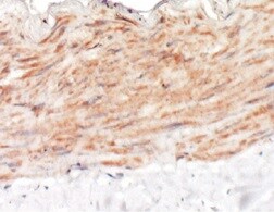
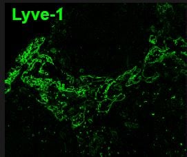
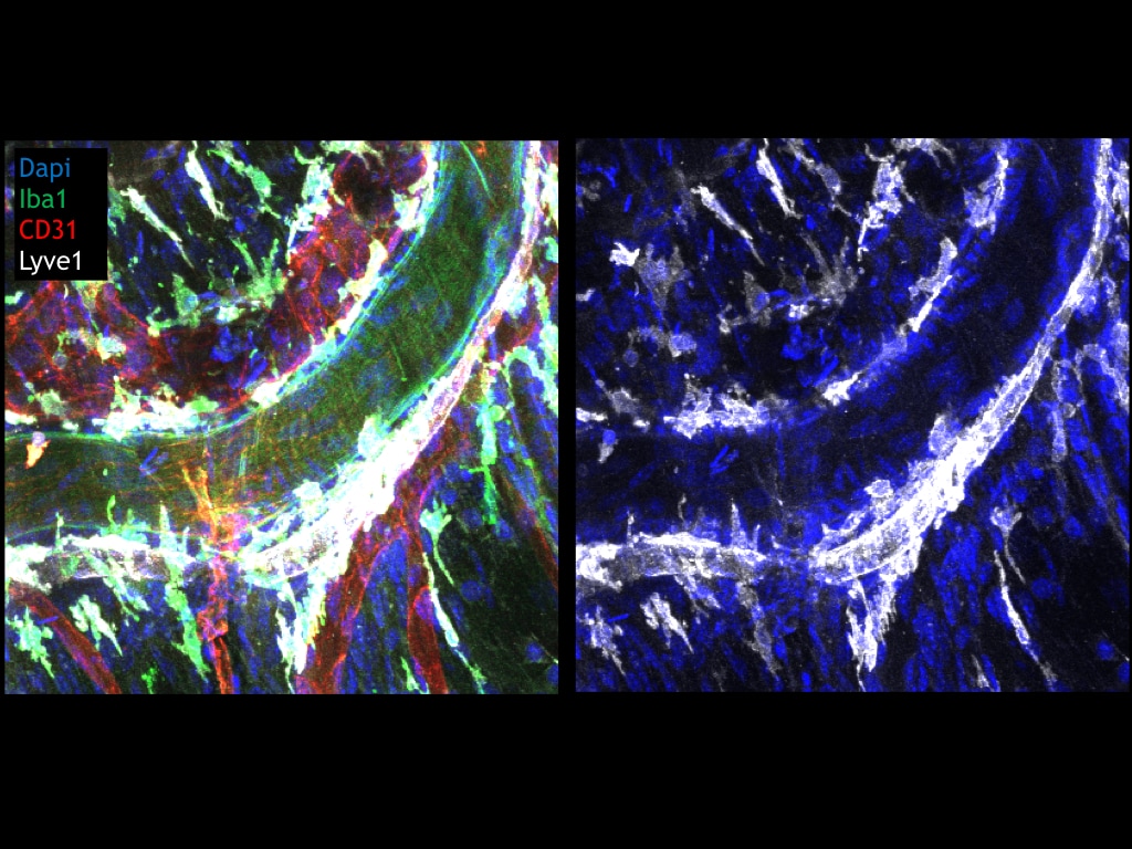
Lymphatic vessel endothelial hyaluronan (HA) receptor-1 (LYVE-1) is a recently identified receptor of HA, a linear high molecular weight polymer composed of alternating units of D-glucuronic acid and N-acetyl-D-glucosamine. HA is found in the extracellular matrix of most animal tissues and in body fluids. It modulates cell behavior and functions during tissue remodeling, development, homeostasis, and disease (1). The turnover of HA (several grams/day in humans) occurs primarily in the lymphatics and liver, the two major clearance systems that catabolize approximately 85% and 15% of HA, respectively (1-3). LYVE-1 shares 41% homology with the other known HA receptor, CD44 (4). The homology between the two proteins increases to 61% within the HA binding domain. The HA binding domain, known as the link module, is a common structural motif found in other HA binding proteins such as link protein, aggrecan and versican (1, 5). Human and mouse LYVE-1 share 69% amino acid sequence identity.

LYVE-1 is primarily expressed on both the luminal and abluminal surfaces of lymphatic vessels (4, 5). In addition, LYVE-1 is also present in normal hepatic blood sinusoidal endothelial cells (6). LYVE-1 mediates the endocytosis of HA and may transport HA from tissue to lymph by transcytosis, delivering HA to lymphatic capillaries for removal and degradation in the regional lymph nodes (5, 7, 8). Because of its restricted expression patterns, LYVE-1, along with other lymphatic proteins such as VEGF R3, podoplanin and the homeobox protein propero-related (Prox-1), constitute a set of markers useful for distinguishing between lymphatic and blood microvasculature (4, 5, 9-11).

References

  1. Knudson, C.B. and W. Knudson (1993) FASEB J. 7:1233.
  2. Evered, D. and J. Whelan (1989) Ciba Found. Symp. 143:1.
  3. Laurent, T.C. and J.R.F. Fraser (1992) FASEB J. 6:2397.
  4. Banerji, S. et al. (1999) J. Cell Biol. 144:789.
  5. Prevo, R. et al. (2001) J. Biol. Chem. 276:19420.
  6. Carreira, C.M. et al. (2001) Cancer Research 61:8079.
  7. Jackson, D.J. et al. (2001) Trends Immunol. 22:317.
  8. Zhou, B. et al. (2000) J. Biol. Chem. 275:37733.
  9. Achen, M. et al. (1998) Proc. Natl. Acad. Sci. USA 95:548.
  10. Breiteneder-Gellef, S. et al. (1999) Am. J. Pathol. 154:385.
  11. Wiggle, J.T. and G. Oliver (1999) Cell 98:769.

Long Name

Lymphatic Vessel Endothelial Hyaluronan Receptor 1

Alternate Names

LYVE1, XLKD1

Entrez Gene IDs

10894 (Human); 114332 (Mouse); 293186 (Rat)

Gene Symbol

LYVE1

UniProt

Additional LYVE-1 Products

Product Documents for Mouse LYVE‑1 Antibody

Certificate of Analysis

To download a Certificate of Analysis, please enter a lot or batch number in the search box below.

Note: Certificate of Analysis not available for kit components.

Product Specific Notices for Mouse LYVE‑1 Antibody

For research use only

Citations for Mouse LYVE‑1 Antibody

Customer Reviews for Mouse LYVE‑1 Antibody (3)

5 out of 5
3 Customer Ratings
5 Stars
100%
4 Stars
0%
3 Stars
0%
2 Stars
0%
1 Stars
0%

Have you used Mouse LYVE‑1 Antibody?

Submit a review and receive an Amazon gift card!

$25/€18/£15/$25CAN/¥2500 Yen for a review with an image

$10/€7/£6/$10CAN/¥1110 Yen for a review without an image

Submit a review
Amazon Gift Card

Customer Images


Showing  1 - 3 of 3 reviews Showing All
Filter By:
  • Mouse LYVE-1 Antibody
    Name: Anonymous
    Application: Immunohistochemistry
    Sample Tested: optic nerve
    Species: Mouse
    Verified Customer | Posted 08/17/2021
    Mouse LYVE‑1 Antibody MAB2125
  • Mouse LYVE-1 Antibody
    Name: Anonymous
    Application: Immunocytochemistry/Immunofluorescence
    Sample Tested: Lymph node tissue
    Species: Mouse
    Verified Customer | Posted 12/07/2020
    LN were fixed in 4% PFA, sectioned and blocked/permeabilized with 0.1% Tx-100, 0.01% NaN3, 1% FBS. Sections were stained overnight (MAB2125) at a dilution of 1:200. Antibody was detected using a secondary FITC-anti rat (1:500).
    Mouse LYVE‑1 Antibody MAB2125
  • Mouse LYVE-1 Antibody
    Name: Anonymous
    Application: Immunohistochemistry
    Sample Tested: Meninges
    Species: Mouse
    Verified Customer | Posted 10/03/2019
    Mouse meninges stained with Lyve1 (1:100, white), CD31 (red), iba1 (green), dapi. Lyve1 labels lymphatic vessels as well as a subset of macrophages.
    Mouse LYVE‑1 Antibody MAB2125

There are no reviews that match your criteria.

Showing  1 - 3 of 3 reviews Showing All

Protocols

Find general support by application which include: protocols, troubleshooting, illustrated assays, videos and webinars.

FAQs

No product specific FAQs exist for this product.

View all FAQs for Antibodies
Loading...