PLUNC (Palate, lung, and nasal epithelium clone; also SPLUNC1) is a 25-30 kDa member of the PLUNC family, BPI/PLUNC/LBP superfamily of proteins. PLUNC is a secreted glycoprotein that is expressed by both adult and embryonic respiratory epithelium in the nose, trachea and bronchi/primary bronchioles. It serves as a natural antibacterial polypeptide in the respiratory system. Mature mouse PLUNC is 259 amino acids (aa) in length. It contains four, six-amino acid repeats (GxxLPL) over aa 23-52. There is reportedly one splice variant that shows a 28 aa substitution for the C-terminal 45 amino acids (aa 234-278). Over aa 59-278, mouse PLUNC shares 91% and 75% aa identity with rat and human PLUNC, respectively.

Key Product Details
Validated by
Biological Validation
Species Reactivity
Validated:
Mouse
Cited:
Mouse, Transgenic Mouse
Applications
Validated:
Immunohistochemistry, Western Blot
Cited:
Immunohistochemistry, Western Blot
Label
Unconjugated
Antibody Source
Polyclonal Sheep IgG
Loading...
Product Specifications
Immunogen
E. coli-derived recombinant mouse PLUNC
Ala59-Val278
Accession # NP_035256
Ala59-Val278
Accession # NP_035256
Specificity
Detects mouse PLUNC in direct ELISAs and Western blots. In these formats, less than 10% cross-reactivity with recombinant human PLUNC is observed.
Clonality
Polyclonal
Host
Sheep
Isotype
IgG
Scientific Data Images for Mouse PLUNC Antibody
Detection of Mouse PLUNC by Western Blot.
Western blot shows lysates of mouse lung tissue. PVDF membrane was probed with 1 µg/mL of Sheep Anti-Mouse PLUNC Antigen Affinity-purified Polyclonal Antibody (Catalog # AF4274) followed by HRP-conjugated Anti-Sheep IgG Secondary Antibody (Catalog # HAF016). A specific band was detected for PLUNC at approximately 30kDa (as indicated). This experiment was conducted under reducing conditions and using Immunoblot Buffer Group 8.
PLUNC in Mouse Embryo.
PLUNC was detected in immersion fixed frozen sections of mouse embryo nose (13 d.p.c.) using Sheep Anti-Mouse PLUNC Antigen Affinity-purified Polyclonal Antibody (Catalog # AF4274) at 1.7 µg/mL overnight at 4 °C. Tissue was stained using the Anti-Sheep HRP-DAB Cell & Tissue Staining Kit (brown; Catalog # CTS019) and counterstained with hematoxylin (blue). Specific staining was localized to olfactory epithelium. View our protocol for Chromogenic IHC Staining of Frozen Tissue Sections.
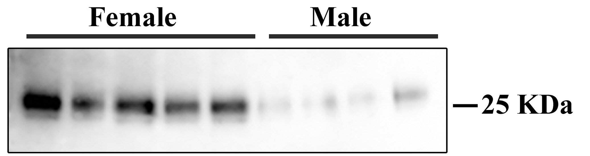
Detection of Mouse PLUNC by Western Blot
A1AT treatment enhances SPLUNC1 and reduces NE activity in bronchoalveolar lavage (BAL) fluid of PA-infected WT mice. PA-infected mice were treated with A1AT for 22 hrs and sacrificed after 24 hrs of infection as described in Materials and Methods. Quantitative analysis (A) and representative image (B) of BAL fluid SPLUNC1 Western blot were shown to demonstrate the therapeutic effect of A1AT treatment. The two lanes of Western blot image under saline or PA treatment represent SPLUNC1 data from two different mice. NE activity (C) was examined by an NE activity assay. N = 4 – 7 mice per group. The vertical dotted red line in Figure 2B separates the saline group from the PA infection group. NS indicates no significant differences. Data are expressed as means ± SEM. Image collected and cropped by CiteAb from the following publication (https://respiratory-research.biomedcentral.com/articles/10.1186/1465-99…), licensed under a CC-BY license. Not internally tested by R&D Systems.
Detection of Mouse PLUNC by Western Blot
Pseudomonas aeruginosa (PA) infection reduces SPLUNC1 and increases neutrophil elastase (NE) activity in bronchoalveolar lavage (BAL) fluid of wild-type (WT) mice. BAL fluid from WT mice was processed for SPLUNC1 protein Western blot. (A) – Quantitative analysis of BAL fluid SPLUNC1 protein expression normalized by albumin. (B) – Representative Western blot image of SPLUNC1 and albumin. The vertical dotted red line separates the saline group from the PA infection group. The two lanes under saline or PA treatment represent SPLUNC1 data from two different mice. (C) – NE activity was examined by an NE activity assay as described in Materials and methods. N = 4 – 5 mice per group. Data are expressed as means ± SEM. Image collected and cropped by CiteAb from the following publication (https://respiratory-research.biomedcentral.com/articles/10.1186/1465-99…), licensed under a CC-BY license. Not internally tested by R&D Systems.Applications for Mouse PLUNC Antibody
Application
Recommended Usage
Immunohistochemistry
5-15 µg/mL
Sample: Immersion fixed frozen cross-sections of mouse embryo nose (13 d.p.c.)
Sample: Immersion fixed frozen cross-sections of mouse embryo nose (13 d.p.c.)
Western Blot
1 µg/mL
Sample: Mouse lung tissue
Sample: Mouse lung tissue
Reviewed Applications
Read 1 review rated 5 using AF4274 in the following applications:
Formulation, Preparation, and Storage
Purification
Antigen Affinity-purified
Reconstitution
Reconstitute at 0.2 mg/mL in sterile PBS. For liquid material, refer to CoA for concentration.
Loading...
Formulation
Lyophilized from a 0.2 μm filtered solution in PBS with Trehalose. *Small pack size (SP) is supplied either lyophilized or as a 0.2 µm filtered solution in PBS.
Shipping
Lyophilized product is shipped at ambient temperature. Liquid small pack size (-SP) is shipped with polar packs. Upon receipt, store immediately at the temperature recommended below.
Stability & Storage
Use a manual defrost freezer and avoid repeated freeze-thaw cycles.
- 12 months from date of receipt, -20 to -70 °C as supplied.
- 1 month, 2 to 8 °C under sterile conditions after reconstitution.
- 6 months, -20 to -70 °C under sterile conditions after reconstitution.
Calculators
Background: PLUNC
Long Name
Palate, Lung, and Nasal Epithelium Associated
Alternate Names
BPIFA1, LUNX, SPLUNC1
Entrez Gene IDs
51297 (Human)
Gene Symbol
BPIFA1
UniProt
Additional PLUNC Products
Product Documents for Mouse PLUNC Antibody
Certificate of Analysis
To download a Certificate of Analysis, please enter a lot or batch number in the search box below.
Note: Certificate of Analysis not available for kit components.
Product Specific Notices for Mouse PLUNC Antibody
For research use only
Related Research Areas
Citations for Mouse PLUNC Antibody
 Powered by Bioz
Customer Reviews for Mouse PLUNC Antibody (1)
5 out of 5
1 Customer Rating
Have you used Mouse PLUNC Antibody?
Submit a review and receive an Amazon gift card!
$25/€18/£15/$25CAN/¥2500 Yen for a review with an image
$10/€7/£6/$10CAN/¥1110 Yen for a review without an image
Submit a review
Customer Images
Showing
1
-
1 of
1 review
Showing All
Filter By:
-
Application: Western BlotSample Tested: Cell LysatesSpecies: MouseVerified Customer | Posted 02/19/2021

There are no reviews that match your criteria.
Protocols
Find general support by application which include: protocols, troubleshooting, illustrated assays, videos and webinars.
- Antigen Retrieval Protocol (PIER)
- Antigen Retrieval for Frozen Sections Protocol
- Appropriate Fixation of IHC/ICC Samples
- Cellular Response to Hypoxia Protocols
- Chromogenic IHC Staining of Formalin-Fixed Paraffin-Embedded (FFPE) Tissue Protocol
- Chromogenic Immunohistochemistry Staining of Frozen Tissue
- ClariTSA™ Fluorophore Kits
- Detection & Visualization of Antibody Binding
- Fluorescent IHC Staining of Frozen Tissue Protocol
- Graphic Protocol for Heat-induced Epitope Retrieval
- Graphic Protocol for the Preparation and Fluorescent IHC Staining of Frozen Tissue Sections
- Graphic Protocol for the Preparation and Fluorescent IHC Staining of Paraffin-embedded Tissue Sections
- Graphic Protocol for the Preparation of Gelatin-coated Slides for Histological Tissue Sections
- IHC Sample Preparation (Frozen sections vs Paraffin)
- Immunofluorescent IHC Staining of Formalin-Fixed Paraffin-Embedded (FFPE) Tissue Protocol
- Immunohistochemistry (IHC) and Immunocytochemistry (ICC) Protocols
- Immunohistochemistry Frozen Troubleshooting
- Immunohistochemistry Paraffin Troubleshooting
- Preparing Samples for IHC/ICC Experiments
- Preventing Non-Specific Staining (Non-Specific Binding)
- Primary Antibody Selection & Optimization
- Protocol for Heat-Induced Epitope Retrieval (HIER)
- Protocol for Making a 4% Formaldehyde Solution in PBS
- Protocol for VisUCyte™ HRP Polymer Detection Reagent
- Protocol for the Preparation & Fixation of Cells on Coverslips
- Protocol for the Preparation and Chromogenic IHC Staining of Frozen Tissue Sections
- Protocol for the Preparation and Chromogenic IHC Staining of Frozen Tissue Sections - Graphic
- Protocol for the Preparation and Chromogenic IHC Staining of Paraffin-embedded Tissue Sections
- Protocol for the Preparation and Chromogenic IHC Staining of Paraffin-embedded Tissue Sections - Graphic
- Protocol for the Preparation and Fluorescent IHC Staining of Frozen Tissue Sections
- Protocol for the Preparation and Fluorescent IHC Staining of Paraffin-embedded Tissue Sections
- Protocol for the Preparation of Gelatin-coated Slides for Histological Tissue Sections
- R&D Systems Quality Control Western Blot Protocol
- TUNEL and Active Caspase-3 Detection by IHC/ICC Protocol
- The Importance of IHC/ICC Controls
- Troubleshooting Guide: Immunohistochemistry
- Troubleshooting Guide: Western Blot Figures
- Western Blot Conditions
- Western Blot Protocol
- Western Blot Protocol for Cell Lysates
- Western Blot Troubleshooting
- Western Blot Troubleshooting Guide
- View all Protocols, Troubleshooting, Illustrated assays and Webinars
Loading...