KLK7 (Kallikrein 7; also Serine protease 6, Thymopsin and Stratum corneum chymotryptic enzyme/SCCE) is a 27 kDa (predicted), secreted glycoprotein member of the kallikrein subfamily, peptidase S1 family of enzymes. It is an IL-4 and IL-13-inducible chymotrypsin-like serine protease that has restricted expression, being associated with stratum spinosum and granulosum keratinocytes, distal and proximal convoluted tubule epithelium, limited pancreatic acinar cells. In contrast, KLK7 is widely expressed by a variety of carcinoma cell types. KLK7 preferentially cleaves peptide bonds that contain an aromatic (Tyr or Phe) residue in the P1 (or postcleavage C-terminal amino acid) position. Molecules known to serve as substrates for KLK7 include chemerin, fibronectin, E-cadherin, LL37, procaspase-14, MMP-9 and desmoglein. The nature of the KLK7 substrates indicates that it plays a key role in intercellular adhesion dissolution with subsequent cell migration. Mouse KLK7 is synthesized as a 249 amino acid (aa) precursor. It contains a 21 aa signal sequence, a four aa prosegment, and a 224 aa mature region (aa 26-249). The mature molecule contains one large protease domain (aa 26-246) and a variable, three aa C-terminus that possesses either a VSW or ASR motif. Over aa 22-249, mouse KLK7 shares 75% and 85% aa sequence identity with human and hamster KLK7, respectively.

Key Product Details
Species Reactivity
Mouse, Rat
Applications
Immunohistochemistry, Western Blot
Label
Unconjugated
Antibody Source
Polyclonal Sheep IgG
Loading...
Product Specifications
Immunogen
Mouse myeloma cell line NS0-derived recombinant mouse Kallikrein 7
Gln22-Arg249 (predicted)
Accession # Q91VE3
Gln22-Arg249 (predicted)
Accession # Q91VE3
Specificity
Detects mouse and rat Kallikrein 7 in Western blots. Detects mouse Kallikrein 7 in direct ELISAs. In direct ELISAs, approximately 10% cross-reactivity with recombinant human Kallikrein 7 is observed, and less than 1% cross-reactivity with recombinant mouse Kallikrein 5 is observed.
Clonality
Polyclonal
Host
Sheep
Isotype
IgG
Scientific Data Images for Kallikrein 7 Antibody
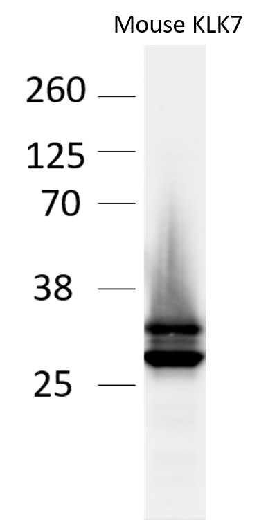
Detection of Mouse Kallikrein 7 by Western Blot.
Western blot shows lysates of mouse skin tissue. PVDF membrane was probed with 2 µg/mL of Sheep Anti-Mouse Kallikrein 7 Antigen Affinity-purified Polyclonal Antibody (Catalog # AF7688) followed by HRP-conjugated Anti-Sheep IgG Secondary Antibody (Catalog # HAF016). A specific band was detected for Kallikrein 7 at approximately 27 kDa (as indicated). This experiment was conducted under reducing conditions and using Immunoblot Buffer Group 1.
Detection of Rat Kallikrein 7 by Western Blot.
Western blot shows lysates of rat skin tissue. PVDF membrane was probed with 1 µg/mL of Sheep Anti-Mouse Kallikrein 7 Antigen Affinity-purified Polyclonal Antibody (Catalog # AF7688) followed by HRP-conjugated Anti-Sheep IgG Secondary Antibody (Catalog # HAF016). A specific band was detected for Kallikrein 7 at approximately 27 kDa (as indicated). This experiment was conducted under reducing conditions and using Immunoblot Buffer Group 1.
Kallikrein 7 in Mouse Skin.
Kallikrein 7 was detected in perfusion fixed frozen sections of mouse skin using Sheep Anti-Mouse Kallikrein 7 Antigen Affinity-purified Polyclonal Antibody (Catalog # AF7688) at 5 µg/mL overnight at 4 °C. Tissue was stained using the NorthernLights™ 557-conjugated Anti-Sheep IgG Secondary Antibody (red; Catalog # NL010) and counterstained with DAPI (blue). Specific staining was localized to keratinocytes. View our protocol for Fluorescent IHC Staining of Frozen Tissue Sections.Applications for Kallikrein 7 Antibody
Application
Recommended Usage
Immunohistochemistry
5-15 µg/mL
Sample: Perfusion fixed frozen sections of mouse skin
Sample: Perfusion fixed frozen sections of mouse skin
Western Blot
1-2 µg/mL
Sample: Mouse skin tissue and rat skin tissue
Sample: Mouse skin tissue and rat skin tissue
Reviewed Applications
Read 1 review rated 3 using AF7688 in the following applications:
Formulation, Preparation, and Storage
Purification
Antigen Affinity-purified
Reconstitution
Reconstitute at 0.2 mg/mL in sterile PBS. For liquid material, refer to CoA for concentration.
Loading...
Formulation
Lyophilized from a 0.2 μm filtered solution in PBS with Trehalose. *Small pack size (SP) is supplied either lyophilized or as a 0.2 µm filtered solution in PBS.
Shipping
Lyophilized product is shipped at ambient temperature. Liquid small pack size (-SP) is shipped with polar packs. Upon receipt, store immediately at the temperature recommended below.
Stability & Storage
Use a manual defrost freezer and avoid repeated freeze-thaw cycles.
- 12 months from date of receipt, -20 to -70 °C as supplied.
- 1 month, 2 to 8 °C under sterile conditions after reconstitution.
- 6 months, -20 to -70 °C under sterile conditions after reconstitution.
Calculators
Background: Kallikrein 7
Alternate Names
KLK7
Gene Symbol
KLK7
UniProt
Additional Kallikrein 7 Products
Product Documents for Kallikrein 7 Antibody
Certificate of Analysis
To download a Certificate of Analysis, please enter a lot or batch number in the search box below.
Note: Certificate of Analysis not available for kit components.
Product Specific Notices for Kallikrein 7 Antibody
For research use only
Customer Reviews for Kallikrein 7 Antibody (1)
3 out of 5
1 Customer Rating
Have you used Kallikrein 7 Antibody?
Submit a review and receive an Amazon gift card!
$25/€18/£15/$25CAN/¥2500 Yen for a review with an image
$10/€7/£6/$10CAN/¥1110 Yen for a review without an image
Submit a review
Customer Images
Showing
1
-
1 of
1 review
Showing All
Filter By:
-
Application: Western BlotSample Tested: mouse tissue and recombinant mouse KLK7 proteinSpecies: MouseVerified Customer | Posted 05/15/2018

There are no reviews that match your criteria.
Protocols
Find general support by application which include: protocols, troubleshooting, illustrated assays, videos and webinars.
- Antigen Retrieval Protocol (PIER)
- Antigen Retrieval for Frozen Sections Protocol
- Appropriate Fixation of IHC/ICC Samples
- Cellular Response to Hypoxia Protocols
- Chromogenic IHC Staining of Formalin-Fixed Paraffin-Embedded (FFPE) Tissue Protocol
- Chromogenic Immunohistochemistry Staining of Frozen Tissue
- ClariTSA™ Fluorophore Kits
- Detection & Visualization of Antibody Binding
- Fluorescent IHC Staining of Frozen Tissue Protocol
- Graphic Protocol for Heat-induced Epitope Retrieval
- Graphic Protocol for the Preparation and Fluorescent IHC Staining of Frozen Tissue Sections
- Graphic Protocol for the Preparation and Fluorescent IHC Staining of Paraffin-embedded Tissue Sections
- Graphic Protocol for the Preparation of Gelatin-coated Slides for Histological Tissue Sections
- IHC Sample Preparation (Frozen sections vs Paraffin)
- Immunofluorescent IHC Staining of Formalin-Fixed Paraffin-Embedded (FFPE) Tissue Protocol
- Immunohistochemistry (IHC) and Immunocytochemistry (ICC) Protocols
- Immunohistochemistry Frozen Troubleshooting
- Immunohistochemistry Paraffin Troubleshooting
- Preparing Samples for IHC/ICC Experiments
- Preventing Non-Specific Staining (Non-Specific Binding)
- Primary Antibody Selection & Optimization
- Protocol for Heat-Induced Epitope Retrieval (HIER)
- Protocol for Making a 4% Formaldehyde Solution in PBS
- Protocol for VisUCyte™ HRP Polymer Detection Reagent
- Protocol for the Preparation & Fixation of Cells on Coverslips
- Protocol for the Preparation and Chromogenic IHC Staining of Frozen Tissue Sections
- Protocol for the Preparation and Chromogenic IHC Staining of Frozen Tissue Sections - Graphic
- Protocol for the Preparation and Chromogenic IHC Staining of Paraffin-embedded Tissue Sections
- Protocol for the Preparation and Chromogenic IHC Staining of Paraffin-embedded Tissue Sections - Graphic
- Protocol for the Preparation and Fluorescent IHC Staining of Frozen Tissue Sections
- Protocol for the Preparation and Fluorescent IHC Staining of Paraffin-embedded Tissue Sections
- Protocol for the Preparation of Gelatin-coated Slides for Histological Tissue Sections
- R&D Systems Quality Control Western Blot Protocol
- TUNEL and Active Caspase-3 Detection by IHC/ICC Protocol
- The Importance of IHC/ICC Controls
- Troubleshooting Guide: Immunohistochemistry
- Troubleshooting Guide: Western Blot Figures
- Western Blot Conditions
- Western Blot Protocol
- Western Blot Protocol for Cell Lysates
- Western Blot Troubleshooting
- Western Blot Troubleshooting Guide
- View all Protocols, Troubleshooting, Illustrated assays and Webinars
Loading...