Uromodulin (also Tamm-Horsfall glycoprotein or THP) is a 105‑120 kDa urinary glycoprotein. It is secreted by renal tubule epithelium, acts as a binding protein for IL‑1, TNF-alpha and C1q, activates resting monocytes, and promotes neutrophil phagocytosis. Uromodulin forms high molecular weight oligomers that line the kidney tubules. Mouse Uromodulin is GPI-linked. Its proprecursor is 619 amino acids (aa) in length. It contains three EGF-like domains (aa 28‑148), a ZP domain that mediates oligomerization (aa 335‑590), and a cleavable C-terminal propeptide (aa 619‑642). There are multiple splice variants. One shows an alternate start site at Met343, and there are two substitutions, a 17 aa substitution for aa 601‑642, and a 166 aa substitution for aa 441‑642. Over aa 25‑618, mouse Uromodulin shares 78% and 89% aa identical to human and rat Uromodulin, respectively.
Mouse Uromodulin Antibody (774056)
R&D Systems | Catalog # MAB5175

Key Product Details
Species Reactivity
Validated:
Mouse
Cited:
Mouse
Applications
Validated:
Immunohistochemistry, Western Blot
Cited:
Immunohistochemistry, Immunohistochemistry-Paraffin, Immunohistochemistry-Frozen, Western Blot
Label
Unconjugated
Antibody Source
Monoclonal Rat IgG2A Clone # 774056
Loading...
Product Specifications
Immunogen
Mouse myeloma cell line NS0-derived recombinant mouse Uromodulin
Ser24-Ala618
Accession # Q91X17
Ser24-Ala618
Accession # Q91X17
Specificity
Detects mouse Uromodulin in direct ELISAs and Western blots. In direct ELISAs, 100% cross-reactivity with
recombinant human Uromodulin is observed.
Clonality
Monoclonal
Host
Rat
Isotype
IgG2A
Scientific Data Images for Mouse Uromodulin Antibody (774056)
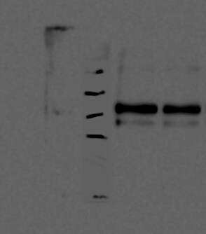
Detection of Mouse Uromodulin by Western Blot.
Western blot shows lysates of mouse kidney tissue. PVDF membrane was probed with 0.5 µg/mL of Rat Anti-Mouse Uromodulin Monoclonal Antibody (Catalog # MAB5175) followed by HRP-conjugated Anti-Rat IgG Secondary Antibody (Catalog # HAF005). A specific band was detected for Uromodulin at approximately 115 kDa (as indicated). This experiment was conducted under reducing conditions and using Immunoblot Buffer Group 8.
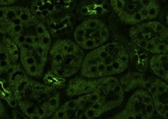
Uromodulin in Mouse Kidney.
Uromodulin was detected in immersion fixed frozen sections of mouse kidney using Rat Anti-Mouse Uromodulin Monoclonal Antibody (Catalog # MAB5175) at 10 µg/mL overnight at 4 °C. Tissue was stained using the NorthernLights™ 557-conjugated Anti-Rat IgG Secondary Antibody (red; Catalog # NL013) and counterstained with DAPI (blue). Specific staining was localized to convoluted tubule epithelial cells. View our protocol for Fluorescent IHC Staining of Frozen Tissue Sections.
Detection of Uromodulin by Western Blot
Tolvaptan induces Nrf2 nuclear translocation and HO-1 expression in vivo. (a) Tolvaptan promotes Nrf2 nuclear translocation in the outer medulla of mouse kidneys. (Top panel) Male C57BL/6J mice aged 8–9-weeks were fed with a diet containing 0.5% tolvaptan for 24 h. The nuclear extract was obtained from the outer medulla. Arrow indicates the band of Nrf2. (Bottom panel) Nrf2 bands were quantified using densitometric analysis. Bar indicates average from four or five experiments (control: n = 4, tolvaptan: n = 5). Student’s t-test, **P < 0.01. C: control, Tol: 0.5% tolvaptan. (b) Tolvaptan increases HO-1 protein expression in the outer medulla of mouse kidneys. (Top panel) Tolvaptan (0.5%) was administered via diet to 8–9-week-old male C57BL/6J mice for 24 h. The kidneys were separated into the cortex and the outer medulla, and the whole protein extraction was performed. The blots of HO-1 and Actin were configured with the blot of cortex and that of outer medulla. Fractionation of the cortex and the outer medulla was verified using UMOD and pNCC antibodies. (Bottom panel) Densitometric analysis of HO-1 is shown. Bar indicates average from four or five experiments (control: n = 4, tolvaptan: n = 5). Student’s t-test, *P < 0.05. C: control, Tol: 0.5% tolvaptan. Image collected and cropped by CiteAb from the following open publication (https://pubmed.ncbi.nlm.nih.gov/31239473), licensed under a CC-BY license. Not internally tested by R&D Systems.Applications for Mouse Uromodulin Antibody (774056)
Application
Recommended Usage
Immunohistochemistry
8-25 µg/mL
Sample: Immersion fixed frozen sections of mouse kidney
Sample: Immersion fixed frozen sections of mouse kidney
Western Blot
0.5 µg/mL
Sample: Mouse kidney tissue
Sample: Mouse kidney tissue
Reviewed Applications
Read 2 reviews rated 5 using MAB5175 in the following applications:
Formulation, Preparation, and Storage
Purification
Protein A or G purified from hybridoma culture supernatant
Reconstitution
Sterile PBS to a final concentration of 0.5 mg/mL. For liquid material, refer to CoA for concentration.
Loading...
Formulation
Lyophilized from a 0.2 μm filtered solution in PBS with Trehalose. *Small pack size (SP) is supplied either lyophilized or as a 0.2 µm filtered solution in PBS.
Shipping
Lyophilized product is shipped at ambient temperature. Liquid small pack size (-SP) is shipped with polar packs. Upon receipt, store immediately at the temperature recommended below.
Stability & Storage
Use a manual defrost freezer and avoid repeated freeze-thaw cycles.
- 12 months from date of receipt, -20 to -70 °C as supplied.
- 1 month, 2 to 8 °C under sterile conditions after reconstitution.
- 6 months, -20 to -70 °C under sterile conditions after reconstitution.
Calculators
Background: Uromodulin
Alternate Names
ADMCKD2, FJHN, HNFJ, MCKD2, THGP, THP, UMOD, Uromucoid
Gene Symbol
UMOD
UniProt
Additional Uromodulin Products
Product Documents for Mouse Uromodulin Antibody (774056)
Certificate of Analysis
To download a Certificate of Analysis, please enter a lot or batch number in the search box below.
Note: Certificate of Analysis not available for kit components.
Product Specific Notices for Mouse Uromodulin Antibody (774056)
For research use only
Citations for Mouse Uromodulin Antibody (774056)
 Powered by Bioz
Customer Reviews for Mouse Uromodulin Antibody (774056) (2)
5 out of 5
2 Customer Ratings
Have you used Mouse Uromodulin Antibody (774056)?
Submit a review and receive an Amazon gift card!
$25/€18/£15/$25CAN/¥2500 Yen for a review with an image
$10/€7/£6/$10CAN/¥1110 Yen for a review without an image
Submit a review
Customer Images
Showing
1
-
2 of
2 reviews
Showing All
Filter By:
-
Application: ImmunofluorescenceSample Tested: Kidney tissueSpecies: MouseVerified Customer | Posted 10/25/2021

-
Application: Western BlotSample Tested: Kidney tissueSpecies: MouseVerified Customer | Posted 05/24/2017- loaded 30 ug of total protein from mouse kidney lysate - wet transfer (100V, 1h) - used buffers as described by datasheet - 1:1500 dilution of UMOD antibody in 0.5% BSA - 1:10000 dilution of rat IgG antibody (HAF005) in 3% BSA
There are no reviews that match your criteria.
Protocols
Find general support by application which include: protocols, troubleshooting, illustrated assays, videos and webinars.
- Antigen Retrieval Protocol (PIER)
- Antigen Retrieval for Frozen Sections Protocol
- Appropriate Fixation of IHC/ICC Samples
- Cellular Response to Hypoxia Protocols
- Chromogenic IHC Staining of Formalin-Fixed Paraffin-Embedded (FFPE) Tissue Protocol
- Chromogenic Immunohistochemistry Staining of Frozen Tissue
- ClariTSA™ Fluorophore Kits
- Detection & Visualization of Antibody Binding
- Fluorescent IHC Staining of Frozen Tissue Protocol
- Graphic Protocol for Heat-induced Epitope Retrieval
- Graphic Protocol for the Preparation and Fluorescent IHC Staining of Frozen Tissue Sections
- Graphic Protocol for the Preparation and Fluorescent IHC Staining of Paraffin-embedded Tissue Sections
- Graphic Protocol for the Preparation of Gelatin-coated Slides for Histological Tissue Sections
- IHC Sample Preparation (Frozen sections vs Paraffin)
- Immunofluorescent IHC Staining of Formalin-Fixed Paraffin-Embedded (FFPE) Tissue Protocol
- Immunohistochemistry (IHC) and Immunocytochemistry (ICC) Protocols
- Immunohistochemistry Frozen Troubleshooting
- Immunohistochemistry Paraffin Troubleshooting
- Preparing Samples for IHC/ICC Experiments
- Preventing Non-Specific Staining (Non-Specific Binding)
- Primary Antibody Selection & Optimization
- Protocol for Heat-Induced Epitope Retrieval (HIER)
- Protocol for Making a 4% Formaldehyde Solution in PBS
- Protocol for VisUCyte™ HRP Polymer Detection Reagent
- Protocol for the Preparation & Fixation of Cells on Coverslips
- Protocol for the Preparation and Chromogenic IHC Staining of Frozen Tissue Sections
- Protocol for the Preparation and Chromogenic IHC Staining of Frozen Tissue Sections - Graphic
- Protocol for the Preparation and Chromogenic IHC Staining of Paraffin-embedded Tissue Sections
- Protocol for the Preparation and Chromogenic IHC Staining of Paraffin-embedded Tissue Sections - Graphic
- Protocol for the Preparation and Fluorescent IHC Staining of Frozen Tissue Sections
- Protocol for the Preparation and Fluorescent IHC Staining of Paraffin-embedded Tissue Sections
- Protocol for the Preparation of Gelatin-coated Slides for Histological Tissue Sections
- R&D Systems Quality Control Western Blot Protocol
- TUNEL and Active Caspase-3 Detection by IHC/ICC Protocol
- The Importance of IHC/ICC Controls
- Troubleshooting Guide: Immunohistochemistry
- Troubleshooting Guide: Western Blot Figures
- Western Blot Conditions
- Western Blot Protocol
- Western Blot Protocol for Cell Lysates
- Western Blot Troubleshooting
- Western Blot Troubleshooting Guide
- View all Protocols, Troubleshooting, Illustrated assays and Webinars
Loading...