N-Cadherin Antibody (5D5)
Novus Biologicals | Catalog # NBP1-51612
![Immunohistochemistry-Paraffin: N-Cadherin Antibody (5D5) - BSA Free [NBP1-51612]](https://resources.rndsystems.com/images/products/N-Cadherin-Antibody-5D5-Immunohistochemistry-Paraffin-NBP1-51612-img0015.jpg "Immunohistochemistry-Paraffin: N-Cadherin Antibody (5D5) - BSA Free [NBP1-51612]")
Loading...
Key Product Details
Species Reactivity
Human, Mouse, Rat
Applications
Validated:
Immunohistochemistry, Immunohistochemistry-Paraffin, Western Blot, ELISA, Flow Cytometry, Immunocytochemistry/ Immunofluorescence
Cited:
Western Blot
Label
Unconjugated
Antibody Source
Monoclonal Mouse IgG1 Clone # 5D5
Loading...
Product Specifications
Immunogen
Purified recombinant fragment of human N-Cadherin expressed in E. Coli.
Reactivity Notes
Please note that this antibody is reactive to Mouse and derived from the same host, Mouse. Mouse-On-Mouse blocking reagent may be needed for IHC and ICC experiments to reduce high background signal. You can find these reagents under catalog numbers PK-2200-NB and MP-2400-NB. Please contact Technical Support if you have any questions.
Marker
Mesenchymal Cells Marker
Clonality
Monoclonal
Host
Mouse
Isotype
IgG1
Theoretical MW
99.8 kDa.
Disclaimer note: The observed molecular weight of the protein may vary from the listed predicted molecular weight due to post translational modifications, post translation cleavages, relative charges, and other experimental factors.
Disclaimer note: The observed molecular weight of the protein may vary from the listed predicted molecular weight due to post translational modifications, post translation cleavages, relative charges, and other experimental factors.
Scientific Data Images for N-Cadherin Antibody (5D5)
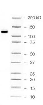
Immunohistochemistry-Paraffin: N-Cadherin Antibody (5D5) - BSA Free [NBP1-51612]
Immunohistochemistry-Paraffin: N-Cadherin Antibody (5D5) [NBP1-51612] - Analysis of human lung cancer (A), colon cancer (B), ovarian cancer (C) and mammary cancer(D), using CDH2 mouse mAb with DAB staining.![Western Blot: N-Cadherin Antibody (5D5)BSA Free [NBP1-51612]](https://resources.rndsystems.com/images/products/N-Cadherin-Antibody-5D5-Western-Blot-NBP1-51612-img0016.jpg "Western Blot: N-Cadherin Antibody (5D5)BSA Free [NBP1-51612]")
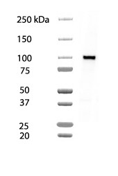
Western Blot: N-Cadherin Antibody (5D5)BSA Free [NBP1-51612]
Western Blot: N-Cadherin Antibody (5D5) [NBP1-51612] - Analysis using CDH2 mouse mAb against A431 (1), NIH/3T3 (2), Hela (3), C6 (4) and LNCap (5) cell lysate.![Immunocytochemistry/ Immunofluorescence: N-Cadherin Antibody (5D5) - BSA Free [NBP1-51612]](https://resources.rndsystems.com/images/products/N-Cadherin-Antibody-5D5-Immunocytochemistry-Immunofluorescence-NBP1-51612-img0012.jpg "Immunocytochemistry/ Immunofluorescence: N-Cadherin Antibody (5D5) - BSA Free [NBP1-51612]")
Immunocytochemistry/ Immunofluorescence: N-Cadherin Antibody (5D5) - BSA Free [NBP1-51612]
Immunocytochemistry/Immunofluorescence: N-Cadherin Antibody (5D5) [NBP1-51612] - Immunofluorescence analysis of A431 cells using CDH2 mouse mAb (green). Blue: DRAQ5 fluorescent DNA dye.![Flow Cytometry: N-Cadherin Antibody (5D5) - BSA Free [NBP1-51612]](https://resources.rndsystems.com/images/products/N-Cadherin-Antibody-5D5-Flow-Cytometry-NBP1-51612-img0011.jpg "Flow Cytometry: N-Cadherin Antibody (5D5) - BSA Free [NBP1-51612]")
Flow Cytometry: N-Cadherin Antibody (5D5) - BSA Free [NBP1-51612]
Flow Cytometry: N-Cadherin Antibody (5D5) [NBP1-51612] - Analysis of PC-2 cells using CDH2 mouse mAb (green) and negative control (purple).![Immunohistochemistry-Paraffin: N-Cadherin Antibody (5D5) - BSA Free [NBP1-51612]](https://resources.rndsystems.com/images/products/N-Cadherin-Antibody-5D5-Immunohistochemistry-Paraffin-NBP1-51612-img0014.jpg "Immunohistochemistry-Paraffin: N-Cadherin Antibody (5D5) - BSA Free [NBP1-51612]")
Immunohistochemistry-Paraffin: N-Cadherin Antibody (5D5) - BSA Free [NBP1-51612]
Immunohistochemistry-Paraffin: N-Cadherin Antibody (5D5) [NBP1-51612] - Analysis of human Placenta tissues using CDH2 mouse mAbApplications for N-Cadherin Antibody (5D5)
Application
Recommended Usage
ELISA
1:10000
Flow Cytometry
1:200 - 1:400
Immunocytochemistry/ Immunofluorescence
1:200 - 1:1000
Immunohistochemistry
1:200-1:400
Immunohistochemistry-Paraffin
1:200 - 1:1000
Western Blot
1:500 - 1:2000
Reviewed Applications
Read 3 reviews rated 4.7 using NBP1-51612 in the following applications:
Flow Cytometry Panel Builder
Bio-Techne Knows Flow Cytometry
Save time and reduce costly mistakes by quickly finding compatible reagents using the Panel Builder Tool.
Advanced Features
- Spectra Viewer - Custom analysis of spectra from multiple fluorochromes
- Spillover Popups - Visualize the spectra of individual fluorochromes
- Antigen Density Selector - Match fluorochrome brightness with antigen density

Formulation, Preparation, and Storage
Purification
Ascites
Formulation
Ascites
Preservative
0.03% Sodium Azide
Concentration
This product is unpurified. The exact concentration of antibody is not quantifiable.
Shipping
The product is shipped with polar packs. Upon receipt, store it immediately at the temperature recommended below.
Stability & Storage
Store at 4C short term. Aliquot and store at -20C long term. Avoid freeze-thaw cycles.
Background: N-Cadherin
N-cadherin is expressed on multiple cell types but is most highly expressed by mesenchymal cells and neural tissue (2). Functionally, N-cadherin has a number of roles including maintaining structural integrity and adhesion, cell signaling, and formation of neuronal synapses and the vascular wall (2). The cytoplasmic tail interacts with beta-catenin which then binds with alpha-catenin, forming the cadherin-catenin adhesion complex, an important component of adhesions junctions (1-3). Given its role in adhesion, N-cadherin serves as an indicator of epithelial-to-mesenchymal transition (EMT) (1-4). The loss of E-cadherin during EMT corresponds with an increase in N-cadherin expression (1-4). This "cadherin-switch" is associated with increased migratory and invasive behavior observed in tumor progress (1-4). Proteases including activity of a disintegrin and metalloprotease 10 (ADAM10), matrix metalloproteinases (MMPs), caspase 3, presenilin, and calpain can cleave N-cadherin as a mechanism for regulating Wnt/beta-catenin signaling and inducing oncogenic signals (3,4). In addition to its expression in solid tumors, N-cadherin has been indicated in hematological disorders such as leukemia and multiple myeloma (1). N-cadherin antagonists are currently being studied as potential therapeutics for a variety of cancer studies (1-2).
References
1. Mrozik, K. M., Blaschuk, O. W., Cheong, C. M., Zannettino, A., & Vandyke, K. (2018). N-cadherin in cancer metastasis, its emerging role in haematological malignancies and potential as a therapeutic target in cancer. BMC Cancer. https://doi.org/10.1186/s12885-018-4845-0
2. Loh, C. Y., Chai, J. Y., Tang, T. F., Wong, W. F., Sethi, G., Shanmugam, M. K., Chong, P. P., & Looi, C. Y. (2019). The E-Cadherin and N-Cadherin Switch in Epithelial-to-Mesenchymal Transition: Signaling, Therapeutic Implications, and Challenges. Cells. https://doi.org/10.3390/cells8101118
3. Derycke, L. D., & Bracke, M. E. (2004). N-cadherin in the spotlight of cell-cell adhesion, differentiation, embryogenesis, invasion and signalling. The International Journal of Developmental Biology. https://doi.org/10.1387/ijdb.041793ld
4. Yu, W., Yang, L., Li, T., & Zhang, Y. (2019). Cadherin Signaling in Cancer: Its Functions and Role as a Therapeutic Target. Frontiers in Oncology. https://doi.org/10.3389/fonc.2019.00989
5. Unitprot (P1903)
Long Name
Neural Cadherin
Alternate Names
Cadherin-2, CD325, CDH2, NCadherin
Gene Symbol
CDH2
UniProt
Additional N-Cadherin Products
Product Documents for N-Cadherin Antibody (5D5)
Certificate of Analysis
To download a Certificate of Analysis, please enter a lot or batch number in the search box below.
Product Specific Notices for N-Cadherin Antibody (5D5)
This product is for research use only and is not approved for use in humans or in clinical diagnosis. Primary Antibodies are guaranteed for 1 year from date of receipt.
Citations for N-Cadherin Antibody (5D5)
 Powered by Bioz
Customer Reviews for N-Cadherin Antibody (5D5) (3)
4.7 out of 5
3 Customer Ratings
Have you used N-Cadherin Antibody (5D5)?
Submit a review and receive an Amazon gift card!
$25/€18/£15/$25CAN/¥2500 Yen for a review with an image
$10/€7/£6/$10CAN/¥1110 Yen for a review without an image
Submit a review
Customer Images
Showing
1
-
3 of
3 reviews
Showing All
Filter By:
-
Application: Western BlotSample Tested: MCF-7 human breast cancer cell lineSpecies: HumanVerified Customer | Posted 01/01/2026Blot of whole lysate of MCF7 cellsUsed at 1 ug/mL

-
Application: Western BlotSample Tested: Adipocytes HCT116 cocultureSpecies: HumanVerified Customer | Posted 07/18/2025N-Cadherin bandCo culture of adipocytes and HCT116 cells. 1:2000 antibody dilution
-
Application: Western BlotSample Tested: Human cancer cellSpecies: HumanVerified Customer | Posted 10/04/2015
There are no reviews that match your criteria.
Protocols
Find general support by application which include: protocols, troubleshooting, illustrated assays, videos and webinars.
- 7-Amino Actinomycin D (7-AAD) Cell Viability Flow Cytometry Protocol
- Antigen Retrieval Protocol (PIER)
- Antigen Retrieval for Frozen Sections Protocol
- Appropriate Fixation of IHC/ICC Samples
- Cellular Response to Hypoxia Protocols
- Chromogenic IHC Staining of Formalin-Fixed Paraffin-Embedded (FFPE) Tissue Protocol
- Chromogenic Immunohistochemistry Staining of Frozen Tissue
- ClariTSA™ Fluorophore Kits
- Detection & Visualization of Antibody Binding
- ELISA Sample Preparation & Collection Guide
- ELISA Troubleshooting Guide
- Extracellular Membrane Flow Cytometry Protocol
- Flow Cytometry Protocol for Cell Surface Markers
- Flow Cytometry Protocol for Staining Membrane Associated Proteins
- Flow Cytometry Staining Protocols
- Flow Cytometry Troubleshooting Guide
- Fluorescent IHC Staining of Frozen Tissue Protocol
- Graphic Protocol for Heat-induced Epitope Retrieval
- Graphic Protocol for the Preparation and Fluorescent IHC Staining of Frozen Tissue Sections
- Graphic Protocol for the Preparation and Fluorescent IHC Staining of Paraffin-embedded Tissue Sections
- Graphic Protocol for the Preparation of Gelatin-coated Slides for Histological Tissue Sections
- How to Run an R&D Systems DuoSet ELISA
- How to Run an R&D Systems Quantikine ELISA
- How to Run an R&D Systems Quantikine™ QuicKit™ ELISA
- ICC Cell Smear Protocol for Suspension Cells
- ICC Immunocytochemistry Protocol Videos
- ICC for Adherent Cells
- IHC Sample Preparation (Frozen sections vs Paraffin)
- Immunocytochemistry (ICC) Protocol
- Immunocytochemistry Troubleshooting
- Immunofluorescence of Organoids Embedded in Cultrex Basement Membrane Extract
- Immunofluorescent IHC Staining of Formalin-Fixed Paraffin-Embedded (FFPE) Tissue Protocol
- Immunohistochemistry (IHC) and Immunocytochemistry (ICC) Protocols
- Immunohistochemistry Frozen Troubleshooting
- Immunohistochemistry Paraffin Troubleshooting
- Intracellular Flow Cytometry Protocol Using Alcohol (Methanol)
- Intracellular Flow Cytometry Protocol Using Detergents
- Intracellular Nuclear Staining Flow Cytometry Protocol Using Detergents
- Intracellular Staining Flow Cytometry Protocol Using Alcohol Permeabilization
- Intracellular Staining Flow Cytometry Protocol Using Detergents to Permeabilize Cells
- Preparing Samples for IHC/ICC Experiments
- Preventing Non-Specific Staining (Non-Specific Binding)
- Primary Antibody Selection & Optimization
- Propidium Iodide Cell Viability Flow Cytometry Protocol
- Protocol for Heat-Induced Epitope Retrieval (HIER)
- Protocol for Liperfluo
- Protocol for Making a 4% Formaldehyde Solution in PBS
- Protocol for VisUCyte™ HRP Polymer Detection Reagent
- Protocol for the Characterization of Human Th22 Cells
- Protocol for the Characterization of Human Th9 Cells
- Protocol for the Fluorescent ICC Staining of Cell Smears - Graphic
- Protocol for the Fluorescent ICC Staining of Cultured Cells on Coverslips - Graphic
- Protocol for the Preparation & Fixation of Cells on Coverslips
- Protocol for the Preparation and Chromogenic IHC Staining of Frozen Tissue Sections
- Protocol for the Preparation and Chromogenic IHC Staining of Frozen Tissue Sections - Graphic
- Protocol for the Preparation and Chromogenic IHC Staining of Paraffin-embedded Tissue Sections
- Protocol for the Preparation and Chromogenic IHC Staining of Paraffin-embedded Tissue Sections - Graphic
- Protocol for the Preparation and Fluorescent ICC Staining of Cells on Coverslips
- Protocol for the Preparation and Fluorescent ICC Staining of Non-adherent Cells
- Protocol for the Preparation and Fluorescent ICC Staining of Stem Cells on Coverslips
- Protocol for the Preparation and Fluorescent IHC Staining of Frozen Tissue Sections
- Protocol for the Preparation and Fluorescent IHC Staining of Paraffin-embedded Tissue Sections
- Protocol for the Preparation of Gelatin-coated Slides for Histological Tissue Sections
- Protocol for the Preparation of a Cell Smear for Non-adherent Cell ICC - Graphic
- Protocol: Annexin V and PI Staining by Flow Cytometry
- Protocol: Annexin V and PI Staining for Apoptosis by Flow Cytometry
- Quantikine HS ELISA Kit Assay Principle, Alkaline Phosphatase
- Quantikine HS ELISA Kit Principle, Streptavidin-HRP Polymer
- R&D Systems Quality Control Western Blot Protocol
- Sandwich ELISA (Colorimetric) – Biotin/Streptavidin Detection Protocol
- Sandwich ELISA (Colorimetric) – Direct Detection Protocol
- TUNEL and Active Caspase-3 Detection by IHC/ICC Protocol
- The Importance of IHC/ICC Controls
- Troubleshooting Guide: ELISA
- Troubleshooting Guide: Fluorokine Flow Cytometry Kits
- Troubleshooting Guide: Immunohistochemistry
- Troubleshooting Guide: Western Blot Figures
- Western Blot Conditions
- Western Blot Protocol
- Western Blot Protocol for Cell Lysates
- Western Blot Troubleshooting
- Western Blot Troubleshooting Guide
- View all Protocols, Troubleshooting, Illustrated assays and Webinars


