O-GlcNAc Antibody (RL2) - BSA Free
Novus Biologicals | Catalog # NB300-524
Key Product Details
Validated by
Species Reactivity
Validated:
Cited:
Applications
Validated:
Cited:
Label
Antibody Source
Format
Product Specifications
Immunogen
Reactivity Notes
Localization
Specificity
Clonality
Host
Isotype
Scientific Data Images for O-GlcNAc Antibody (RL2) - BSA Free
![Immunohistochemistry-Paraffin: O-GlcNAc Antibody (RL2) - BSA Free [NB300-524]](https://resources.rndsystems.com/images/products/O-GlcNAc-Antibody-RL2-Immunohistochemistry-Paraffin-NB300-524-img0011.jpg "Immunohistochemistry-Paraffin: O-GlcNAc Antibody (RL2) - BSA Free [NB300-524]")
Immunohistochemistry-Paraffin: O-GlcNAc Antibody (RL2) - BSA Free [NB300-524]
Immunohistochemistry-Paraffin: O-GlcNAc Antibody (RL2) [NB300-524] - Analysis of a FFPE tissue section of the mouse colon using 1:200 dilution of O-GlcNAc [RL2] antibody (NB300-524). The signal was developed using HRP-DAB method which followed counterstaining of the cells with hematoxylin.![Western Blot: O-GlcNAc Antibody (RL2)BSA Free [NB300-524]](https://resources.rndsystems.com/images/products/O-GlcNAc-Antibody-RL2-Western-Blot-NB300-524-img0016.jpg "Western Blot: O-GlcNAc Antibody (RL2)BSA Free [NB300-524]")
![Immunocytochemistry/ Immunofluorescence: O-GlcNAc Antibody (RL2) - BSA Free [NB300-524]](https://resources.rndsystems.com/images/products/O-GlcNAc-Antibody-RL2-Immunocytochemistry-Immunofluorescence-NB300-524-img0013.jpg "Immunocytochemistry/ Immunofluorescence: O-GlcNAc Antibody (RL2) - BSA Free [NB300-524]")
Immunocytochemistry/ Immunofluorescence: O-GlcNAc Antibody (RL2) - BSA Free [NB300-524]
Immunocytochemistry/Immunofluorescence: O-GlcNAc Antibody (RL2) [NB300-524] - Neuro2a cells were fixed for 10 minutes using 10% formalin and then permeabilized for 5 minutes using 1X PBS + 0.5% Triton X-100. The cells were incubated with anti-O-GlcNAc (RL2) at 5 ug/mL overnight at 4C and detected with an anti-mouse Dylight 488 (Green) at a 1:500 dilution. Nuclei were counterstained with DAPI (Blue). Cells were imaged using a 40X objective.![Flow Cytometry: O-GlcNAc Antibody (RL2) - BSA Free [NB300-524]](https://resources.rndsystems.com/images/products/O-GlcNAc-Antibody-RL2-Flow-Cytometry-NB300-524-img0018.jpg "Flow Cytometry: O-GlcNAc Antibody (RL2) - BSA Free [NB300-524]")
Flow Cytometry: O-GlcNAc Antibody (RL2) - BSA Free [NB300-524]
Flow Cytometry: O-GlcNAc Antibody (RL2) [NB300-524] - An intracellular stain was performed on Neuro2a cells with O-GlcNAc Antibody [RL2] NB300-524 (blue) and a matched isotype control (orange). Cells were fixed with 4% PFA and then permeabilized with 0.1% saponin. Cells were incubated in an antibody dilution of 1.0 ug/mL for 30 minutes at room temperature, followed by Mouse IgG (H+L) Cross-Adsorbed Secondary Antibody, Dylight 550 (35503, Thermo Fisher).![Western Blot: O-GlcNAc Antibody (RL2)BSA Free [NB300-524]](https://resources.rndsystems.com/images/products/O-GlcNAc-Antibody-RL2-Western-Blot-NB300-524-img0005.jpg "Western Blot: O-GlcNAc Antibody (RL2)BSA Free [NB300-524]")
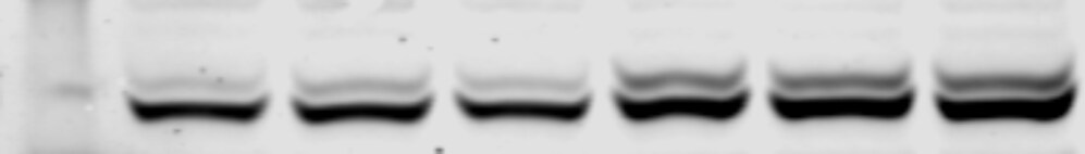
Western Blot: O-GlcNAc Antibody (RL2)BSA Free [NB300-524]
Western Blot: O-GlcNAc Antibody (RL2) [NB300-524] - Analysis of mouse cortical brain lysates using O-Linked N-Acetylglucosamine Monoclonal Antibody. Blots containing cortical extracts from 4 individual C57BL/6 mice (Lanes 1-4) were blocked with 5% milk in TBST, and probed with MA1-072 at 1:1000, followed by a fluorophore-conjugated goat anti-mouse IgG secondary antibody. Data courtesy of the Innovators Program.![Immunocytochemistry/ Immunofluorescence: O-GlcNAc Antibody (RL2) - BSA Free [NB300-524]](https://resources.rndsystems.com/images/products/O-GlcNAc-Antibody-RL2-Immunocytochemistry-Immunofluorescence-NB300-524-img0012.jpg "Immunocytochemistry/ Immunofluorescence: O-GlcNAc Antibody (RL2) - BSA Free [NB300-524]")
Immunocytochemistry/ Immunofluorescence: O-GlcNAc Antibody (RL2) - BSA Free [NB300-524]
Immunocytochemistry/Immunofluorescence: O-GlcNAc Antibody (RL2) [NB300-524] - HeLa cells were fixed for 10 minutes using 10% formalin and then permeabilized for 5 minutes using 1X PBS + 0.5% Triton X-100. The cells were incubated with anti-O-GlcNAc (RL2) at 5 ug/mL overnight at 4C and detected with an anti-mouse Dylight 488 (Green) at a 1:500 dilution. Nuclei were counterstained with DAPI (Blue). Cells were imaged using a 40X objective.![Flow Cytometry: O-GlcNAc Antibody (RL2) - BSA Free [NB300-524]](https://resources.rndsystems.com/images/products/O-GlcNAc-Antibody-RL2-Flow-Cytometry-NB300-524-img0006.jpg "Flow Cytometry: O-GlcNAc Antibody (RL2) - BSA Free [NB300-524]")
Flow Cytometry: O-GlcNAc Antibody (RL2) - BSA Free [NB300-524]
Flow Cytometry: O-GlcNAc Antibody (RL2) [NB300-524] - Analysis using Alexa Fluor (R) 647 conjugate of NB300-524. An intracellular stain was performed on Jurkat cells with O-GlcNAc antibody (RL2) NB300-524 (blue) and a matched isotype control NBP2-27287 (orange). Cells were fixed with 4% PFA and then permeablized with 0.1% saponin. 1 ug of antibody was added to 100 uL of staining buffer and cells were incubated for 30 minutes at room temperature. Both antibodies were conjugated to Alexa Fluor 647.![Flow Cytometry: O-GlcNAc Antibody (RL2) - BSA Free [NB300-524]](https://resources.rndsystems.com/images/products/O-GlcNAc-Antibody-RL2-Flow-Cytometry-NB300-524-img0007.jpg "Flow Cytometry: O-GlcNAc Antibody (RL2) - BSA Free [NB300-524]")
Flow Cytometry: O-GlcNAc Antibody (RL2) - BSA Free [NB300-524]
Flow Cytometry: O-GlcNAc Antibody (RL2) [NB300-524] - An intracellular stain was performed on U-937 cells with O-GlcNAc antibody (RL2) NB300-524AF647 (blue) and a matched isotype control. Cells were fixed with 4% PFA and then permeablized with 0.1% saponin. Cells were incubated in an antibody dilution of 2.5 ug/mL for 30 minutes at room temperature. Both antibodies were conjugated to Alexa Fluor 647.![Flow Cytometry: O-GlcNAc Antibody (RL2) - BSA Free [NB300-524]](https://resources.rndsystems.com/images/products/O-GlcNAc-Antibody-RL2-Flow-Cytometry-NB300-524-img0008.jpg "Flow Cytometry: O-GlcNAc Antibody (RL2) - BSA Free [NB300-524]")
Flow Cytometry: O-GlcNAc Antibody (RL2) - BSA Free [NB300-524]
Flow Cytometry: O-GlcNAc Antibody (RL2) [NB300-524] - An intracellular stain was performed on Jurkat cells with O-GlcNAc antibody (RL2) NB300-524PE (blue) and a matched isotype control. Cells were fixed with 4% PFA and then permeablized with 0.1% saponin. Cells were incubated in an antibody dilution of 2.5 ug/mL for 30 minutes at room temperature. Both antibodies were conjugated to Phycoerythrin.![Flow Cytometry: O-GlcNAc Antibody (RL2) - BSA Free [NB300-524]](https://resources.rndsystems.com/images/products/O-GlcNAc-Antibody-RL2-Flow-Cytometry-NB300-524-img0009.jpg "Flow Cytometry: O-GlcNAc Antibody (RL2) - BSA Free [NB300-524]")
Flow Cytometry: O-GlcNAc Antibody (RL2) - BSA Free [NB300-524]
Flow Cytometry: O-GlcNAc Antibody (RL2) [NB300-524] - An intracellular stain was performed on SK-MEL-28 cells with O-GlcNAc antibody (RL2) NB300-524AF647 (blue) and a matched isotype control. Cells were fixed with 4% PFA and then permeablized with 0.1% saponin. Cells were incubated in an antibody dilution of 2.5 ug/mL for 30 minutes at room temperature. Both antibodies were conjugated to Alexa Fluor 647.![Flow Cytometry: O-GlcNAc Antibody (RL2) - BSA Free [NB300-524]](https://resources.rndsystems.com/images/products/O-GlcNAc-Antibody-RL2-Flow-Cytometry-NB300-524-img0010.jpg "Flow Cytometry: O-GlcNAc Antibody (RL2) - BSA Free [NB300-524]")
Flow Cytometry: O-GlcNAc Antibody (RL2) - BSA Free [NB300-524]
Flow Cytometry: O-GlcNAc Antibody (RL2) [NB300-524] - An intracellular stain was performed on HeLa cells with O-GlcNAc Antibody [RL2] Antibody NB300-524AF647 (blue) and a matched isotype control (orange). Cells were fixed with 4% PFA and then permeabilized with 0.1% saponin. Cells were incubated in an antibody dilution of 2.5 ug/mL for 30 minutes at room temperature. Both antibodies were conjugated to Alexa Fluor 647.![Flow Cytometry: O-GlcNAc Antibody (RL2) - BSA Free [NB300-524]](https://resources.rndsystems.com/images/products/O-GlcNAc-Antibody-RL2-Flow-Cytometry-NB300-524-img0014.jpg "Flow Cytometry: O-GlcNAc Antibody (RL2) - BSA Free [NB300-524]")
Flow Cytometry: O-GlcNAc Antibody (RL2) - BSA Free [NB300-524]
Flow Cytometry: O-GlcNAc Antibody (RL2) [NB300-524] - An intracellular stain was performed on Neuro2a cells with O-GlcNAc Antibody [RL2] NB300-524AF647 (blue) and a matched isotype control (orange). Cells were fixed with 4% PFA and then permeabilized with 0.1% saponin. Cells were incubated in an antibody dilution of 2.5 ug/mL for 30 minutes at room temperature. Both antibodies were conjugated to Alexa Fluor 647.![Flow Cytometry: O-GlcNAc Antibody (RL2) - BSA Free [NB300-524]](https://resources.rndsystems.com/images/products/O-GlcNAc-Antibody-RL2-Flow-Cytometry-NB300-524-img0015.jpg "Flow Cytometry: O-GlcNAc Antibody (RL2) - BSA Free [NB300-524]")
Flow Cytometry: O-GlcNAc Antibody (RL2) - BSA Free [NB300-524]
Flow Cytometry: O-GlcNAc Antibody (RL2) [NB300-524] - An intracellular stain was performed on RH30 cells with O-GlcNAc [RL2] Antibody NB300-524AF647 (blue) and a matched isotype control (orange). Cells were fixed with 4% PFA and then permeabilized with 0.1% saponin. Cells were incubated in an antibody dilution of 2.5 ug/mL for 30 minutes at room temperature. Both antibodies were conjugated to Alexa Fluor 647.![Flow Cytometry: O-GlcNAc Antibody (RL2) - BSA Free [NB300-524]](https://resources.rndsystems.com/images/products/O-GlcNAc-Antibody-RL2-Flow-Cytometry-NB300-524-img0017.jpg "Flow Cytometry: O-GlcNAc Antibody (RL2) - BSA Free [NB300-524]")
Flow Cytometry: O-GlcNAc Antibody (RL2) - BSA Free [NB300-524]
Flow Cytometry: O-GlcNAc Antibody (RL2) [NB300-524] - An intracellular stain was performed on Jurkat cells with O-GlcNAc Antibody [RL2] NB300-524 (blue) and a matched isotype control (orange). Cells were fixed with 4% PFA and then permeabilized with 0.1% saponin. Cells were incubated in an antibody dilution of 1.0 ug/mL for 30 minutes at room temperature, followed by Mouse IgG (H+L) Cross-Adsorbed Secondary Antibody, Dylight 550 (35503, Thermo Fisher). - BSA Free [NB300-524] -")
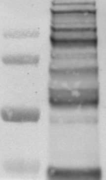
Western Blot: O-GlcNAc Antibody (RL2) - BSA Free [NB300-524] -
Western Blot: O-GlcNAc Antibody (RL2) - BSA Free [NB300-524] - Analysis of O-GlcNAcylated LV proteins by Western blot & WGA-SDS-PAGE gel electrophoresis. (A) Red ponceau staining (left panel) & western blot (right panel) of O-GlcNAcylated proteins (50 μg) extracted from sham- & HF-rats treated or not with thiamet G. The positions of molecular weight are indicated as kilodalton (kDa) on the left. (B) Red ponceau staining (left panel) & WGA-SDS-PAGE of O-GlcNAcylated proteins levels (middle panel) of O-GlcNAcylated desmin levels (right panel) from the same samples. The arrow in desmin WGA gels indicates the non-O-GlcNAcylated form. Image collected & cropped by CiteAb from the following publication (https://pubmed.ncbi.nlm.nih.gov/30344511), licensed under a CC-BY license. Not internally tested by Novus Biologicals. - BSA Free [NB300-524] -")
Western Blot: O-GlcNAc Antibody (RL2) - BSA Free [NB300-524] -
Western Blot: O-GlcNAc Antibody (RL2) - BSA Free [NB300-524] - Effect of OGA inhibition by thiamet G in isolated perfused heart. (A) Description of the protocol designed for thiamet G (TG) perfusion in sham- (n = 6) & HF- (n = 7) rats 6 weeks post-MI. (B) Western blot (left panel) & quantification (right panel) of O-GlcNAcylated proteins levels measured in proteins extracted from LVs of isolated perfused sham- & HF-rat hearts treated or not with 100 μM thiamet G for 2 h (n = 7 in each group). (C) Western blots (upper panel) & quantification (lower panel) of total desmin levels in the same samples. (D) Phosphorylation profiles of desmin were analyzed in the same samples by Phos-tag™ gel. Graphs show mean ± SEM values expressed in arbitrary units (A.U.). The positions of molecular weight are indicated as kilodalton (kDa) on the left. *P < 0.05; ** < 0.01. Image collected & cropped by CiteAb from the following publication (https://pubmed.ncbi.nlm.nih.gov/30344511), licensed under a CC-BY license. Not internally tested by Novus Biologicals.Applications for O-GlcNAc Antibody (RL2) - BSA Free
Chromatin Immunoprecipitation
Chromatin Immunoprecipitation (ChIP)
Dot Blot
ELISA
Flow Cytometry
Immunohistochemistry
Immunohistochemistry-Paraffin
Immunoprecipitation
Western Blot
Reviewed Applications
Read 2 reviews rated 4 using NB300-524 in the following applications:
Flow Cytometry Panel Builder
Bio-Techne Knows Flow Cytometry
Save time and reduce costly mistakes by quickly finding compatible reagents using the Panel Builder Tool.
Advanced Features
- Spectra Viewer - Custom analysis of spectra from multiple fluorochromes
- Spillover Popups - Visualize the spectra of individual fluorochromes
- Antigen Density Selector - Match fluorochrome brightness with antigen density

Formulation, Preparation, and Storage
Purification
Formulation
Format
Preservative
Concentration
Shipping
Stability & Storage
Background: O-GlcNAc
Alternate Names
Additional O-GlcNAc Products
Product Documents for O-GlcNAc Antibody (RL2) - BSA Free
Certificate of Analysis
To download a Certificate of Analysis, please enter a lot or batch number in the search box below.
Product Specific Notices for O-GlcNAc Antibody (RL2) - BSA Free
This product is for research use only and is not approved for use in humans or in clinical diagnosis. Primary Antibodies are guaranteed for 1 year from date of receipt.
Citations for O-GlcNAc Antibody (RL2) - BSA Free
 Powered by Bioz
Customer Reviews for O-GlcNAc Antibody (RL2) - BSA Free (2)
Have you used O-GlcNAc Antibody (RL2) - BSA Free?
Submit a review and receive an Amazon gift card!
$25/€18/£15/$25CAN/¥2500 Yen for a review with an image
$10/€7/£6/$10CAN/¥1110 Yen for a review without an image
Submit a review
Customer Images
-
Application: Western BlotSample Tested: Mouse Embryonic Stem CellsSpecies: MouseVerified Customer | Posted 04/17/2020Glut2 expression in two different culture conditions. The nitrocellulose membrane was incubated overnight with Glut2 ab at a concentration of 1:1000 in 5% BSA in PBS-Tween (0.01%)

-
Application: Western BlotSample Tested: Mouse Embryonic Stem CellsSpecies: MouseVerified Customer | Posted 01/16/2020O-GlcNAc Western blot from 20 ug of mESCs lysateNitrocellulose membrane, Blocked in 5% BSA in PBS-T, 1:1000 ab overnight at 4 degrees.
There are no reviews that match your criteria.
Protocols
View specific protocols for O-GlcNAc Antibody (RL2) - BSA Free (NB300-524):
Immunohistochemistry-Paraffin Embedded Sections
Antigen Unmasking:
Bring slides to a boil in 10 mM sodium citrate buffer (pH 6.0) then maintain at a sub-boiling temperature for 10 minutes. Cool slides on bench-top for 30 minutes.
Staining:
1. Wash sections in deionized water three times for 5 minutes each.
2. Wash sections in wash buffer for 5 minutes.
3. Block each section with 100-400 ul blocking solution for 1 hour at room temperature.
4. Remove blocking solution and add 100-400 ul diluted primary antibody. Incubate overnight at 4 C.
5. Remove antibody solution and wash sections in wash buffer three times for 5 minutes each.
6. Add 100-400 ul biotinylated diluted secondary antibody. Incubate 30 minutes at room temperature.
7. Remove secondary antibody solution and wash sections three times with wash buffer for 5 minutes each.
8. Add 100-400 ul Streptavidin-HRP reagent to each section and incubate for 30 minutes at room temperature.
9. Wash sections three times in wash buffer for 5 minutes each.
10. Add 100-400 ul DAB substrate to each section and monitor staining closely.
11. As soon as the sections develop, immerse slides in deionized water.
12. Counterstain sections in hematoxylin.
13. Wash sections in deionized water two times for 5 minutes each.
14. Dehydrate sections.
15. Mount coverslips.
Find general support by application which include: protocols, troubleshooting, illustrated assays, videos and webinars.
- 7-Amino Actinomycin D (7-AAD) Cell Viability Flow Cytometry Protocol
- Antigen Retrieval Protocol (PIER)
- Antigen Retrieval for Frozen Sections Protocol
- Appropriate Fixation of IHC/ICC Samples
- Cellular Response to Hypoxia Protocols
- ChIP Protocol Video
- Chromatin Immunoprecipitation (ChIP) Protocol
- Chromatin Immunoprecipitation Protocol
- Chromogenic IHC Staining of Formalin-Fixed Paraffin-Embedded (FFPE) Tissue Protocol
- Chromogenic Immunohistochemistry Staining of Frozen Tissue
- ClariTSA™ Fluorophore Kits
- Detection & Visualization of Antibody Binding
- ELISA Sample Preparation & Collection Guide
- ELISA Troubleshooting Guide
- Extracellular Membrane Flow Cytometry Protocol
- Flow Cytometry Protocol for Cell Surface Markers
- Flow Cytometry Protocol for Staining Membrane Associated Proteins
- Flow Cytometry Staining Protocols
- Flow Cytometry Troubleshooting Guide
- Fluorescent IHC Staining of Frozen Tissue Protocol
- Graphic Protocol for Heat-induced Epitope Retrieval
- Graphic Protocol for the Preparation and Fluorescent IHC Staining of Frozen Tissue Sections
- Graphic Protocol for the Preparation and Fluorescent IHC Staining of Paraffin-embedded Tissue Sections
- Graphic Protocol for the Preparation of Gelatin-coated Slides for Histological Tissue Sections
- How to Run an R&D Systems DuoSet ELISA
- How to Run an R&D Systems Quantikine ELISA
- How to Run an R&D Systems Quantikine™ QuicKit™ ELISA
- ICC Cell Smear Protocol for Suspension Cells
- ICC Immunocytochemistry Protocol Videos
- ICC for Adherent Cells
- IHC Sample Preparation (Frozen sections vs Paraffin)
- Immunocytochemistry (ICC) Protocol
- Immunocytochemistry Troubleshooting
- Immunofluorescence of Organoids Embedded in Cultrex Basement Membrane Extract
- Immunofluorescent IHC Staining of Formalin-Fixed Paraffin-Embedded (FFPE) Tissue Protocol
- Immunohistochemistry (IHC) and Immunocytochemistry (ICC) Protocols
- Immunohistochemistry Frozen Troubleshooting
- Immunohistochemistry Paraffin Troubleshooting
- Immunoprecipitation Protocol
- Intracellular Flow Cytometry Protocol Using Alcohol (Methanol)
- Intracellular Flow Cytometry Protocol Using Detergents
- Intracellular Nuclear Staining Flow Cytometry Protocol Using Detergents
- Intracellular Staining Flow Cytometry Protocol Using Alcohol Permeabilization
- Intracellular Staining Flow Cytometry Protocol Using Detergents to Permeabilize Cells
- Preparing Samples for IHC/ICC Experiments
- Preventing Non-Specific Staining (Non-Specific Binding)
- Primary Antibody Selection & Optimization
- Propidium Iodide Cell Viability Flow Cytometry Protocol
- Protocol for Heat-Induced Epitope Retrieval (HIER)
- Protocol for Liperfluo
- Protocol for Making a 4% Formaldehyde Solution in PBS
- Protocol for VisUCyte™ HRP Polymer Detection Reagent
- Protocol for the Characterization of Human Th22 Cells
- Protocol for the Characterization of Human Th9 Cells
- Protocol for the Fluorescent ICC Staining of Cell Smears - Graphic
- Protocol for the Fluorescent ICC Staining of Cultured Cells on Coverslips - Graphic
- Protocol for the Preparation & Fixation of Cells on Coverslips
- Protocol for the Preparation and Chromogenic IHC Staining of Frozen Tissue Sections
- Protocol for the Preparation and Chromogenic IHC Staining of Frozen Tissue Sections - Graphic
- Protocol for the Preparation and Chromogenic IHC Staining of Paraffin-embedded Tissue Sections
- Protocol for the Preparation and Chromogenic IHC Staining of Paraffin-embedded Tissue Sections - Graphic
- Protocol for the Preparation and Fluorescent ICC Staining of Cells on Coverslips
- Protocol for the Preparation and Fluorescent ICC Staining of Non-adherent Cells
- Protocol for the Preparation and Fluorescent ICC Staining of Stem Cells on Coverslips
- Protocol for the Preparation and Fluorescent IHC Staining of Frozen Tissue Sections
- Protocol for the Preparation and Fluorescent IHC Staining of Paraffin-embedded Tissue Sections
- Protocol for the Preparation of Gelatin-coated Slides for Histological Tissue Sections
- Protocol for the Preparation of a Cell Smear for Non-adherent Cell ICC - Graphic
- Protocol: Annexin V and PI Staining by Flow Cytometry
- Protocol: Annexin V and PI Staining for Apoptosis by Flow Cytometry
- Quantikine HS ELISA Kit Assay Principle, Alkaline Phosphatase
- Quantikine HS ELISA Kit Principle, Streptavidin-HRP Polymer
- R&D Systems Quality Control Western Blot Protocol
- Sandwich ELISA (Colorimetric) – Biotin/Streptavidin Detection Protocol
- Sandwich ELISA (Colorimetric) – Direct Detection Protocol
- TUNEL and Active Caspase-3 Detection by IHC/ICC Protocol
- The Importance of IHC/ICC Controls
- Troubleshooting Guide: ELISA
- Troubleshooting Guide: Fluorokine Flow Cytometry Kits
- Troubleshooting Guide: Immunohistochemistry
- Troubleshooting Guide: Western Blot Figures
- Western Blot Conditions
- Western Blot Protocol
- Western Blot Protocol for Cell Lysates
- Western Blot Troubleshooting
- Western Blot Troubleshooting Guide
- View all Protocols, Troubleshooting, Illustrated assays and Webinars
FAQs for O-GlcNAc Antibody (RL2) - BSA Free
-
Q: Is it possible to get your mAb without BSA or sodium azide? I am interested in NB300-614 and NB300-524.
A:
These antibodies are supplied in Sodium Azide, but if you are interested, we do provide kits to clean up the antibodies. Here is the link to our AbSelect Antibody Purification Kits .