Osteopontin/OPN Antibody (1B20) - BSA Free
Novus Biologicals | Catalog # NB110-89062
Key Product Details
Validated by
Species Reactivity
Validated:
Cited:
Applications
Validated:
Cited:
Label
Antibody Source
Format
Product Specifications
Immunogen
Reactivity Notes
Localization
Clonality
Host
Isotype
Scientific Data Images for Osteopontin/OPN Antibody (1B20) - BSA Free
![Western Blot: Osteopontin/OPN Antibody (1B20)BSA Free [NB110-89062]](https://resources.rndsystems.com/images/products/Osteopontin-OPN-Antibody-1B20-BSA-Free-Western-Blot-NB110-89062-img0007.jpg "Western Blot: Osteopontin/OPN Antibody (1B20)BSA Free [NB110-89062]")
Western Blot: Osteopontin/OPN Antibody (1B20)BSA Free [NB110-89062]
Western Blot: Osteopontin/OPN Antibody (1B20) - BSA Free [NB110-89062] - Analysis of Osteopontin expression in U2OS whole cell lysate.![Immunocytochemistry/ Immunofluorescence: Osteopontin/OPN Antibody (1B20) - BSA Free [NB110-89062]](https://resources.rndsystems.com/images/products/Osteopontin-OPN-Antibody-1B20-BSA-Free-Immunocytochemistry-Immunofluorescence-NB110-89062-img0008.jpg "Immunocytochemistry/ Immunofluorescence: Osteopontin/OPN Antibody (1B20) - BSA Free [NB110-89062]")
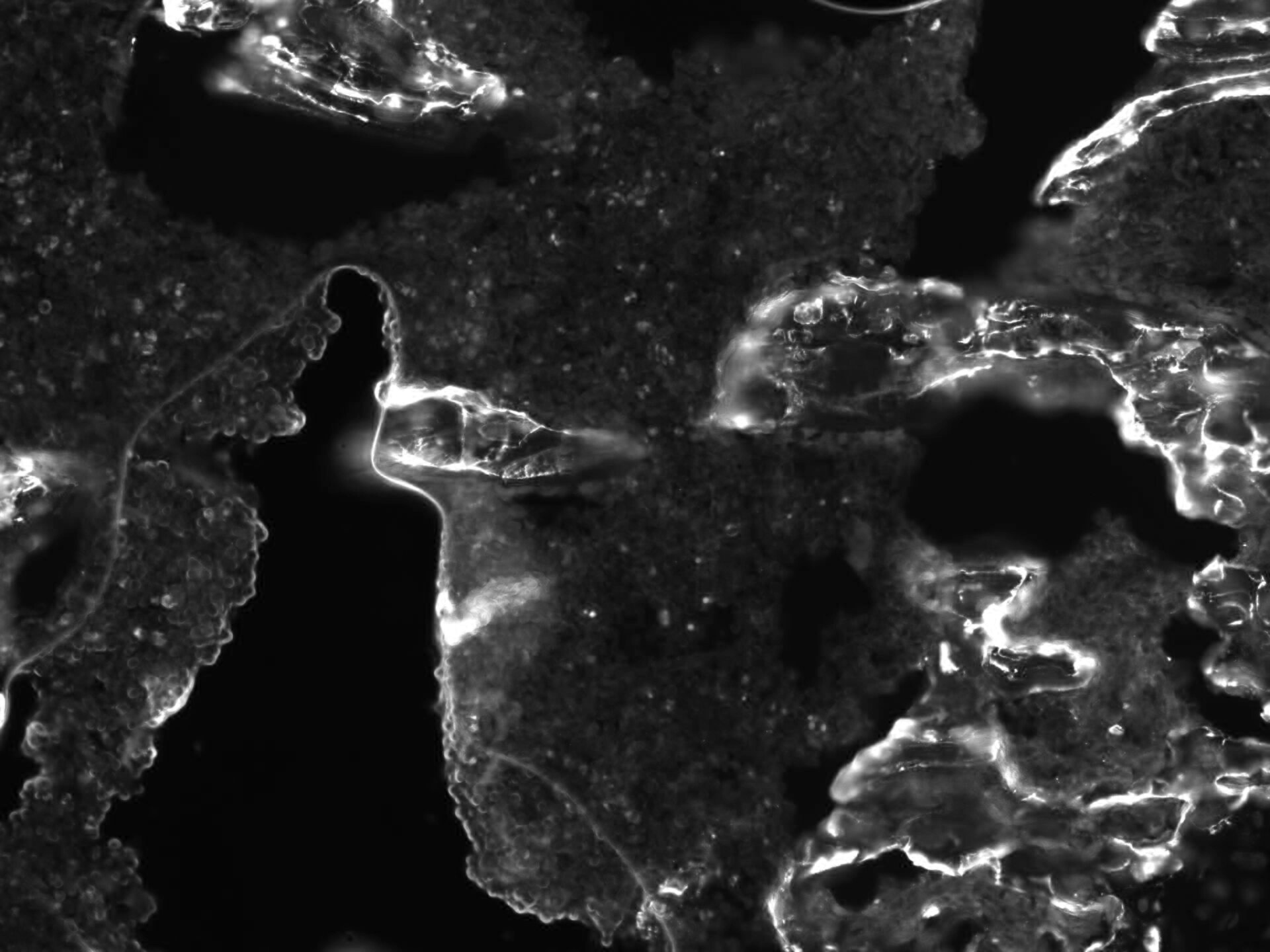
Immunocytochemistry/ Immunofluorescence: Osteopontin/OPN Antibody (1B20) - BSA Free [NB110-89062]
Immunocytochemistry/Immunofluorescence: Osteopontin/OPN Antibody (1B20) - BSA Free [NB110-89062] - Osteopontin antibody was tested at 1:50 in U2OS cells with FITC (green). Nuclei and actin were counterstained with Dapi (blue) and Phalloidin (red).![Immunohistochemistry: Osteopontin/OPN Antibody (1B20) - BSA Free [NB110-89062]](https://resources.rndsystems.com/images/products/Osteopontin-OPN-Antibody-1B20-BSA-Free-Immunohistochemistry-NB110-89062-img0012.jpg "Immunohistochemistry: Osteopontin/OPN Antibody (1B20) - BSA Free [NB110-89062]")
![Immunohistochemistry: Osteopontin/OPN Antibody (1B20) - BSA Free [NB110-89062]](https://resources.rndsystems.com/images/products/Osteopontin-OPN-Antibody-1B20-BSA-Free-Immunohistochemistry-NB110-89062-img0013.jpg "Immunohistochemistry: Osteopontin/OPN Antibody (1B20) - BSA Free [NB110-89062]")
Immunohistochemistry: Osteopontin/OPN Antibody (1B20) - BSA Free [NB110-89062]
Osteopontin-OPN-Antibody-1B20-BSA-Free-Immunohistochemistry-NB110-89062-img0013.jpg![Immunohistochemistry: Osteopontin/OPN Antibody (1B20) - BSA Free [NB110-89062]](https://resources.rndsystems.com/images/products/Osteopontin-OPN-Antibody-1B20-BSA-Free-Immunohistochemistry-NB110-89062-img0009.jpg "Immunohistochemistry: Osteopontin/OPN Antibody (1B20) - BSA Free [NB110-89062]")
Immunohistochemistry: Osteopontin/OPN Antibody (1B20) - BSA Free [NB110-89062]
Immunohistochemistry: Osteopontin/OPN Antibody (1B20) - BSA Free [NB110-89062] - Analysis of Osteopontin on human lung adenocarcinoma.![Immunohistochemistry: Osteopontin/OPN Antibody (1B20) - BSA Free [NB110-89062]](https://resources.rndsystems.com/images/products/Osteopontin-OPN-Antibody-1B20-BSA-Free-Immunohistochemistry-NB110-89062-img0011.jpg "Immunohistochemistry: Osteopontin/OPN Antibody (1B20) - BSA Free [NB110-89062]")
![Simple Western: Osteopontin/OPN Antibody (1B20)BSA Free [NB110-89062]](https://resources.rndsystems.com/images/products/Osteopontin-OPN-Antibody-1B20-BSA-Free-Simple-Western-NB110-89062-img0010.jpg "Simple Western: Osteopontin/OPN Antibody (1B20)BSA Free [NB110-89062]")
Simple Western: Osteopontin/OPN Antibody (1B20)BSA Free [NB110-89062]
Simple Western: Osteopontin/OPN Antibody (1B20) - BSA Free [NB110-89062] - Simple Western lane view shows a specific band for Osteopontin in 0.5 mg/ml of Human Breast Cancer lysate. This experiment was performed under reducing conditions using the 12-230 kDa separation system. * Non-specific interaction with the 230 kDa Simple Western standard may be seen with this antibody - BSA Free [NB110-89062] -")
Immunohistochemistry: Osteopontin/OPN Antibody (1B20) - BSA Free [NB110-89062] -
Immunohistochemistry: Osteopontin/OPN Antibody (1B20) - BSA Free [NB110-89062] - Photomicrographs of immunostaining for osteocalcin & osteopontin in (A) Group I, (B) Group II (C) Group III at 6 weeks.The pictures are arranged by staining technique (columns) & the investigated treatment (rows). Areas that stained positive for osteocalcin & osteopontin are indicated by red arrowheads. NB, new bone; OS, osteoid; FM, fatty marrow. Image collected & cropped by CiteAb from the following publication (https://peerj.com/articles/3513), licensed under a CC-BY license. Not internally tested by Novus Biologicals. - BSA Free [NB110-89062] -")
Western Blot: Osteopontin/OPN Antibody (1B20) - BSA Free [NB110-89062] -
Western Blot: Osteopontin/OPN Antibody (1B20) - BSA Free [NB110-89062] - Photomicrographs of immunostaining for osteocalcin & osteopontin in (A) Group I, (B) Group II (C) Group III at 6 weeks.The pictures are arranged by staining technique (columns) & the investigated treatment (rows). Areas that stained positive for osteocalcin & osteopontin are indicated by red arrowheads. NB, new bone; OS, osteoid; FM, fatty marrow. Image collected & cropped by CiteAb from the following publication (https://peerj.com/articles/3513), licensed under a CC-BY license. Not internally tested by Novus Biologicals. in HepG2 Human Cell Line.")
Osteopontin/OPN (1B20) in HepG2 Human Cell Line.
Osteopontin/OPN (1B20) was detected in immersion fixed HepG2 human hepatocellular carcinoma cell line using Rat anti- Osteopontin/OPN (1B20) Protein G Purified Monoclonal Antibody conjugated to Alexa Fluor® 488 (Catalog # NB110-89062AF488) (green) at 10 µg/mL overnight at 4C. Cells were counterstained with DAPI (dark blue). Cells were imaged using a 100X objective and digitally deconvolved.Applications for Osteopontin/OPN Antibody (1B20) - BSA Free
Immunocytochemistry/ Immunofluorescence
Immunohistochemistry
Immunohistochemistry-Paraffin
Immunoprecipitation
Simple Western
Western Blot
Reviewed Applications
Read 2 reviews rated 5 using NB110-89062 in the following applications:
Formulation, Preparation, and Storage
Purification
Formulation
Format
Preservative
Concentration
Shipping
Stability & Storage
Background: Osteopontin/OPN
Long Name
Alternate Names
Gene Symbol
UniProt
Additional Osteopontin/OPN Products
Product Documents for Osteopontin/OPN Antibody (1B20) - BSA Free
Certificate of Analysis
To download a Certificate of Analysis, please enter a lot or batch number in the search box below.
Product Specific Notices for Osteopontin/OPN Antibody (1B20) - BSA Free
This product is for research use only and is not approved for use in humans or in clinical diagnosis. Primary Antibodies are guaranteed for 1 year from date of receipt.
Citations for Osteopontin/OPN Antibody (1B20) - BSA Free
 Powered by Bioz
Customer Reviews for Osteopontin/OPN Antibody (1B20) - BSA Free (2)
Have you used Osteopontin/OPN Antibody (1B20) - BSA Free?
Submit a review and receive an Amazon gift card!
$25/€18/£15/$25CAN/¥2500 Yen for a review with an image
$10/€7/£6/$10CAN/¥1110 Yen for a review without an image
Submit a review
Customer Images
-
Application: Immunofluorescence - fixed-frozenSample Tested: bone marrowSpecies: MouseVerified Customer | Posted 02/29/2020Mouse OPN 1:100Mouse bones were fixed in PFA 4% for 4h on ice, decalcified in EDTA for 24h at 4°C, cryoprotected in 30% sucrose for 24 at 4°C, and embedded in OCT

-
Application: ELISASample Tested: Human breast carcinoma and Human breast cancer plasmaSpecies: HumanVerified Customer | Posted 08/11/2017It will be nice if using PBS buffer for product. Removing Gly-Tris buffer is a pain.
There are no reviews that match your criteria.
Protocols
View specific protocols for Osteopontin/OPN Antibody (1B20) - BSA Free (NB110-89062):
Immunohistochemistry-Paraffin Embedded Sections
Antigen Unmasking:
Bring slides to a boil in 10 mM sodium citrate buffer (pH 6.0) then maintain at a sub-boiling temperature for 10 minutes. Cool slides on bench-top for 30 minutes.
Staining:
1. Wash sections in deionized water three times for 5 minutes each.
2. Wash sections in wash buffer for 5 minutes.
3. Block each section with 100-400 ul blocking solution for 1 hour at room temperature.
4. Remove blocking solution and add 100-400 ul diluted primary antibody. Incubate overnight at 4C.
5. Remove antibody solution and wash sections in wash buffer three times for 5 minutes each.
6. Add 100-400 ul biotinylated diluted secondary antibody. Incubate 30 minutes at room temperature.
7. Remove secondary antibody solution and wash sections three times with wash buffer for 5 minutes each.
8. Add 100-400 ul Streptavidin-HRP reagent to each section and incubate for 30 minutes at room temperature.
9. Wash sections three times in wash buffer for 5 minutes each.
10. Add 100-400 ul DAB substrate to each section and monitor staining closely.
11. As soon as the sections develop, immerse slides in deionized water.
12. Counterstain sections in hematoxylin.
13. Wash sections in deionized water two times for 5 minutes each.
14. Dehydrate sections.
15. Mount coverslips.
Immunocytochemistry Protocol
Culture cells to appropriate density in 35 mm culture dishes or 6-well plates.
1. Remove culture medium and add 10% formalin to the dish. Fix at room temperature for 30 minutes.
2. Remove the formalin and add ice cold methanol. Incubate for 5-10 minutes.
3. Remove methanol and add washing solution (i.e. PBS). Be sure to not let the specimen dry out. Wash three times for 10 minutes.
4. To block nonspecific antibody binding incubate in 10% normal goat serum from 1 hour to overnight at room temperature.
5. Add primary antibody at appropriate dilution and incubate at room temperature from 2 hours to overnight at room temperature.
6. Remove primary antibody and replace with washing solution. Wash three times for 10 minutes.
7. Add secondary antibody at appropriate dilution. Incubate for 1 hour at room temperature.
8. Remove antibody and replace with wash solution, then wash for 10 minutes. Add Hoechst 33258 to wash solution at 1:25,0000 and incubate for 10 minutes. Wash a third time for 10 minutes.
9. Cells can be viewed directly after washing. The plates can also be stored in PBS containing Azide covered in Parafilm (TM). Cells can also be cover-slipped using Fluoromount, with appropriate sealing.
*The above information is only intended as a guide. The researcher should determine what protocol best meets their needs. Please follow safe laboratory procedures."
Western Blot Protocol
1. Perform SDS-PAGE on samples to be analyzed, loading 30 ug of total protein per lane.
2. Transfer proteins to membrane according to the instructions provided by the manufacturer of the membrane and transfer apparatus.
3. Stain according to standard Ponceau S procedure (or similar product) to assess transfer success, and mark molecular weight standards where appropriate.
4. Rinse the blot.
5. Block the membrane using standard blocking buffer for at least 1 hour.
6. Wash the membrane in wash buffer three times for 10 minutes each.
7. Dilute primary antibody in blocking buffer and incubate 1 hour at room temperature.
8. Wash the membrane in wash buffer three times for 10 minutes each.
9. Apply the diluted HRP conjugated secondary antibody in blocking buffer (as per manufacturers instructions) and incubate 1 hour at room temperature.
10. Wash the blot in wash buffer three times for 10 minutes each (this step can be repeated as required to reduce background).
11. Apply the detection reagent of choice in accordance with the manufacturers instructions.
Note: Tween-20 can be added to the blocking or antibody dilution buffer at a final concentration of 0.05-0.2%.
Find general support by application which include: protocols, troubleshooting, illustrated assays, videos and webinars.
- Antigen Retrieval Protocol (PIER)
- Antigen Retrieval for Frozen Sections Protocol
- Appropriate Fixation of IHC/ICC Samples
- Cellular Response to Hypoxia Protocols
- Chromogenic IHC Staining of Formalin-Fixed Paraffin-Embedded (FFPE) Tissue Protocol
- Chromogenic Immunohistochemistry Staining of Frozen Tissue
- ClariTSA™ Fluorophore Kits
- Detection & Visualization of Antibody Binding
- Fluorescent IHC Staining of Frozen Tissue Protocol
- Graphic Protocol for Heat-induced Epitope Retrieval
- Graphic Protocol for the Preparation and Fluorescent IHC Staining of Frozen Tissue Sections
- Graphic Protocol for the Preparation and Fluorescent IHC Staining of Paraffin-embedded Tissue Sections
- Graphic Protocol for the Preparation of Gelatin-coated Slides for Histological Tissue Sections
- ICC Cell Smear Protocol for Suspension Cells
- ICC Immunocytochemistry Protocol Videos
- ICC for Adherent Cells
- IHC Sample Preparation (Frozen sections vs Paraffin)
- Immunocytochemistry (ICC) Protocol
- Immunocytochemistry Troubleshooting
- Immunofluorescence of Organoids Embedded in Cultrex Basement Membrane Extract
- Immunofluorescent IHC Staining of Formalin-Fixed Paraffin-Embedded (FFPE) Tissue Protocol
- Immunohistochemistry (IHC) and Immunocytochemistry (ICC) Protocols
- Immunohistochemistry Frozen Troubleshooting
- Immunohistochemistry Paraffin Troubleshooting
- Immunoprecipitation Protocol
- Preparing Samples for IHC/ICC Experiments
- Preventing Non-Specific Staining (Non-Specific Binding)
- Primary Antibody Selection & Optimization
- Protocol for Heat-Induced Epitope Retrieval (HIER)
- Protocol for Making a 4% Formaldehyde Solution in PBS
- Protocol for VisUCyte™ HRP Polymer Detection Reagent
- Protocol for the Fluorescent ICC Staining of Cell Smears - Graphic
- Protocol for the Fluorescent ICC Staining of Cultured Cells on Coverslips - Graphic
- Protocol for the Preparation & Fixation of Cells on Coverslips
- Protocol for the Preparation and Chromogenic IHC Staining of Frozen Tissue Sections
- Protocol for the Preparation and Chromogenic IHC Staining of Frozen Tissue Sections - Graphic
- Protocol for the Preparation and Chromogenic IHC Staining of Paraffin-embedded Tissue Sections
- Protocol for the Preparation and Chromogenic IHC Staining of Paraffin-embedded Tissue Sections - Graphic
- Protocol for the Preparation and Fluorescent ICC Staining of Cells on Coverslips
- Protocol for the Preparation and Fluorescent ICC Staining of Non-adherent Cells
- Protocol for the Preparation and Fluorescent ICC Staining of Stem Cells on Coverslips
- Protocol for the Preparation and Fluorescent IHC Staining of Frozen Tissue Sections
- Protocol for the Preparation and Fluorescent IHC Staining of Paraffin-embedded Tissue Sections
- Protocol for the Preparation of Gelatin-coated Slides for Histological Tissue Sections
- Protocol for the Preparation of a Cell Smear for Non-adherent Cell ICC - Graphic
- R&D Systems Quality Control Western Blot Protocol
- TUNEL and Active Caspase-3 Detection by IHC/ICC Protocol
- The Importance of IHC/ICC Controls
- Troubleshooting Guide: Immunohistochemistry
- Troubleshooting Guide: Western Blot Figures
- Western Blot Conditions
- Western Blot Protocol
- Western Blot Protocol for Cell Lysates
- Western Blot Troubleshooting
- Western Blot Troubleshooting Guide
- View all Protocols, Troubleshooting, Illustrated assays and Webinars
FAQs for Osteopontin/OPN Antibody (1B20) - BSA Free
-
Q: I am very interested in your anti-human osteopontin (SPP1) (cat #NB110-89062). I work with bovine and would like to use the antibody in the blot. According to your description, the immunogen is a synthetic peptide which binds to the C-terminus of the human. The sequences of the C-termini are very similar in humans and cattle. Could you kindly tell me what amino acids against the C-terminus of the human antibody is directed exactly?
A:
The immunogen sequence for NB110-89062 lies within amino acids 284-314 of isoform A, UniProt P10451. I have carried out a UniProt Blast analysis of the exact immunogen sequence versus Bos taurus (bovine) proteins for you, and the result was a 95% match for both osteopontin and osteopontin-K. This is a high percentage strongly suggesting that the antibody will be suitable for bovine, although we cannot guarantee it as this has not been tested in the lab. Because you would be using NB110-89062 with a species not stated on the web page / datasheet for this product, you would not be covered by the Novus Guarantee but you would be eligible for our special Innovator's Reward program, where as a thank you for sharing your results with us (whether the antibody proved suitable or not) by completing a review, we would give you a voucher code for the price of the product. Please contact us at innovators@novusbio.com for more information.
Associated Pathways
