PCNA Antibody (PC10) - BSA Free
Novus Biologicals | Catalog # NB500-106
Key Product Details
Species Reactivity
Validated:
Cited:
Applications
Validated:
Cited:
Label
Antibody Source
Format
Product Specifications
Immunogen
Reactivity Notes
Marker
Specificity
Clonality
Host
Isotype
Theoretical MW
Disclaimer note: The observed molecular weight of the protein may vary from the listed predicted molecular weight due to post translational modifications, post translation cleavages, relative charges, and other experimental factors.
Scientific Data Images for PCNA Antibody (PC10) - BSA Free
![Simple Western: PCNA Antibody (PC10) [NB500-106]](https://resources.rndsystems.com/images/products/PCNA-Antibody-PC10-Simple-Western-NB500-106-img0008.jpg "Simple Western: PCNA Antibody (PC10) [NB500-106]")
Simple Western: PCNA Antibody (PC10) [NB500-106]
Simple Western: PCNA Antibody (PC10) [NB500-106] - Image shows a specific band for PCNA in 0.5 mg/mL of HeLa lysate. This experiment was performed under reducing conditions using the 12-230 kDa separation system.![Immunocytochemistry/ Immunofluorescence: PCNA Antibody (PC10) [NB500-106]](https://resources.rndsystems.com/images/products/PCNA-Antibody-PC10-Immunocytochemistry-Immunofluorescence-NB500-106-img0015.jpg "Immunocytochemistry/ Immunofluorescence: PCNA Antibody (PC10) [NB500-106]")
Immunocytochemistry/ Immunofluorescence: PCNA Antibody (PC10) [NB500-106]
Immunocytochemistry/Immunofluorescence: PCNA Antibody (PC10) [NB500-106] - NIH3T3 cells were fixed and permeabilized for 10 minutes using -20C MeOH. The cells were incubated with anti-PCNA Antibody (PC10) NB500-106 at 1 ug/ml overnight at 4C and detected with an anti-mouse Dylight 488 (Green) at a 1:1000 dilution for 60 minutes. Nuclei were counterstained with DAPI (Blue). Cells were imaged using a 100X objective and digitally deconvolved.![Immunohistochemistry-Paraffin: PCNA Antibody (PC10) [NB500-106]](https://resources.rndsystems.com/images/products/PCNA-Antibody-PC10-Immunohistochemistry-Paraffin-NB500-106-img0007.jpg "Immunohistochemistry-Paraffin: PCNA Antibody (PC10) [NB500-106]")
Immunohistochemistry-Paraffin: PCNA Antibody (PC10) [NB500-106]
Immunohistochemistry-Paraffin: PCNA Antibody (PC10) [NB500-106] - Analysis of paraffin-embedded human colon carcinoma, showing nuclear localization.![Western Blot: PCNA Antibody (PC10) [NB500-106]](https://resources.rndsystems.com/images/products/PCNA-Antibody-PC10-Western-Blot-NB500-106-img0006.jpg "Western Blot: PCNA Antibody (PC10) [NB500-106]")
Western Blot: PCNA Antibody (PC10) [NB500-106]
Western Blot: PCNA Antibody (PC10) [NB500-106] - Analysis of human (HeLa lysate), murine (SV-T2 lysate), bovine (BAEC lysate), and porcine (PAE lysate) cell extracts.![Western Blot: PCNA Antibody (PC10) [NB500-106]](https://resources.rndsystems.com/images/products/PCNA-Antibody-PC10-Western-Blot-NB500-106-img0014.jpg "Western Blot: PCNA Antibody (PC10) [NB500-106]")
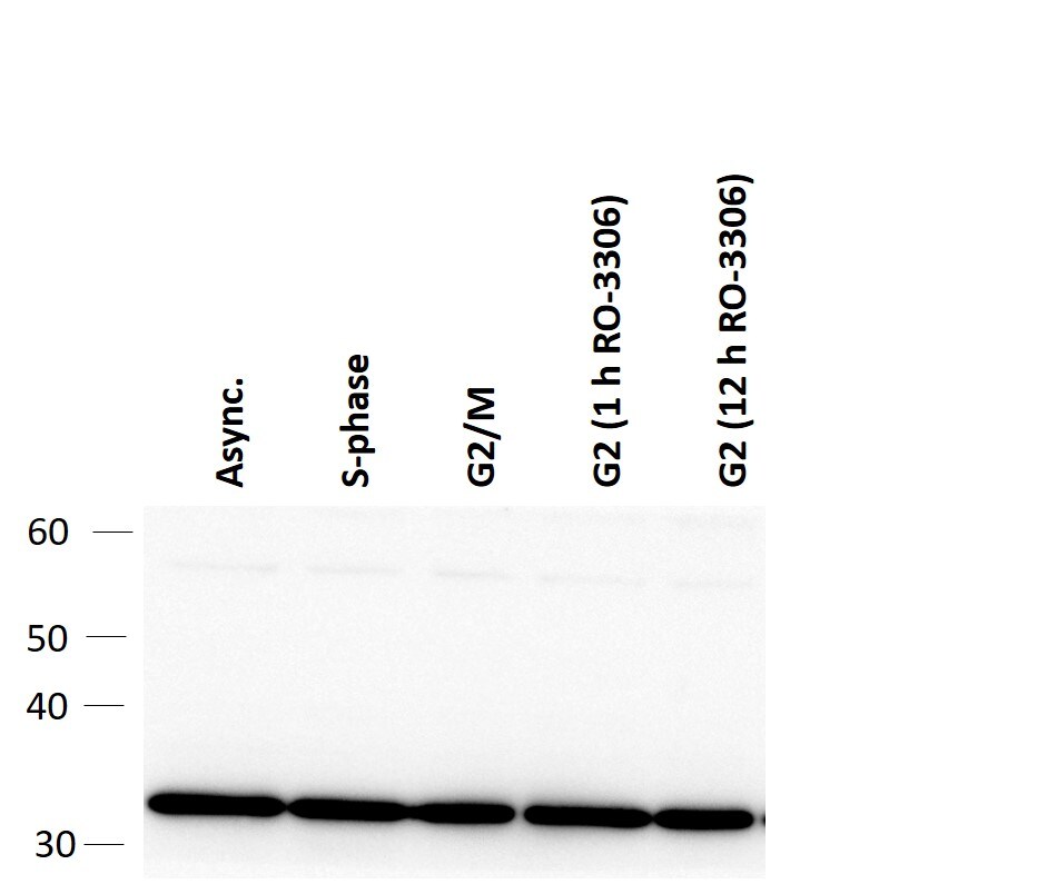
Western Blot: PCNA Antibody (PC10) [NB500-106]
Western Blot: PCNA Antibody (PC10) [NB500-106] - Western blot of PCNA levels across different stages of the cell cycle. U-2 OS cells were synchronised at G1/S with 2 mM thymidine an released for 10 hours for G2/M or re-arrested in G2 for extended duration with 10uM RO-3306. Antibody at 1:2000. WB image submitted by a verified customer review.![Immunohistochemistry-Frozen: PCNA Antibody (PC10) [NB500-106]](https://resources.rndsystems.com/images/products/PCNA-Antibody-PC10-Immunohistochemistry-Frozen-NB500-106-img0009.jpg "Immunohistochemistry-Frozen: PCNA Antibody (PC10) [NB500-106]")
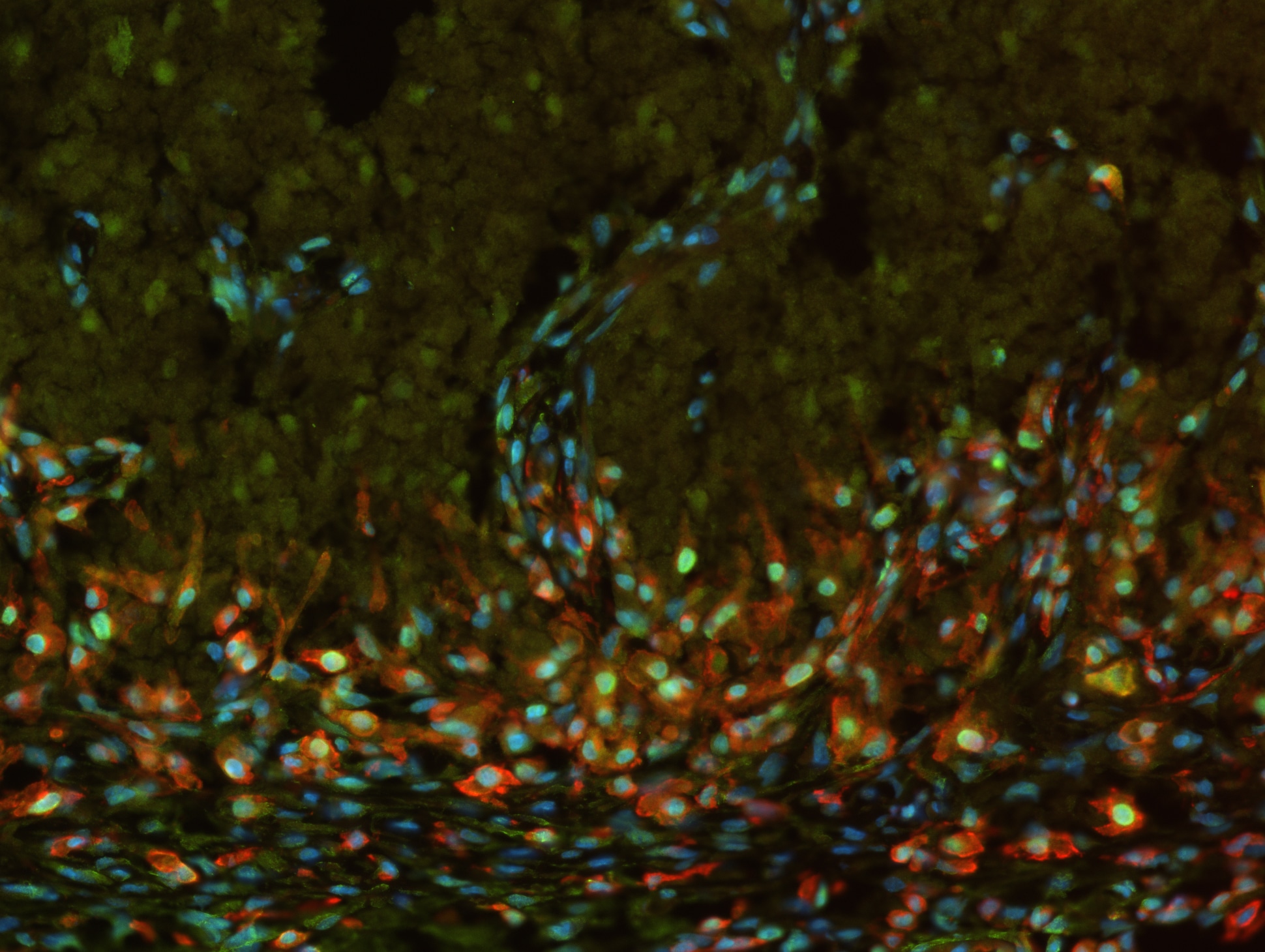
Immunohistochemistry-Frozen: PCNA Antibody (PC10) [NB500-106]
Immunohistochemistry-Frozen: PCNA Antibody (PC10) [NB500-106] - Paraformaldehyde fixed frozen section of brain from murine embryo using PCNA antibody clone PC10 (green), an IB4 antibody (red) and DAPI. Image provided by Dr. Siegenthaler via product review.![Immunocytochemistry/ Immunofluorescence: PCNA Antibody (PC10) [NB500-106]](https://resources.rndsystems.com/images/products/PCNA-Antibody-PC10-Immunocytochemistry-Immunofluorescence-NB500-106-img0016.jpg "Immunocytochemistry/ Immunofluorescence: PCNA Antibody (PC10) [NB500-106]")
Immunocytochemistry/ Immunofluorescence: PCNA Antibody (PC10) [NB500-106]
Immunocytochemistry/Immunofluorescence: PCNA Antibody (PC10) [NB500-106] - PC12 cells were fixed and permeabilized for 10 minutes using -20C MeOH. The cells were incubated with anti-PCNA Antibody (PC10) NB500-106 at 1 ug/ml overnight at 4C and detected with an anti-mouse Dylight 488 (Green) at a 1:1000 dilution for 60 minutes. Nuclei were counterstained with DAPI (Blue). Cells were imaged using a 100X objective and digitally deconvolved.![Immunocytochemistry/ Immunofluorescence: PCNA Antibody (PC10) [NB500-106]](https://resources.rndsystems.com/images/products/PCNA-Antibody-PC10-Immunocytochemistry-Immunofluorescence-NB500-106-img0010.jpg "Immunocytochemistry/ Immunofluorescence: PCNA Antibody (PC10) [NB500-106]")
Immunocytochemistry/ Immunofluorescence: PCNA Antibody (PC10) [NB500-106]
Immunocytochemistry/Immunofluorescence: PCNA Antibody (PC10) [NB500-106] - HeLa cells were fixed and permeabilized for 10 minutes using cold (-20C) MeOH. The cells were incubated with anti-PCNA [PC10] at 5 ug/mL overnight at 4C and detected with an anti-mouse IgG Dylight 488 (Green) at a 1:500 dilution. Nuclei were counterstained with DAPI (Blue). Cells were imaged using a 40X objective.![Immunohistochemistry: PCNA Antibody (PC10) [NB500-106]](https://resources.rndsystems.com/images/products/PCNA-Antibody-PC10-Immunohistochemistry-NB500-106-img0013.jpg "Immunohistochemistry: PCNA Antibody (PC10) [NB500-106]")
Immunohistochemistry: PCNA Antibody (PC10) [NB500-106]
PCNA-Antibody-PC10-Immunohistochemistry-NB500-106-img0013.jpg![Flow Cytometry: PCNA Antibody (PC10) [NB500-106]](https://resources.rndsystems.com/images/products/PCNA-Antibody-PC10-Flow-Cytometry-NB500-106-img0012.jpg "Flow Cytometry: PCNA Antibody (PC10) [NB500-106]")
Flow Cytometry: PCNA Antibody (PC10) [NB500-106]
Flow Cytometry: PCNA Antibody (PC10) [NB500-106] - An intracellular stain was performed on HepG2 cells with PCNA Antibody (PC10) NB500-106G (blue) and a matched isotype control (orange). Cells were fixed with 4% PFA and then permeabilized with 0.1% saponin. Cells were incubated in an antibody dilution of 10 ug/mL for 30 minutes at room temperature. Both antibodies were conjugated to DyLight 488.![Western Blot: PCNA Antibody (PC10) [NB500-106]](https://resources.rndsystems.com/images/products/PCNA-Antibody-PC10-Western-Blot-NB500-106-img0005.jpg "Western Blot: PCNA Antibody (PC10) [NB500-106]")
Western Blot: PCNA Antibody (PC10) [NB500-106]
Western Blot: PCNA Antibody (PC10) [NB500-106] - HeLa cell nuclear extract was resolved by electrophoresis, transferred to nitrocellulose and probed with monoclonal anti-PCNA antibody. Proteins were visualized using a goat anti-mouse secondary conjugated to HRP and a chemiluminescence detection system.![Western Blot: PCNA Antibody (PC10) [NB500-106]](https://resources.rndsystems.com/images/products/PCNA-Antibody-PC10-Western-Blot-NB500-106-img0011.jpg "Western Blot: PCNA Antibody (PC10) [NB500-106]")
Western Blot: PCNA Antibody (PC10) [NB500-106]
Western Blot: PCNA Antibody (PC10) [NB500-106] - Whole cell protein from human HeLa, A431, mouse Neuro2A and rat PC12 cells was separated on a 4-20% gel by SDS-PAGE, transferred to 0.2 um PVDF membrane and blocked in 5% non-fat milk in TBST. The membrane was probed with 1.0 ug/mL anti-Histone h3 in block buffer and detected with an anti-mouse HRP secondary antibody using chemiluminescence.![Immunohistochemistry: PCNA Antibody (PC10) [NB500-106]](https://resources.rndsystems.com/images/products/PCNA-Antibody-PC10-Immunohistochemistry-NB500-106-img0001.jpg "Immunohistochemistry: PCNA Antibody (PC10) [NB500-106]")
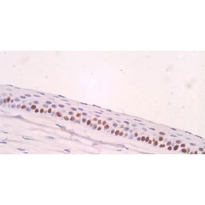
Immunohistochemistry: PCNA Antibody (PC10) [NB500-106]
Immunohistochemistry: PCNA Antibody (PC10) [NB500-106] - Analysis of PCNA in mouse cornea. Image courtesy of product review by Bo-Yie Chen. [NB500-106] -")
Chromatin Immunoprecipitation: PCNA Antibody (PC10) [NB500-106] -
Chromatin Immunoprecipitation: PCNA Antibody (PC10) [NB500-106] - Events taking place during a transient telomere dysfunction. All strains were cdc13–1, incubated for 160 min at 36°C to induce telomere uncapping, followed by 90 min at 23°C to allow telomere re-capping. Nocodazole & BrdU were added to the cultures at time 0. (A) Dynamics of ssDNA loss in sub-telomeres. (B) BrdU incorporation in sub-telomeres, minus incorporation at ‘CEN’ (e.g. a centromere-proximal locus, in this case ERG26). (C–F) Dynamics of the protein association with sub-telomeres, measured for Rap1 & major DNA synthesis factors (indicated above each graph). ChIP (%) was calculated as the fraction of immunoprecipitated sub-telomeric DNA, minus the fraction precipitated at ‘CEN’ (G) Dynamics of ssDNA loss at YER188W. (H) BrdU incorporation at YER188W, minus incorporation at ‘CEN’. (I–L) The protein association with YER188W was analyzed as in C–F. (M) ssDNA at ‘CEN’. (N) BrdU incorporation at ‘CEN’. (O–R) Association of proteins with ‘CEN’. (S) Growth of serial dilution of wild-type (first row) & cdc13–1 cells with or without additional mutations (indicated on the left of each row) at temperatures indicated above each plate. The plate shown on the right was cycled three times between 4 h at 36°C (to accumulate ssDNA) & 4 h at 21°C (to re-synthesize DNA), followed by incubation at 21°C for another two days. Image collected & cropped by CiteAb from the following publication (https://academic.oup.com/nar/article-lookup/doi/10.1093/nar/gkw071), licensed under a CC-BY license. Not internally tested by Novus Biologicals. [NB500-106] -")
Immunocytochemistry/ Immunofluorescence: PCNA Antibody (PC10) [NB500-106] -
Immunocytochemistry/ Immunofluorescence: PCNA Antibody (PC10) [NB500-106] - Development of the germinal zones in the tree shrew neocortex. (A–D) Immunofluorescence for PCNA (yellow) & DAPI staining (blue) on 30 μm-cryosections of E32–P1 tree shrew neocortex. The top margin of the image corresponds to the transition zone SVZ/intermediate zone. Scale bars, 50 μm. VZ, ventricular zone; SVZ, subventricular zone; iSVZ, inner SVZ; oSVZ, outer SVZ. (E) Quantification of the VZ thickness of the E32–P1 tree shrew neocortex. (F) Quantification of the SVZ thickness of the E32–P1 tree shrew neocortex. (G) Quantification of the VZ & SVZ thickness of the E32–P1 tree shrew neocortex, expressed as percentage of the sum of VZ & SVZ. (H) Quantification of the iSVZ & oSVZ thickness of the E37 tree shrew neocortex, expressed as percentage of the sum of iSVZ & oSVZ. (E–H) Data represent mean ± SD & were obtained from two consecutive sections of two brains each. Image collected & cropped by CiteAb from the following publication (https://pubmed.ncbi.nlm.nih.gov/29725291), licensed under a CC-BY license. Not internally tested by Novus Biologicals. - BSA Free [NB500-106] -")
Western Blot: PCNA Antibody (PC10) - BSA Free [NB500-106] -
Nuclear accumulation of SIRT4 is increased in renal tubules after injury.(A) Western blots analysis of SIRT4, FN1, COL1A1, and Tubulin in the kidney of UUO, uIRI, and sham mice (control group). (B) Nuclear fractions were prepared from the kidney of UUO, uIRI, and sham mice. Nuclear PCNA and cytoplasmic tubulin were used as controls. (C, D) Representative images of immunohistochemical staining of SIRT4 (scale bar, 50 μm) in the kidneys from mice that underwent sham surgery, UUO surgery on day 10 post surgery or uIRI surgery on day 28 post surgery. (E, F) Representative images of Masson’s trichrome staining (the upper panel; scale bar = 50 μm) and SIRT4 immumohistochemical staining (the bottom panel; scale bar = 20 μm) in the kidney sections from patients with CKD (n=8) and minimal change disease (control group, n=1).Figure 1—source data 1.Original files for western blot analysis displayed in Figure 1A, B.Figure 1—source data 2.The uncropped gels or blots with the relevant bands clearly labeled in Figure 1A and B.Original files for western blot analysis displayed in Figure 1A, B.The uncropped gels or blots with the relevant bands clearly labeled in Figure 1A and B. Image collected and cropped by CiteAb from the following open publication (https://elifesciences.org/articles/98524), licensed under a CC-BY license. Not internally tested by Novus Biologicals. - BSA Free [NB500-106] -")
Western Blot: PCNA Antibody (PC10) - BSA Free [NB500-106] -
U0126 prevents SIRT4 overexpression induced kidney fibrosis in UUO mice.(A, B) AAV9-Ksp-Sirt4 was injected into kidneys of mice in situ at three independent points in situ. After 2-week transfection, the mice received UUO surgery, and then mice were treated with U0126 (10 mg/kg body weight) or vehicle for 10 days (n=6 per group). (A) Organelle separation experiment and immunoblot analysis detected the localization of SIRT4 and Tubulin in the kidneys of mice. (B) Representative images of immunohistochemical staining of SIRT4 (scale bar, 50 μm) in the kidney sections of mice.Figure 8—figure supplement 1—source data 1.Original files for western blot analysis displayed in Figure 8—figure supplement 1A.Figure 8—figure supplement 1—source data 2.The uncropped gels or blots with the relevant bands clearly labeled in Figure 8—figure supplement 1A.Original files for western blot analysis displayed in Figure 8—figure supplement 1A.The uncropped gels or blots with the relevant bands clearly labeled in Figure 8—figure supplement 1A. Image collected and cropped by CiteAb from the following open publication (https://elifesciences.org/articles/98524), licensed under a CC-BY license. Not internally tested by Novus Biologicals. - BSA Free [NB500-106] -")
Western Blot: PCNA Antibody (PC10) - BSA Free [NB500-106] -
Inhibition of nucleus accumulation of beta -catenin failed to suppresses kidney fibrosis induced by SIRT4 overexpression in UUO mice.(A–C) AAV9-Ksp-Sirt4 or AAV-Ctrl was injected into kidneys of mice in situ at three independent points in situ. After 2-week transfection, the mice received UUO surgery. After UUO surgery, mice were treated with vehicle or MSAB for 10 days (n=6 per group). (A) Western blot analysis of the expression of SIRT4, CCN2, FN1, COL3A1, alpha -SMA, and Tubulin in the kidneys from mice. (B) Representative images of Masson’s trichrome staining and Sirius red staining in kidney sections of mice (scale bar, 100 μm). (C) The mRNA level of Col1a1, Fn1, Eln, Ccn2, Acta2, and Col3a1 in the kidney of mice. For all panels, data are presented as mean +/- SD. ns: not significant difference, *p<0.05, **p<0.01, ***p<0.001 by one-way ANOVA with Bonferroni correction test.Figure 8—figure supplement 3—source data 1.Original files for western blot analysis displayed in Figure 8—figure supplement 3A.Figure 8—figure supplement 3—source data 2.The uncropped gels or blots with the relevant bands clearly labeled in Figure 8—figure supplement 3A.Original files for western blot analysis displayed in Figure 8—figure supplement 3A.The uncropped gels or blots with the relevant bands clearly labeled in Figure 8—figure supplement 3A. Image collected and cropped by CiteAb from the following open publication (https://elifesciences.org/articles/98524), licensed under a CC-BY license. Not internally tested by Novus Biologicals. - BSA Free [NB500-106] -")
Western Blot: PCNA Antibody (PC10) - BSA Free [NB500-106] -
Exosomes contain anti-SIRT4 antibody alleviated UUO-induced kidney fibrosis.(A–D) After sham or UUO surgery, WT mice were treated with exosomes null or contain anti-SIRT4 for 10 days (n=6 per group). (A) Western blot analysis of the expression of CCN2, FN1, COL1A1, COL3A1, E-cadherin. alpha -SMA and Tubulin in the kidney from mice. (B) Representative images of Masson’s trichrome staining and Sirius red staining in kidneys sections of mice (scale bar, 100 μm). (C) The mRNA level of Col1a1, Fn1, Eln, Ccn2, Acta2, and Col3a1 in the kidney of mice. (D) Organelle separation experiment and immunoblot analysis detected the localization of SIRT4 in the kidneys of mice. For all panels, data are presented as mean +/- SD. *p<0.05, **p<0.01, ***p<0.001 by one-way ANOVA with Bonferroni correction test.Figure 8—figure supplement 2—source data 1.Original files for western blot analysis displayed in Figure 8—figure supplement 2A, D.Figure 8—figure supplement 2—source data 2.The uncropped gels or blots with the relevant bands clearly labeled in Figure 8—figure supplement 2A, D.Original files for western blot analysis displayed in Figure 8—figure supplement 2A, D.The uncropped gels or blots with the relevant bands clearly labeled in Figure 8—figure supplement 2A, D. Image collected and cropped by CiteAb from the following open publication (https://elifesciences.org/articles/98524), licensed under a CC-BY license. Not internally tested by Novus Biologicals. - BSA Free [NB500-106] -")
Western Blot: PCNA Antibody (PC10) - BSA Free [NB500-106] -
(a) Western blot analysis of PARP1 and PCNA protein levels. (b) Representative images of immunoreactive bands. Data are presented as means +/- SEM of densitometric analysis of immunoreactive bands normalized to internal reference protein (GAPDH). One-way ANOVA p < 0.05; a,bp < 0.05, Holm–Sidak post hoc multiple comparison. Image collected and cropped by CiteAb from the following open publication (https://pubmed.ncbi.nlm.nih.gov/33802807), licensed under a CC-BY license. Not internally tested by Novus Biologicals.Applications for PCNA Antibody (PC10) - BSA Free
Chromatin Immunoprecipitation
Chromatin Immunoprecipitation (ChIP)
ELISA
Flow Cytometry
Immunocytochemistry/ Immunofluorescence
Immunohistochemistry
Immunohistochemistry Free-Floating
Immunohistochemistry-Frozen
Immunohistochemistry-Paraffin
Immunoprecipitation
Simple Western
Western Blot
In Simple Western only 10 - 15 uL of the recommended dilution is used per data point.
See Simple Western Antibody Database for Simple Western validation: Tested in HeLa lysate 0.5 mg/mL, separated by Size, antibody dilution of 1:200, apparent MW was 36 kDa. Separated by Size-Wes, Sally Sue/Peggy Sue.
The observed molecular weight of the protein may vary from the listed predicted molecular weight due to post translational modifications, post translation cleavages, relative charges, and other experimental factors.
Reviewed Applications
Read 5 reviews rated 4.8 using NB500-106 in the following applications:
Flow Cytometry Panel Builder
Bio-Techne Knows Flow Cytometry
Save time and reduce costly mistakes by quickly finding compatible reagents using the Panel Builder Tool.
Advanced Features
- Spectra Viewer - Custom analysis of spectra from multiple fluorochromes
- Spillover Popups - Visualize the spectra of individual fluorochromes
- Antigen Density Selector - Match fluorochrome brightness with antigen density

Formulation, Preparation, and Storage
Purification
Formulation
Format
Preservative
Concentration
Shipping
Stability & Storage
Background: PCNA
Long Name
Alternate Names
Gene Symbol
UniProt
Additional PCNA Products
Product Documents for PCNA Antibody (PC10) - BSA Free
Certificate of Analysis
To download a Certificate of Analysis, please enter a lot or batch number in the search box below.
Product Specific Notices for PCNA Antibody (PC10) - BSA Free
This product is for research use only and is not approved for use in humans or in clinical diagnosis. Primary Antibodies are guaranteed for 1 year from date of receipt.
Citations for PCNA Antibody (PC10) - BSA Free
 Powered by Bioz
Customer Reviews for PCNA Antibody (PC10) - BSA Free (5)
Have you used PCNA Antibody (PC10) - BSA Free?
Submit a review and receive an Amazon gift card!
$25/€18/£15/$25CAN/¥2500 Yen for a review with an image
$10/€7/£6/$10CAN/¥1110 Yen for a review without an image
Submit a review
Customer Images
-
Application: Western BlotSample Tested: U-2 OS cellsSpecies: HumanVerified Customer | Posted 12/17/2020Western blot of PCNA levels across different stages of the cell cycle. U-2 OS cells were synchronised at G1/S with 2mM thymidine an released for 10 hours for G2/M or re-arrested in G2 for extended duration with 10uM RO-3306.Used at 1:2000 dilution in 0.5% milk PBST. Incubated overnight at 4C.

-
Application: Western BlotSample Tested: Human cancer cell whole cell lysateSpecies: HumanVerified Customer | Posted 08/23/2015
-
Application: Immunohistochemistry-ParaffinSample Tested: Chicken intestineSpecies: OtherVerified Customer | Posted 03/28/2014
-
Application: ImmunofluorescenceVerified Customer | Posted 02/24/2014PCNA - IB4 & DAPI
-
Application: ImmunohistochemistrySample Tested: Mouse corneaSpecies: MouseVerified Customer | Posted 02/06/2012
There are no reviews that match your criteria.
Protocols
Find general support by application which include: protocols, troubleshooting, illustrated assays, videos and webinars.
- 7-Amino Actinomycin D (7-AAD) Cell Viability Flow Cytometry Protocol
- Antigen Retrieval Protocol (PIER)
- Antigen Retrieval for Frozen Sections Protocol
- Appropriate Fixation of IHC/ICC Samples
- Cellular Response to Hypoxia Protocols
- ChIP Protocol Video
- Chromatin Immunoprecipitation (ChIP) Protocol
- Chromatin Immunoprecipitation Protocol
- Chromogenic IHC Staining of Formalin-Fixed Paraffin-Embedded (FFPE) Tissue Protocol
- Chromogenic Immunohistochemistry Staining of Frozen Tissue
- ClariTSA™ Fluorophore Kits
- Detection & Visualization of Antibody Binding
- ELISA Sample Preparation & Collection Guide
- ELISA Troubleshooting Guide
- Extracellular Membrane Flow Cytometry Protocol
- Flow Cytometry Protocol for Cell Surface Markers
- Flow Cytometry Protocol for Staining Membrane Associated Proteins
- Flow Cytometry Staining Protocols
- Flow Cytometry Troubleshooting Guide
- Fluorescent IHC Staining of Frozen Tissue Protocol
- Graphic Protocol for Heat-induced Epitope Retrieval
- Graphic Protocol for the Preparation and Fluorescent IHC Staining of Frozen Tissue Sections
- Graphic Protocol for the Preparation and Fluorescent IHC Staining of Paraffin-embedded Tissue Sections
- Graphic Protocol for the Preparation of Gelatin-coated Slides for Histological Tissue Sections
- How to Run an R&D Systems DuoSet ELISA
- How to Run an R&D Systems Quantikine ELISA
- How to Run an R&D Systems Quantikine™ QuicKit™ ELISA
- ICC Cell Smear Protocol for Suspension Cells
- ICC Immunocytochemistry Protocol Videos
- ICC for Adherent Cells
- IHC Sample Preparation (Frozen sections vs Paraffin)
- Immunocytochemistry (ICC) Protocol
- Immunocytochemistry Troubleshooting
- Immunofluorescence of Organoids Embedded in Cultrex Basement Membrane Extract
- Immunofluorescent IHC Staining of Formalin-Fixed Paraffin-Embedded (FFPE) Tissue Protocol
- Immunohistochemistry (IHC) and Immunocytochemistry (ICC) Protocols
- Immunohistochemistry Frozen Troubleshooting
- Immunohistochemistry Paraffin Troubleshooting
- Immunoprecipitation Protocol
- Intracellular Flow Cytometry Protocol Using Alcohol (Methanol)
- Intracellular Flow Cytometry Protocol Using Detergents
- Intracellular Nuclear Staining Flow Cytometry Protocol Using Detergents
- Intracellular Staining Flow Cytometry Protocol Using Alcohol Permeabilization
- Intracellular Staining Flow Cytometry Protocol Using Detergents to Permeabilize Cells
- Preparing Samples for IHC/ICC Experiments
- Preventing Non-Specific Staining (Non-Specific Binding)
- Primary Antibody Selection & Optimization
- Propidium Iodide Cell Viability Flow Cytometry Protocol
- Protocol for Heat-Induced Epitope Retrieval (HIER)
- Protocol for Liperfluo
- Protocol for Making a 4% Formaldehyde Solution in PBS
- Protocol for VisUCyte™ HRP Polymer Detection Reagent
- Protocol for the Characterization of Human Th22 Cells
- Protocol for the Characterization of Human Th9 Cells
- Protocol for the Fluorescent ICC Staining of Cell Smears - Graphic
- Protocol for the Fluorescent ICC Staining of Cultured Cells on Coverslips - Graphic
- Protocol for the Preparation & Fixation of Cells on Coverslips
- Protocol for the Preparation and Chromogenic IHC Staining of Frozen Tissue Sections
- Protocol for the Preparation and Chromogenic IHC Staining of Frozen Tissue Sections - Graphic
- Protocol for the Preparation and Chromogenic IHC Staining of Paraffin-embedded Tissue Sections
- Protocol for the Preparation and Chromogenic IHC Staining of Paraffin-embedded Tissue Sections - Graphic
- Protocol for the Preparation and Fluorescent ICC Staining of Cells on Coverslips
- Protocol for the Preparation and Fluorescent ICC Staining of Non-adherent Cells
- Protocol for the Preparation and Fluorescent ICC Staining of Stem Cells on Coverslips
- Protocol for the Preparation and Fluorescent IHC Staining of Frozen Tissue Sections
- Protocol for the Preparation and Fluorescent IHC Staining of Paraffin-embedded Tissue Sections
- Protocol for the Preparation of Gelatin-coated Slides for Histological Tissue Sections
- Protocol for the Preparation of a Cell Smear for Non-adherent Cell ICC - Graphic
- Protocol: Annexin V and PI Staining by Flow Cytometry
- Protocol: Annexin V and PI Staining for Apoptosis by Flow Cytometry
- Quantikine HS ELISA Kit Assay Principle, Alkaline Phosphatase
- Quantikine HS ELISA Kit Principle, Streptavidin-HRP Polymer
- R&D Systems Quality Control Western Blot Protocol
- Sandwich ELISA (Colorimetric) – Biotin/Streptavidin Detection Protocol
- Sandwich ELISA (Colorimetric) – Direct Detection Protocol
- TUNEL and Active Caspase-3 Detection by IHC/ICC Protocol
- The Importance of IHC/ICC Controls
- Troubleshooting Guide: ELISA
- Troubleshooting Guide: Fluorokine Flow Cytometry Kits
- Troubleshooting Guide: Immunohistochemistry
- Troubleshooting Guide: Western Blot Figures
- Western Blot Conditions
- Western Blot Protocol
- Western Blot Protocol for Cell Lysates
- Western Blot Troubleshooting
- Western Blot Troubleshooting Guide
- View all Protocols, Troubleshooting, Illustrated assays and Webinars
FAQs for PCNA Antibody (PC10) - BSA Free
-
Q: Can you please tell me the specific region of immunogen of this product?
A: This antibody was raised against whole rat PCNA protein fused to Protein A. As we have not yet epitope mapped this product, we do not know the exact immunogen recognized by this antibody.
-
Q: Do you have data/information on the use of this antibody for Drosophila (mentioned on your website as possible target species)? I would like to know if any specific treatment is needed with regard to this type of sample and the use of the antibody in WB, IP in ICC.
A:
This particular product is a widely licensed monoclonal clone. We obtained validation of its ability to detect the Drosophila protein from the following publication: PMID 22253867. You should be able to follow our general protocols for WB, IP, and ICC to detect this protein in their samples. Please see the following link for protocols.
-
Q: I am looking into preparing an exercise for an undergraduate lab that would immunostain drosophila testes. I think that PCNA Antibody (PC10) (cat # NB500-106) would be used as the primary antibody. What would you suggest to be used for the secondary? We have a fluorescent microscope and have used FITC in the past for some of our labs. Just trying to give the students a chance to visualize stained testes.
A:
After looking at the applications of this antibody, I just found that we have not established this antibody for its Immunofluorescence application in our lab yet, however, as it has been tested successfully in IHC-P by our lab and by other researchers; there is a very strong chance that it should work in IHC-P/IF application also. If you would be interested in testing this novel application, please take a look at our Innovator's Reward Program. Regarding your specific query on secondary antibody, our FITC conjugated Mouse IgG antibody with catalog # NB720-F would be a perfect choice for primary PCNA Antibody (PC10) with catalog # NB500-106.
-
Q: Can you please tell me the specific region of immunogen of this product?
A: This antibody was raised against whole rat PCNA protein fused to Protein A. As we have not yet epitope mapped this product, we do not know the exact immunogen recognized by this antibody.
-
Q: Do you have data/information on the use of this antibody for Drosophila (mentioned on your website as possible target species)? I would like to know if any specific treatment is needed with regard to this type of sample and the use of the antibody in WB, IP in ICC.
A:
This particular product is a widely licensed monoclonal clone. We obtained validation of its ability to detect the Drosophila protein from the following publication: PMID 22253867. You should be able to follow our general protocols for WB, IP, and ICC to detect this protein in their samples. Please see the following link for protocols.
-
Q: I am looking into preparing an exercise for an undergraduate lab that would immunostain drosophila testes. I think that PCNA Antibody (PC10) (cat # NB500-106) would be used as the primary antibody. What would you suggest to be used for the secondary? We have a fluorescent microscope and have used FITC in the past for some of our labs. Just trying to give the students a chance to visualize stained testes.
A:
After looking at the applications of this antibody, I just found that we have not established this antibody for its Immunofluorescence application in our lab yet, however, as it has been tested successfully in IHC-P by our lab and by other researchers; there is a very strong chance that it should work in IHC-P/IF application also. If you would be interested in testing this novel application, please take a look at our Innovator's Reward Program. Regarding your specific query on secondary antibody, our FITC conjugated Mouse IgG antibody with catalog # NB720-F would be a perfect choice for primary PCNA Antibody (PC10) with catalog # NB500-106.