Perilipin Antibody - BSA Free
Novus Biologicals | Catalog # NB110-40760
![Western Blot: Perilipin Antibody [NB110-40760]](https://resources.rndsystems.com/images/products/Perilipin-Antibody-Western-Blot-NB110-40760-img0002.jpg "Western Blot: Perilipin Antibody [NB110-40760]")
Key Product Details
Species Reactivity
Validated:
Human, Mouse, Rat, Porcine, Fish
Cited:
Human, Mouse, Rat
Predicted:
Bovine (92%), Sheep (92%). Backed by our 100% Guarantee.
Applications
Validated:
Immunohistochemistry, Immunohistochemistry-Paraffin, Western Blot, Flow Cytometry, Immunocytochemistry/ Immunofluorescence, Imaging Mass Cytometry
Cited:
Immunohistochemistry, Immunohistochemistry-Paraffin, Western Blot, Immunocytochemistry/ Immunofluorescence, IF/IHC
Label
Unconjugated
Antibody Source
Polyclonal Rabbit IgG
Format
BSA Free
Loading...
Product Specifications
Immunogen
This Perilipin Antibody was developed against a synthetic peptide made to a region between residues 450-522 (C-terminus) of the human Perilipin protein. [Swiss-Prot# O60240]
Reactivity Notes
Fish reactivity reported in scientific literature (PMID: 27012897).
Localization
Endoplasmic reticulum, Lipid droplet (surface-associated).
Clonality
Polyclonal
Host
Rabbit
Isotype
IgG
Theoretical MW
60 kDa.
Disclaimer note: The observed molecular weight of the protein may vary from the listed predicted molecular weight due to post translational modifications, post translation cleavages, relative charges, and other experimental factors.
Disclaimer note: The observed molecular weight of the protein may vary from the listed predicted molecular weight due to post translational modifications, post translation cleavages, relative charges, and other experimental factors.
Scientific Data Images for Perilipin Antibody - BSA Free
Western Blot: Perilipin Antibody [NB110-40760]
Western Blot: Perilipin Antibody [NB110-40760] - Total protein from Human breast (lane 1), Human adipose membrane fraction (lane 2) and Human adipose tissue (lane 3) were separated on a 12% gel by SDS-PAGE. Protein was transferred to PVDF membrane, probed with 2 ug/mL anti-perilipin antibody followed by anti-rabbit HRP conjugated secondary antibody and detected with chemiluminescence.![Immunohistochemistry-Paraffin: Perilipin Antibody [NB110-40760]](https://resources.rndsystems.com/images/products/Perilipin-Antibody-Immunohistochemistry-Paraffin-NB110-40760-img0005.jpg "Immunohistochemistry-Paraffin: Perilipin Antibody [NB110-40760]")
Immunohistochemistry-Paraffin: Perilipin Antibody [NB110-40760]
Immunohistochemistry-Paraffin: Perilipin Antibody [NB110-40760] - IHC analysis of formalin fixed paraffin embedded section of liver tissue from mouse with Perilipin antibody at 1:200 dilution. The hepatocytes developed specific staining in the cytoplasm and around the nuclei of some cells, the immunostaining reflected a punctate appearance which is potentially the ER of the cells.![Flow Cytometry: Perilipin Antibody [NB110-40760]](https://resources.rndsystems.com/images/products/Perilipin-Antibody-Flow-Cytometry-NB110-40760-img0008.jpg "Flow Cytometry: Perilipin Antibody [NB110-40760]")
Flow Cytometry: Perilipin Antibody [NB110-40760]
Flow Cytometry: Perilipin Antibody [NB110-40760] - An intracellular stain was performed on U2OS cells with Perilipin Antibody NB110-40760AF488 (blue) and a matched isotype control (orange). Cells were fixed with 4% PFA and then permeabilized with 0.1% saponin. Cells were incubated in an antibody dilution of 5 ug/mL for 30 minutes at room temperature. Both antibodies were conjugated to Alexa Fluor 488.![Immunohistochemistry-Paraffin: Perilipin Antibody [NB110-40760]](https://resources.rndsystems.com/images/products/Perilipin-Antibody-Immunohistochemistry-Paraffin-NB110-40760-img0003.jpg "Immunohistochemistry-Paraffin: Perilipin Antibody [NB110-40760]")
Immunohistochemistry-Paraffin: Perilipin Antibody [NB110-40760]
Immunohistochemistry-Paraffin: Perilipin Antibody [NB110-40760] - IHC analysis of formalin fixed paraffin embedded section of fat tissue from mouse with Perilipin antibody at 1:200 dilution. The antibody generated an expected staining in the adipocytes towards periphery of the cells.![Immunohistochemistry-Paraffin: Perilipin Antibody [NB110-40760]](https://resources.rndsystems.com/images/products/Perilipin-Antibody-Immunohistochemistry-Paraffin-NB110-40760-img0006.jpg "Immunohistochemistry-Paraffin: Perilipin Antibody [NB110-40760]")
Immunohistochemistry-Paraffin: Perilipin Antibody [NB110-40760]
Immunohistochemistry-Paraffin: Perilipin Antibody [NB110-40760] - IHC analysis of formalin fixed paraffin embedded section of liver tissue from mouse with Perilipin antibody at 1:200 dilution. The hepatocytes developed specific staining in the cytoplasm and around the nuclei of some cells, the immunostaining reflected a punctate appearance which is potentially the ER of the cells.![Flow Cytometry: Perilipin Antibody [NB110-40760]](https://resources.rndsystems.com/images/products/Perilipin-Antibody-Flow-Cytometry-NB110-40760-img0007.jpg "Flow Cytometry: Perilipin Antibody [NB110-40760]")
Flow Cytometry: Perilipin Antibody [NB110-40760]
Flow Cytometry: Perilipin Antibody [NB110-40760] - An intracellular stain was performed on MCF7 cells with Perilipin Antibody NB110-40760AF488 (blue) and a matched isotype control (orange). Cells were fixed with 4% PFA and then permeabilized with 0.1% saponin. Cells were incubated in an antibody dilution of 10 ug/mL for 30 minutes at room temperature. Both antibodies were conjugated to Alexa Fluor 488.![Imaging Mass Cytometry: Perilipin Antibody [NB110-40760]](https://resources.rndsystems.com/images/products/Perilipin-Antibody-Imaging-Mass-Cytometry-NB110-40760-img0009.jpg "Imaging Mass Cytometry: Perilipin Antibody [NB110-40760]")
Imaging Mass Cytometry: Perilipin Antibody [NB110-40760]
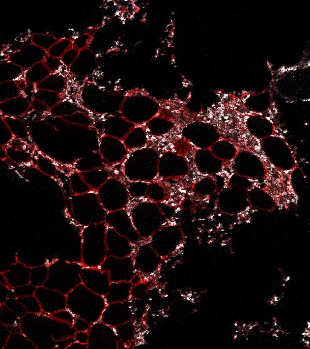
Imaging Mass Cytometry: Perilipin Antibody [NB110-40760] - Human bone marrow analyzed by IMC. Perilipin antibody conjugated with 151Eu and used 1:50 on human bone marrow FFPE. Perilipin - red. DNA- white. IMC image submitted by a verified customer review.![Perilipin Antibody - BSA Free Immunocytochemistry/ Immunofluorescence: Perilipin Antibody [NB110-40760]](https://resources.rndsystems.com/images/products/antibody/nb110-40760_rabbit-polyclonal-perilipin-antibody-231202682452.jpg "Immunocytochemistry/ Immunofluorescence: Perilipin Antibody [NB110-40760]")
Immunocytochemistry/ Immunofluorescence: Perilipin Antibody [NB110-40760]
Perilipin was detected in immersion fixed HepG2 human hepatocellular carcinoma cell line using Rabbit anti-Perilipin Antigen Affinity Purified Polyclonal Antibody (Catalog # NB110-40760) at 1.0 µg/mL overnight at 4C. Cells were stained using DyLight 488-conjugated Anti-Rabbit IgG (H+L) Cross-Absorbed Secondary Antibody (green), and counterstained with DAPI (blue). Cells were imaged using a 100X objective and digitally deconvolved.
Immunohistochemistry: Perilipin Antibody - BSA Free [NB110-40760] -
Basic CFO structure. Basic CFO structure was investigated by determining the presence of an enveloping surface membrane and absence of a cell nucleus-like structure. (A) Some CFOs displayed a wrinkled surface (Control, CFO only) suggesting the presence of an enveloping membrane. The enveloping surface membrane and absence of a nucleus-like structure were investigated with positive staining by membrane-specific CellMask green and negative staining of DNA-binding Hoechst 33,342, respectively. K562 cells were added to indicate positive staining (200×). CellTrace CFSE failed completely to stain CFOs. (B) An indirect detection method for CFO membrane was tested by detecting membrane perilipin and perilipin-2 proteins with biotin-conjugated specific antibodies, which were revealed by secondary binding of streptavidin-conjugated nanoparticles, each of which was 4.5 μm in diameter. During microscopic imaging (400×), when the focus was set on bottom of a CFO, nanoparticles binding specifically on the CFO could be seen. Image collected and cropped by CiteAb from the following open publication (https://pubmed.ncbi.nlm.nih.gov/35237181), licensed under a CC-BY license. Not internally tested by Novus Biologicals.Applications for Perilipin Antibody - BSA Free
Application
Recommended Usage
Flow Cytometry
5 - 10 ug/mL
Immunocytochemistry/ Immunofluorescence
reported in scientific literature
Immunohistochemistry
1:200
Immunohistochemistry-Paraffin
1:200
Western Blot
2 ug/mL
Application Notes
This Perilipin antibody is useful for Western blot anlaysis where a band at ~60 kDa is seen. This Perilipin Antibody is validated for Imaging Mass Cytometry from a verified customer review.
Reviewed Applications
Read 1 review rated 5 using NB110-40760 in the following applications:
Flow Cytometry Panel Builder
Bio-Techne Knows Flow Cytometry
Save time and reduce costly mistakes by quickly finding compatible reagents using the Panel Builder Tool.
Advanced Features
- Spectra Viewer - Custom analysis of spectra from multiple fluorochromes
- Spillover Popups - Visualize the spectra of individual fluorochromes
- Antigen Density Selector - Match fluorochrome brightness with antigen density

Formulation, Preparation, and Storage
Purification
Immunogen affinity purified
Formulation
PBS
Format
BSA Free
Preservative
0.02% Sodium Azide
Concentration
1.0 mg/ml
Shipping
The product is shipped with polar packs. Upon receipt, store it immediately at the temperature recommended below.
Stability & Storage
Store at 4C short term. Aliquot and store at -20C long term. Avoid freeze-thaw cycles.
Background: Perilipin
Alternate Names
PERI, PLIN, PLIN1
Gene Symbol
PLIN1
Additional Perilipin Products
Product Documents for Perilipin Antibody - BSA Free
Certificate of Analysis
To download a Certificate of Analysis, please enter a lot or batch number in the search box below.
Product Specific Notices for Perilipin Antibody - BSA Free
This product is for research use only and is not approved for use in humans or in clinical diagnosis. Primary Antibodies are guaranteed for 1 year from date of receipt.
Citations for Perilipin Antibody - BSA Free
 Powered by Bioz
Customer Reviews for Perilipin Antibody - BSA Free (1)
5 out of 5
1 Customer Rating
Have you used Perilipin Antibody - BSA Free?
Submit a review and receive an Amazon gift card!
$25/€18/£15/$25CAN/¥2500 Yen for a review with an image
$10/€7/£6/$10CAN/¥1110 Yen for a review without an image
Submit a review
Customer Images
Showing
1
-
1 of
1 review
Showing All
Filter By:
-
Application: Imaging Mass CytometrySample Tested: bone marrowSpecies: HumanVerified Customer | Posted 02/22/2021Human bone marrow analyzed by IMC. Perilipin - red DNA- whiteConjugated with 151Eu and used 1:50 on human bone marrow FFPE. Perilipin - red DNA- white
 Bio-Techne ResponseThis review was submitted through the legacy Novus Innovators Program, reflecting a new species or application tested on a primary antibody.
There are no reviews that match your criteria.
Protocols
View specific protocols for Perilipin Antibody - BSA Free (NB110-40760):
Immunocytochemistry Protocol
Culture cells to appropriate density in 35 mm culture dishes or 6-well plates.
1. Remove culture medium and wash the cells briefly in PBS. Add 4% paraformaldehyde to the dish and fix at room temperature for 10 minutes.
2. Remove the paraformaldehyde and wash the cells in PBS.
3. Permeabilize the cells with 0.1% Triton X100 or other suitable detergent for 2 min.
4. Remove the permeabilization buffer and wash three times for 5 minutes each in PBS. Be sure to not let the specimen dry out.
5. To block nonspecific antibody binding, incubate in 10% normal goat serum from 1 hour to overnight at room temperature.
6. Add primary antibody at appropriate dilution and incubate overnight at 4C.
7. Remove primary antibody and replace with PBS. Wash three times for 5 minutes each.
8. Add secondary antibody at appropriate dilution. Incubate for 1 hour at room temperature.
9. Remove secondary antibody and replace with PBS. Wash three times for 5 minutes each.
10. Counter stain DNA with DAPI if required.
Immunohistochemistry-Paraffin Embedded Sections
Antigen Unmasking:
Bring slides to a boil in 10 mM sodium citrate buffer (pH 6.0) then maintain at a sub-boiling temperature for 10 minutes. Cool slides on bench-top for 30 minutes (keep slides in the sodium citrate buffer at all times).
Staining:
1. Wash sections in deionized water three times for 5 minutes each.
2. Wash sections in PBS for 5 minutes.
3. Block each section with 100-400 ul blocking solution (1% BSA in PBS) for 1 hour at room temperature.
4. Remove blocking solution and add 100-400 ul diluted primary antibody. Incubate overnight at 4 C.
5. Remove antibody solution and wash sections in wash buffer three times for 5 minutes each.
6. Add 100-400 ul HRP polymer conjugated secondary antibody. Incubate 30 minutes at room temperature.
7. Wash sections three times in wash buffer for 5 minutes each.
8. Add 100-400 ul DAB substrate to each section and monitor staining closely.
9. As soon as the sections develop, immerse slides in deionized water.
10. Counterstain sections in hematoxylin.
11. Wash sections in deionized water two times for 5 minutes each.
12. Dehydrate sections.
13. Mount coverslips.
Western Blot Protocol
1. Perform SDS-PAGE on samples to be analyzed, loading 10-25 ug of total protein per lane.
2. Transfer proteins to PVDF membrane according to the instructions provided by the manufacturer of the membrane and transfer apparatus.
3. Stain the membrane with Ponceau S (or similar product) to assess transfer success, and mark molecular weight standards where appropriate.
4. Rinse the blot TBS -0.05% Tween 20 (TBST).
5. Block the membrane in 5% Non-fat milk in TBST (blocking buffer) for at least 1 hour.
6. Wash the membrane in TBST three times for 10 minutes each.
7. Dilute primary antibody in blocking buffer and incubate overnight at 4C with gentle rocking.
8. Wash the membrane in TBST three times for 10 minutes each.
9. Incubate the membrane in diluted HRP conjugated secondary antibody in blocking buffer (as per manufacturer's instructions) for 1 hour at room temperature.
10. Wash the blot in TBST three times for 10 minutes each (this step can be repeated as required to reduce background).
11. Apply the detection reagent of choice in accordance with the manufacturer's instructions.
Find general support by application which include: protocols, troubleshooting, illustrated assays, videos and webinars.
- 7-Amino Actinomycin D (7-AAD) Cell Viability Flow Cytometry Protocol
- Antigen Retrieval Protocol (PIER)
- Antigen Retrieval for Frozen Sections Protocol
- Appropriate Fixation of IHC/ICC Samples
- Cellular Response to Hypoxia Protocols
- Chromogenic IHC Staining of Formalin-Fixed Paraffin-Embedded (FFPE) Tissue Protocol
- Chromogenic Immunohistochemistry Staining of Frozen Tissue
- ClariTSA™ Fluorophore Kits
- Detection & Visualization of Antibody Binding
- Extracellular Membrane Flow Cytometry Protocol
- Flow Cytometry Protocol for Cell Surface Markers
- Flow Cytometry Protocol for Staining Membrane Associated Proteins
- Flow Cytometry Staining Protocols
- Flow Cytometry Troubleshooting Guide
- Fluorescent IHC Staining of Frozen Tissue Protocol
- Graphic Protocol for Heat-induced Epitope Retrieval
- Graphic Protocol for the Preparation and Fluorescent IHC Staining of Frozen Tissue Sections
- Graphic Protocol for the Preparation and Fluorescent IHC Staining of Paraffin-embedded Tissue Sections
- Graphic Protocol for the Preparation of Gelatin-coated Slides for Histological Tissue Sections
- ICC Cell Smear Protocol for Suspension Cells
- ICC Immunocytochemistry Protocol Videos
- ICC for Adherent Cells
- IHC Sample Preparation (Frozen sections vs Paraffin)
- Immunocytochemistry (ICC) Protocol
- Immunocytochemistry Troubleshooting
- Immunofluorescence of Organoids Embedded in Cultrex Basement Membrane Extract
- Immunofluorescent IHC Staining of Formalin-Fixed Paraffin-Embedded (FFPE) Tissue Protocol
- Immunohistochemistry (IHC) and Immunocytochemistry (ICC) Protocols
- Immunohistochemistry Frozen Troubleshooting
- Immunohistochemistry Paraffin Troubleshooting
- Intracellular Flow Cytometry Protocol Using Alcohol (Methanol)
- Intracellular Flow Cytometry Protocol Using Detergents
- Intracellular Nuclear Staining Flow Cytometry Protocol Using Detergents
- Intracellular Staining Flow Cytometry Protocol Using Alcohol Permeabilization
- Intracellular Staining Flow Cytometry Protocol Using Detergents to Permeabilize Cells
- Preparing Samples for IHC/ICC Experiments
- Preventing Non-Specific Staining (Non-Specific Binding)
- Primary Antibody Selection & Optimization
- Propidium Iodide Cell Viability Flow Cytometry Protocol
- Protocol for Heat-Induced Epitope Retrieval (HIER)
- Protocol for Liperfluo
- Protocol for Making a 4% Formaldehyde Solution in PBS
- Protocol for VisUCyte™ HRP Polymer Detection Reagent
- Protocol for the Characterization of Human Th22 Cells
- Protocol for the Characterization of Human Th9 Cells
- Protocol for the Fluorescent ICC Staining of Cell Smears - Graphic
- Protocol for the Fluorescent ICC Staining of Cultured Cells on Coverslips - Graphic
- Protocol for the Preparation & Fixation of Cells on Coverslips
- Protocol for the Preparation and Chromogenic IHC Staining of Frozen Tissue Sections
- Protocol for the Preparation and Chromogenic IHC Staining of Frozen Tissue Sections - Graphic
- Protocol for the Preparation and Chromogenic IHC Staining of Paraffin-embedded Tissue Sections
- Protocol for the Preparation and Chromogenic IHC Staining of Paraffin-embedded Tissue Sections - Graphic
- Protocol for the Preparation and Fluorescent ICC Staining of Cells on Coverslips
- Protocol for the Preparation and Fluorescent ICC Staining of Non-adherent Cells
- Protocol for the Preparation and Fluorescent ICC Staining of Stem Cells on Coverslips
- Protocol for the Preparation and Fluorescent IHC Staining of Frozen Tissue Sections
- Protocol for the Preparation and Fluorescent IHC Staining of Paraffin-embedded Tissue Sections
- Protocol for the Preparation of Gelatin-coated Slides for Histological Tissue Sections
- Protocol for the Preparation of a Cell Smear for Non-adherent Cell ICC - Graphic
- Protocol: Annexin V and PI Staining by Flow Cytometry
- Protocol: Annexin V and PI Staining for Apoptosis by Flow Cytometry
- R&D Systems Quality Control Western Blot Protocol
- TUNEL and Active Caspase-3 Detection by IHC/ICC Protocol
- The Importance of IHC/ICC Controls
- Troubleshooting Guide: Fluorokine Flow Cytometry Kits
- Troubleshooting Guide: Immunohistochemistry
- Troubleshooting Guide: Western Blot Figures
- Western Blot Conditions
- Western Blot Protocol
- Western Blot Protocol for Cell Lysates
- Western Blot Troubleshooting
- Western Blot Troubleshooting Guide
- View all Protocols, Troubleshooting, Illustrated assays and Webinars
Loading...