Peroxiredoxin 2 Antibody (4E10-2D2) - Azide and BSA Free
Novus Biologicals | Catalog # H00007001-M01
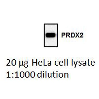
![Western Blot: Peroxiredoxin 2 Antibody (4E10-2D2) [H00007001-M01]](https://resources.rndsystems.com/images/products/Peroxiredoxin-2-Antibody-4E10-2D2-Western-Blot-H00007001-M01-img0011.jpg "Western Blot: Peroxiredoxin 2 Antibody (4E10-2D2) [H00007001-M01]")
Loading...
Key Product Details
Validated by
Knockout/Knockdown, Biological Validation
Species Reactivity
Human
Applications
Validated:
Immunohistochemistry, Immunohistochemistry-Paraffin, Western Blot, ELISA, Immunocytochemistry/ Immunofluorescence, Knockdown Validated
Cited:
Western Blot
Label
Unconjugated
Antibody Source
Monoclonal Mouse IgG1 kappa Clone # 4E10-2D2
Format
Azide and BSA Free
Loading...
Product Specifications
Immunogen
PRDX2 (AAH00452, 1 a.a. ~ 198 a.a) full-length recombinant protein with GST tag. MW of the GST tag alone is 26 KDa. MASGNARIGKPAPDFKATAVVDGAFKEVKLSDYKGKYVVLFFYPLDFTFVCPTEIIAFSNRAEDFRKLGCEVLGVSVDSQFTHLAWINTPRKEGGLGPLNIPLLADVTRRLSEDYGVLKTDEGIAYRGLFIIDGKGVLRQITVNDLPVGRSVDEALRLVQAFQYTDEHGEVCPAGWKPGSDTIKPNVDDSKEYFSKHN
Localization
Cytoplasmic
Specificity
PRDX2 - peroxiredoxin 2
Clonality
Monoclonal
Host
Mouse
Isotype
IgG1 kappa
Description
Quality control test: Antibody Reactive Against Recombinant Protein.
Scientific Data Images for Peroxiredoxin 2 Antibody (4E10-2D2) - Azide and BSA Free
Western Blot: Peroxiredoxin 2 Antibody (4E10-2D2) [H00007001-M01]
Western Blot: Peroxiredoxin 2 Antibody (4E10-2D2) [H00007001-M01] - PRDX2 monoclonal antibody (M01), clone 4E10-2D2 Analysis of PRDX2 expression in Hela.![Immunocytochemistry/ Immunofluorescence: Peroxiredoxin 2 Antibody (4E10-2D2) [H00007001-M01]](https://resources.rndsystems.com/images/products/Peroxiredoxin-2-Antibody-4E10-2D2-Immunocytochemistry-Immunofluorescence-H00007001-M01-img0007.jpg "Immunocytochemistry/ Immunofluorescence: Peroxiredoxin 2 Antibody (4E10-2D2) [H00007001-M01]")
Immunocytochemistry/ Immunofluorescence: Peroxiredoxin 2 Antibody (4E10-2D2) [H00007001-M01]
Immunocytochemistry/Immunofluorescence: Peroxiredoxin 2 Antibody (4E10-2D2) [H00007001-M01] - Analysis of monoclonal antibody to PRDX2 on HeLa cell. Antibody concentration 10 ug/ml.![Immunohistochemistry-Paraffin: Peroxiredoxin 2 Antibody (4E10-2D2) [H00007001-M01]](https://resources.rndsystems.com/images/products/Peroxiredoxin-2-Antibody-4E10-2D2-Immunohistochemistry-Paraffin-H00007001-M01-img0009.jpg "Immunohistochemistry-Paraffin: Peroxiredoxin 2 Antibody (4E10-2D2) [H00007001-M01]")
Immunohistochemistry-Paraffin: Peroxiredoxin 2 Antibody (4E10-2D2) [H00007001-M01]
Immunohistochemistry-Paraffin: Peroxiredoxin 2 Antibody (4E10-2D2) [H00007001-M01] - Analysis of monoclonal antibody to PRDX2 on formalin-fixed paraffin-embedded human malignant lymphoma, diffuse large B tissue. Antibody concentration 1 ug/ml.![Western Blot: Peroxiredoxin 2 Antibody (4E10-2D2) [H00007001-M01]](https://resources.rndsystems.com/images/products/Peroxiredoxin-2-Antibody-4E10-2D2-Western-Blot-H00007001-M01-img0012.jpg "Western Blot: Peroxiredoxin 2 Antibody (4E10-2D2) [H00007001-M01]")
Western Blot: Peroxiredoxin 2 Antibody (4E10-2D2) [H00007001-M01]
Western Blot: Peroxiredoxin 2 Antibody (4E10-2D2) [H00007001-M01] - Analysis of PRDX2 expression in transfected 293T cell line by PRDX2 monoclonal antibody (M01), clone 4E10-2D2.Lane 1: PRDX2 transfected lysate(21.9 KDa).Lane 2: Non-transfected lysate.![ELISA: Peroxiredoxin 2 Antibody (4E10-2D2) [H00007001-M01]](https://resources.rndsystems.com/images/products/Peroxiredoxin-2-Antibody-4E1-0-2D2-ELISA-H00007001-M01-img0013.jpg "ELISA: Peroxiredoxin 2 Antibody (4E10-2D2) [H00007001-M01]")
ELISA: Peroxiredoxin 2 Antibody (4E10-2D2) [H00007001-M01]
ELISA: Peroxiredoxin 2 Antibody (4E1.0-2D2) [H00007001-M01] - Peroxiredoxin 2 Antibody (4E10-2D2) [H00007001-M01] - Detection limit for recombinant GST tagged PRDX2 is approximately 1ng/ml as a capture antibody.![Knockdown Validated: Peroxiredoxin 2 Antibody (4E10-2D2) [H00007001-M01]](https://resources.rndsystems.com/images/products/Peroxiredoxin-2-Antibody-4E10-2D2-Knockdown-Validated-H00007001-M01-img0014.jpg "Western Blot: Peroxiredoxin 2 Antibody (4E10-2D2) [H00007001-M01]")
 [H00007001-M01] -")
Western Blot: Peroxiredoxin 2 Antibody (4E10-2D2) [H00007001-M01] -
Western Blot: Peroxiredoxin 2 Antibody (4E10-2D2) [H00007001-M01] - Expression of PRDX2 or PRDX4 does not affect HIF-1 alpha or HIF-2 alpha protein levelsA. & B. HeLa cells were transfected with EV or vector encoding PRDX2-V5 (A, P2) or PRDX4-V5 (B, P4), & exposed to 20% or 1% O2 for 24 h. WCL was subject to immunoblot assays with antibody against HIF-1 alpha, HIF-2 alpha, V5, or acin. C. HeLa-shSC (sc) & HeLa-shPRDX(2+4) (2+4) cells were exposed to 20% or 1% O2 for 24 h in the presence of doxycycline. WCL was subject to immunoblot assays with antibodies against HIF-1 alpha, HIF-2 alpha, PRDX2, PRDX4, & actin. Image collected & cropped by CiteAb from the following publication (https://www.oncotarget.com/lookup/doi/10.18632/oncotarget.7142), licensed under a CC-BY license. Not internally tested by Novus Biologicals. [H00007001-M01] -")
Western Blot: Peroxiredoxin 2 Antibody (4E10-2D2) [H00007001-M01] -
Western Blot: Peroxiredoxin 2 Antibody (4E10-2D2) [H00007001-M01] - PRDX2 knockdown increases PRDX4 protein levels in HeLa cellsHeLa subclones were exposed to 20% or 1% O2 for indicated time. Each WCL was subject to immunoblot assays with the indicated antibodies. SC, scrambled control shRNA. P2, PRDX2 shRNA. Image collected & cropped by CiteAb from the following publication (https://www.oncotarget.com/lookup/doi/10.18632/oncotarget.7142), licensed under a CC-BY license. Not internally tested by Novus Biologicals. [H00007001-M01] -")
Western Blot: Peroxiredoxin 2 Antibody (4E10-2D2) [H00007001-M01] -
Western Blot: Peroxiredoxin 2 Antibody (4E10-2D2) [H00007001-M01] - Hypoxia induces the nuclear translocation of PRDX2 & PRDX4HeLa cells were transfected with vector encoding PRDX2-V5 (P2) or PRDX4-V5 (P4), or empty vector (EV), & exposed to 20% or 1% O2 for 48 h. Nuclear & cytosolic fractions were isolated & subject to immunoblot assays with antibodies against HIF-1 alpha, HIF-2 alpha, V5, alpha -tubulin, & histone H3. Image collected & cropped by CiteAb from the following publication (https://www.oncotarget.com/lookup/doi/10.18632/oncotarget.7142), licensed under a CC-BY license. Not internally tested by Novus Biologicals. [H00007001-M01] -")
Western Blot: Peroxiredoxin 2 Antibody (4E10-2D2) [H00007001-M01] -
Western Blot: Peroxiredoxin 2 Antibody (4E10-2D2) [H00007001-M01] - Mapping the PRDX2 & PRDX4 binding domains of HIF-1 alpha A. & B. HeLa cells were transfected with PRDX2-V5 (A) or PRDX4-V5 (B) vector & WCL was incubated with purified GST or GST-HIF-1 alpha fusion protein in the presence of glutathione-Sepharose beads, followed by immunoblot assays with anti-V5 antibody (upper panels) or Ponceau S staining (lower panels). Image collected & cropped by CiteAb from the following publication (https://www.oncotarget.com/lookup/doi/10.18632/oncotarget.7142), licensed under a CC-BY license. Not internally tested by Novus Biologicals. [H00007001-M01] -")
Western Blot: Peroxiredoxin 2 Antibody (4E10-2D2) [H00007001-M01] -
Western Blot: Peroxiredoxin 2 Antibody (4E10-2D2) [H00007001-M01] - Expression of PRDX2 or PRDX4 does not affect HIF-1 alpha or HIF-2 alpha protein levelsA. & B. HeLa cells were transfected with EV or vector encoding PRDX2-V5 (A, P2) or PRDX4-V5 (B, P4), & exposed to 20% or 1% O2 for 24 h. WCL was subject to immunoblot assays with antibody against HIF-1 alpha, HIF-2 alpha, V5, or acin. C. HeLa-shSC (sc) & HeLa-shPRDX(2+4) (2+4) cells were exposed to 20% or 1% O2 for 24 h in the presence of doxycycline. WCL was subject to immunoblot assays with antibodies against HIF-1 alpha, HIF-2 alpha, PRDX2, PRDX4, & actin. Image collected & cropped by CiteAb from the following publication (https://www.oncotarget.com/lookup/doi/10.18632/oncotarget.7142), licensed under a CC-BY license. Not internally tested by Novus Biologicals. [H00007001-M01] -")
Western Blot: Peroxiredoxin 2 Antibody (4E10-2D2) [H00007001-M01] -
Western Blot: Peroxiredoxin 2 Antibody (4E10-2D2) [H00007001-M01] - Effect of PRDX2 & PRDX4 on HIF-1 alpha -p300 interactionHeLa cells were transfected with empty vector (EV) or vector encoding PRDX2-V5 or PRDX4-V5, & exposed to 1% O2 for 24 h. WCL was subject to IP with anti-p300 antibody, followed by immunoblot assays using antibodies against HIF-1 alpha, V5, & p300. Image collected & cropped by CiteAb from the following publication (https://www.oncotarget.com/lookup/doi/10.18632/oncotarget.7142), licensed under a CC-BY license. Not internally tested by Novus Biologicals. [H00007001-M01] -")
Western Blot: Peroxiredoxin 2 Antibody (4E10-2D2) [H00007001-M01] -
Western Blot: Peroxiredoxin 2 Antibody (4E10-2D2) [H00007001-M01] - PRDX2 expression is regulated by HIF-1 & HIF-2A. HeLa cells were exposed to 20% or 1% O2 for 24 h. RT-qPCR assays were performed using primers specific for the indicated mRNAs. Data are shown as mean ± SEM, n = 3. ***p < 0.001 versus 20% O2. B. HeLa cells were exposed to 20% or 1% O2 for the indicated time. WCLs were subject to immunoblot assays with antibodies against PRDX2, HIF-1 alpha, HIF-2 alpha, & actin. The PRDX2 & actin bands were quantified by densitometry & normalized to 0 h (20% O2). Normalized data are shown as mean ± SEM, n = 3. *p < 0.05, ***p < 0.001 versus 20% O2. C. HeLa-shSC (SC), HeLa-shHIF-1 alpha (1 alpha ), HeLa-shHIF-2 alpha (2 alpha ), & HeLa-sh1 alpha +2 alpha (DKD) cells were exposed to 20% or 1% O2 for 72 h. WCLs were subject to immunoblot assays with antibodies against PRDX2, HIF-1 alpha, HIF-2 alpha, & actin. Image collected & cropped by CiteAb from the following publication (https://www.oncotarget.com/lookup/doi/10.18632/oncotarget.7142), licensed under a CC-BY license. Not internally tested by Novus Biologicals. [H00007001-M01] -")
Western Blot: Peroxiredoxin 2 Antibody (4E10-2D2) [H00007001-M01] -
Western Blot: Peroxiredoxin 2 Antibody (4E10-2D2) [H00007001-M01] - PRDX proteins bind to HIF-1 alpha & HIF-2 alpha A. HeLa cells were transfected with an expression vector encoding V5-epitope-tagged PRDX2 (PRDX2-V5) & exposed to 1% O2 for 24 h. Whole cell lysate (WCL) was subject to immunoprecipitation (IP) using anti-HIF-1 alpha antibody or control IgG, followed by immunoblot assays with antibody against V5 epitope or HIF-1 alpha. B. HeLa cells were transfected with PRDX2-V5 vector & exposed to 1% O2 for 24 h. The WCL was subject to IP using anti-V5 antibody or control IgG, followed by immunoblot assays with antibody against V5 or HIF-1 alpha. Light IgG: immunoglobulin light chain from the secondary antibody. C. HeLa cells were transfected with vector encoding a V5-tagged PRDX family member & exposed to 1% O2 for 24 h. WCL was subject to IP using anti-HIF-1 alpha antibody, followed by immunoblot assays with antibody against V5 or HIF-1 alpha. D. HeLa cells were transfected with empty vector (EV) or vector encoding a V5-tagged PRDX family member & exposed to 1% O2 for 24 h. WCL was subject to IP using anti-HIF-2 alpha antibody, followed by immunoblot assays with antibody against V5 or HIF-2 alpha. Image collected & cropped by CiteAb from the following publication (https://www.oncotarget.com/lookup/doi/10.18632/oncotarget.7142), licensed under a CC-BY license. Not internally tested by Novus Biologicals. [H00007001-M01] -")
Western Blot: Peroxiredoxin 2 Antibody (4E10-2D2) [H00007001-M01] -
Western Blot: Peroxiredoxin 2 Antibody (4E10-2D2) [H00007001-M01] - PRDX2 expression is regulated by HIF-1 & HIF-2A. HeLa cells were exposed to 20% or 1% O2 for 24 h. RT-qPCR assays were performed using primers specific for the indicated mRNAs. Data are shown as mean ± SEM, n = 3. ***p < 0.001 versus 20% O2. B. HeLa cells were exposed to 20% or 1% O2 for the indicated time. WCLs were subject to immunoblot assays with antibodies against PRDX2, HIF-1 alpha, HIF-2 alpha, & actin. The PRDX2 & actin bands were quantified by densitometry & normalized to 0 h (20% O2). Normalized data are shown as mean ± SEM, n = 3. *p < 0.05, ***p < 0.001 versus 20% O2. C. HeLa-shSC (SC), HeLa-shHIF-1 alpha (1 alpha ), HeLa-shHIF-2 alpha (2 alpha ), & HeLa-sh1 alpha +2 alpha (DKD) cells were exposed to 20% or 1% O2 for 72 h. WCLs were subject to immunoblot assays with antibodies against PRDX2, HIF-1 alpha, HIF-2 alpha, & actin. Image collected & cropped by CiteAb from the following publication (https://www.oncotarget.com/lookup/doi/10.18632/oncotarget.7142), licensed under a CC-BY license. Not internally tested by Novus Biologicals.Applications for Peroxiredoxin 2 Antibody (4E10-2D2) - Azide and BSA Free
Application
Recommended Usage
ELISA
Optimal dilutions of this antibody should be experimentally determined.
Immunocytochemistry/ Immunofluorescence
Optimal dilutions of this antibody should be experimentally determined.
Immunohistochemistry
Optimal dilutions of this antibody should be experimentally determined.
Immunohistochemistry-Paraffin
Optimal dilutions of this antibody should be experimentally determined.
Knockdown Validated
Optimal dilutions of this antibody should be experimentally determined.
Western Blot
Optimal dilutions of this antibody should be experimentally determined.
Application Notes
Antibody reactivity against cell lysate and recombinant protein for WB. It has also been used for IF, IHC-P and ELISA.
Reviewed Applications
Read 1 review rated 5 using H00007001-M01 in the following applications:
Formulation, Preparation, and Storage
Purification
IgG purified
Formulation
In 1x PBS, pH 7.4
Format
Azide and BSA Free
Preservative
No Preservative
Concentration
Concentrations vary lot to lot. See vial label for concentration. If unlisted please contact technical services.
Shipping
The product is shipped with polar packs. Upon receipt, store it immediately at the temperature recommended below.
Stability & Storage
Aliquot and store at -20C or -80C. Avoid freeze-thaw cycles.
Background: Peroxiredoxin 2
Alternate Names
NKEF-B, PRDX2, PRX2, TDPX1, TSA
Entrez Gene IDs
7001 (Human)
Gene Symbol
PRDX2
OMIM
600538 (Human)
UniProt
Additional Peroxiredoxin 2 Products
Product Documents for Peroxiredoxin 2 Antibody (4E10-2D2) - Azide and BSA Free
Certificate of Analysis
To download a Certificate of Analysis, please enter a lot or batch number in the search box below.
Product Specific Notices for Peroxiredoxin 2 Antibody (4E10-2D2) - Azide and BSA Free
This product is produced by and distributed for Abnova, a company based in Taiwan.
This product is for research use only and is not approved for use in humans or in clinical diagnosis. Primary Antibodies are guaranteed for 1 year from date of receipt.
Related Research Areas
Citations for Peroxiredoxin 2 Antibody (4E10-2D2) - Azide and BSA Free
 Powered by Bioz
Customer Reviews for Peroxiredoxin 2 Antibody (4E10-2D2) - Azide and BSA Free (1)
5 out of 5
1 Customer Rating
Have you used Peroxiredoxin 2 Antibody (4E10-2D2) - Azide and BSA Free?
Submit a review and receive an Amazon gift card!
$25/€18/£15/$25CAN/¥2500 Yen for a review with an image
$10/€7/£6/$10CAN/¥1110 Yen for a review without an image
Submit a review
Customer Images
Showing
1
-
1 of
1 review
Showing All
Filter By:
-
Application: Western BlotSample Tested: Human HeLa cell lysateSpecies: HumanVerified Customer | Posted 09/26/2013Western Blot analysis of PRDX2 in human HeLa cell lysate

There are no reviews that match your criteria.
Protocols
Find general support by application which include: protocols, troubleshooting, illustrated assays, videos and webinars.
- Antigen Retrieval Protocol (PIER)
- Antigen Retrieval for Frozen Sections Protocol
- Appropriate Fixation of IHC/ICC Samples
- Cellular Response to Hypoxia Protocols
- Chromogenic IHC Staining of Formalin-Fixed Paraffin-Embedded (FFPE) Tissue Protocol
- Chromogenic Immunohistochemistry Staining of Frozen Tissue
- ClariTSA™ Fluorophore Kits
- Detection & Visualization of Antibody Binding
- ELISA Sample Preparation & Collection Guide
- ELISA Troubleshooting Guide
- Fluorescent IHC Staining of Frozen Tissue Protocol
- Graphic Protocol for Heat-induced Epitope Retrieval
- Graphic Protocol for the Preparation and Fluorescent IHC Staining of Frozen Tissue Sections
- Graphic Protocol for the Preparation and Fluorescent IHC Staining of Paraffin-embedded Tissue Sections
- Graphic Protocol for the Preparation of Gelatin-coated Slides for Histological Tissue Sections
- How to Run an R&D Systems DuoSet ELISA
- How to Run an R&D Systems Quantikine ELISA
- How to Run an R&D Systems Quantikine™ QuicKit™ ELISA
- ICC Cell Smear Protocol for Suspension Cells
- ICC Immunocytochemistry Protocol Videos
- ICC for Adherent Cells
- IHC Sample Preparation (Frozen sections vs Paraffin)
- Immunocytochemistry (ICC) Protocol
- Immunocytochemistry Troubleshooting
- Immunofluorescence of Organoids Embedded in Cultrex Basement Membrane Extract
- Immunofluorescent IHC Staining of Formalin-Fixed Paraffin-Embedded (FFPE) Tissue Protocol
- Immunohistochemistry (IHC) and Immunocytochemistry (ICC) Protocols
- Immunohistochemistry Frozen Troubleshooting
- Immunohistochemistry Paraffin Troubleshooting
- Preparing Samples for IHC/ICC Experiments
- Preventing Non-Specific Staining (Non-Specific Binding)
- Primary Antibody Selection & Optimization
- Protocol for Heat-Induced Epitope Retrieval (HIER)
- Protocol for Making a 4% Formaldehyde Solution in PBS
- Protocol for VisUCyte™ HRP Polymer Detection Reagent
- Protocol for the Fluorescent ICC Staining of Cell Smears - Graphic
- Protocol for the Fluorescent ICC Staining of Cultured Cells on Coverslips - Graphic
- Protocol for the Preparation & Fixation of Cells on Coverslips
- Protocol for the Preparation and Chromogenic IHC Staining of Frozen Tissue Sections
- Protocol for the Preparation and Chromogenic IHC Staining of Frozen Tissue Sections - Graphic
- Protocol for the Preparation and Chromogenic IHC Staining of Paraffin-embedded Tissue Sections
- Protocol for the Preparation and Chromogenic IHC Staining of Paraffin-embedded Tissue Sections - Graphic
- Protocol for the Preparation and Fluorescent ICC Staining of Cells on Coverslips
- Protocol for the Preparation and Fluorescent ICC Staining of Non-adherent Cells
- Protocol for the Preparation and Fluorescent ICC Staining of Stem Cells on Coverslips
- Protocol for the Preparation and Fluorescent IHC Staining of Frozen Tissue Sections
- Protocol for the Preparation and Fluorescent IHC Staining of Paraffin-embedded Tissue Sections
- Protocol for the Preparation of Gelatin-coated Slides for Histological Tissue Sections
- Protocol for the Preparation of a Cell Smear for Non-adherent Cell ICC - Graphic
- Quantikine HS ELISA Kit Assay Principle, Alkaline Phosphatase
- Quantikine HS ELISA Kit Principle, Streptavidin-HRP Polymer
- R&D Systems Quality Control Western Blot Protocol
- Sandwich ELISA (Colorimetric) – Biotin/Streptavidin Detection Protocol
- Sandwich ELISA (Colorimetric) – Direct Detection Protocol
- TUNEL and Active Caspase-3 Detection by IHC/ICC Protocol
- The Importance of IHC/ICC Controls
- Troubleshooting Guide: ELISA
- Troubleshooting Guide: Immunohistochemistry
- Troubleshooting Guide: Western Blot Figures
- Western Blot Conditions
- Western Blot Protocol
- Western Blot Protocol for Cell Lysates
- Western Blot Troubleshooting
- Western Blot Troubleshooting Guide
- View all Protocols, Troubleshooting, Illustrated assays and Webinars
Loading...