PGC1 alpha Antibody - BSA Free
Novus Biologicals | Catalog # NBP1-04676

Key Product Details
Validated by
Knockout/Knockdown, Biological Validation
Species Reactivity
Validated:
Human, Mouse, Rat, Porcine, Goat, Hamster, Sheep, Squirrel
Cited:
Human, Mouse, Rat, Porcine, Avian - Chicken, Canine, Fish - Danio rerio (Zebrafish), Goat, Hamster, Ovine, Squirrel
Predicted:
Canine (93%), Equine (94%), Primate (98%). Backed by our 100% Guarantee.
Applications
Validated:
Knockout Validated, Immunohistochemistry, Immunohistochemistry-Paraffin, Immunohistochemistry-Frozen, Western Blot, Flow Cytometry, Flow (Intracellular), Immunocytochemistry/ Immunofluorescence, Simple Western, Immunoprecipitation, Chromatin Immunoprecipitation, Chromatin Immunoprecipitation (ChIP), Knockdown Validated
Cited:
Knockout Validated, Immunohistochemistry, Immunohistochemistry-Paraffin, Immunohistochemistry-Frozen, Western Blot, Immunoblotting, Flow Cytometry, Immunocytochemistry/ Immunofluorescence, Simple Western, Immunoprecipitation, Chromatin Immunoprecipitation (ChIP), Chemotaxis, In vivo assay, IF/IHC, Knockdown Validated
Label
Unconjugated
Antibody Source
Polyclonal Rabbit IgG
Format
BSA Free
Loading...
Product Specifications
Immunogen
This PGC1 alpha Antibody was developed against a recombinant protein made to an internal portion of the human PGC-1 alpha protein (within residues 400-550). [Swiss-Prot# Q9UBK2].
Reactivity Notes
Use in Rat reported in scientific literature (PMID:35174626, 34573421). Use in Mouse reported in scientific literature (PMID:33719499). Use in Sheep reported in scientific literature (PMID:32403966). Expected from sequence similarity: Mouse
Localization
Nucleus when activated, can be cytoplasmic when inactive (PMID: PMC2253697).
Clonality
Polyclonal
Host
Rabbit
Isotype
IgG
Theoretical MW
91 kDa.
Disclaimer note: The observed molecular weight of the protein may vary from the listed predicted molecular weight due to post translational modifications, post translation cleavages, relative charges, and other experimental factors.
Disclaimer note: The observed molecular weight of the protein may vary from the listed predicted molecular weight due to post translational modifications, post translation cleavages, relative charges, and other experimental factors.
Scientific Data Images for PGC1 alpha Antibody - BSA Free
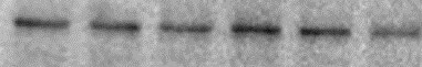
Western Blot Detection of PGC1 alpha in MEFs
PGC1-alpha-Antibody---BSA-Free-Western-Blot-NBP1-04676-img0020.jpg
Fluorescent Staining of PGC1 alpha in HeLa Cells
HeLa cells were fixed in 4% paraformaldehyde for 10 minutes and permeabilized in 0.5% Triton X-100 in PBS for 5 minutes. The cells were incubated with anti-PGC1 alpha Antibody NBP1-04676 at 2 ug/ml overnight at 4C and detected with an anti-rabbit Dylight 488 (Green) at a 1:1000 dilution for 60 minutes. Nuclei were counterstained with DAPI (Blue). Cells were imaged using a 100X objective and digitally deconvolved.
Immunohistological Staining of PGC1 alpha in Paraffin Embedded Mouse Prostate
Analysis of a FFPE section of mouse prostate using rabbit polyclonal PGC1 alpha Antibody at 1:200 dilution. The antibody generated an expected strong nuclear (punctate in some cells) and a weak cytoplasmic staining in the glandular cells lining of tubule-alveolar gland.
Flow Cytometry of A431 Cells Stained with PGC1 alpha Antibody
An intracellular stain was performed on A431 cells with PGC1 alpha Antibody NBP1-04676 (blue) and matched isotype control NBP2-24891 (orange). Cells were fixed with 4% PFA and then permeabilized with 0.1% saponin. Cells were incubated in an antibody dilution of 1.0 ug/mL for 30 minutes at room temperature, followed by Rabbit IgG (H+L) Cross-Adsorbed Secondary Antibody, Dylight 550 (SA5-10033, Thermo Fisher).

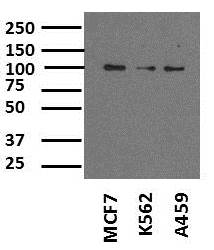
Western Blot Detection of PGC1 alpha in Multiple Tissues and Cell Lines
Total protein from human adipose and skeletal muscle tissue, HeLa and A431 cells lines was separated on a 7.5% gel by SDS-PAGE, transferred to PVDF membrane and blocked in 5% non-fat milk in TBST. The membrane was probed with 2.0 ug/mL PGC1 alpha Antibody (Molecular weight: 91 KDa) in 1% non-fat milk in TBST and detected with an anti-rabbit HRP secondary antibody using chemiluminescence
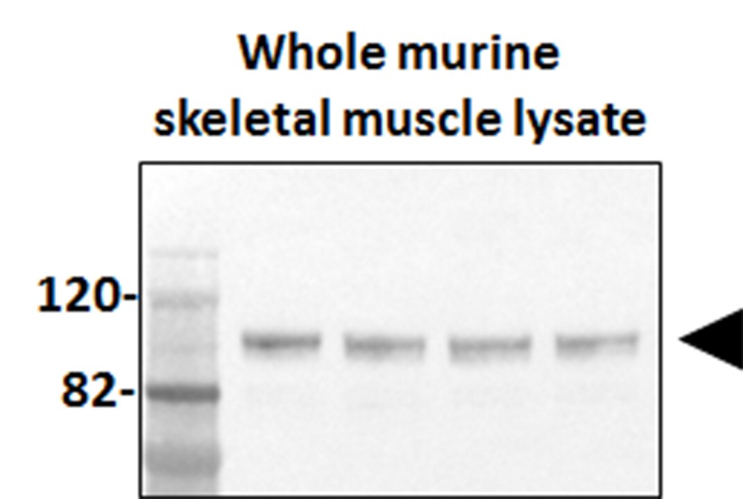
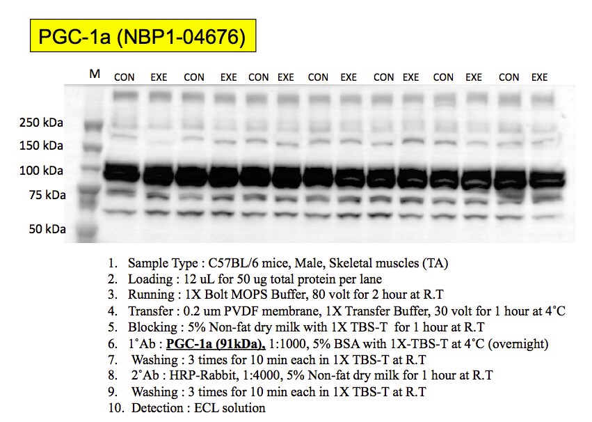
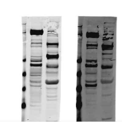
Detection of PGC1 alpha in Murine Skeletal Muscle by Western Blot
Western Blot of total protein from murine skeletal muscle tissue using PGC1 alpha Antibody. Image from verified customer review.
Immunocytochemistry/Immunofluorescence Staining of PGC1 alpha in HeLa Cells
HeLa cells were fixed for 10 minutes using 10% formalin and then permeabilized for 5 minutes using 1X TBS + 0.5% Triton X-100. The cells were incubated with PGC1 alpha Antibody [NBP1-04676] at a 1:200 dilution overnight at 4C and detected with an anti-rabbit DyLight 488 (Green) at a 1:500 dilution. Alpha tubulin (DM1A) [NB100-690] was used as a co-stain at a 1:1000 dilution and detected with an anti-mouse DyLight 550 (Red) at a 1:500 dilution. Nuclei were counterstained with DAPI (Blue). Cells were imaged using a 40X objective.
Immunohistological Staining of PGC1 alpha in Paraffin Embedded Mouse Prostate
Analysis of PGC1 alpha Antibody in mouse prostate using DAB with hematoxylin counterstain.
Flow Cytometry of HeLa Cells Stained with Alexa Fluor 647 Conjugated PGC1 alpha Antibody
An intracellular stain was performed on HeLa cells with NBP1-04676AF647 (blue) and a matched isotype control (orange). Cells were fixed with 4% PFA and then permeabilized with 0.1% saponin. Cells were incubated in an antibody dilution of 2.5 ug/mL for 30 minutes at room temperature. Both antibodies were conjugated to Alexa Fluor 647.
Flow Cytometry of HepG2 Cells Stained with PGC1 alpha Antibody
An intracellular stain was performed on HepG2 cells with PGC1 alpha Antibody and a matched isotype control. Cells were fixed with 4% PFA and then permeablized with 0.1% saponin. Cells were incubated in an antibody dilution of 2.5 ug/mL for 30 minutes at room temperature, followed by Rabbit IgG (H+L) Cross-Adsorbed Secondary Antibody.
Knockout Validation of PGC1 alpha Antibody in Human Squamous Carcinoma Cells and PGC1 alpha Knockout A431 Cells
Western blot shows lysates of A431 human squamous carcinoma parental cell line and PGC1 alpha knockout (KO) A431 cell line. PVDF membrane was probed with 1:1000 of Rabbit Polyclonal PGC1 alpha Antibody (Catalog # NBP1-04676) followed by HRP-conjugated Anti-Rabbit IgG Secondary Antibody (Catalog #HAF008). Specific band was detected for PGC1 alpha at approximately 105 kDa (as indicated) in the parental A431 cell line, but is not detectable in the knockout A431 cell line. This experiment was conducted under reducing conditions.
Immunocytochemistry/Immunofluorescence: PGC1 alpha Antibody - BSA Free [NBP1-04676] -
DM1-derived fibroblasts have no changes in mitochondria biogenesis. (A, B) Representative images of immunofluorescence of TOMM20, and PGC1-alpha in DM1 and control fibroblasts (n=3). (C) Medium fluorescence intensity of MitoTracker Red FM in control (n=3) and DM1 cells (n=5) and (D) of Rhodamine 123 in DM1 and control fibroblasts (n=3). (E) mRNA levels of TFAM transcription factor (n=3). (F) mRNA levels of OPA1, MFN1, MFN2, DRP1 and PARKIN in DM1 and control fibroblasts (n≥2).
Detection of PGC1 alpha in A431 Human Cell Line by Flow Cytometry.
A431 human skin carcinoma cell line was stained with Rabbit anti-PGC1 alpha Affinity-purified Polyclonal Antibody conjugated to Phycoerythrin (Catalog # NBP1-04676PE, blue histogram) or matched control antibody (Catalog # NBP2-24983, orange histogram).
Western Blot: PGC1 alpha Antibody - BSA Free [NBP1-04676] -
Western Blot: PGC1 alpha Antibody - BSA Free [NBP1-04676] - Effect of butein T on SC adipose tissue browning in the ThermoMouse. SC fat was obtained from ThermoMouse exposed for 4 days to butein (10 or 20 mg/kg; n = 4) or vehicle (n = 2). A BAT lysate of a ThermoMouse was used as a positive control for UCP-1 protein levels. Tissues were homogenized & protein lysates (50 µg for SC fat & 10 µg for BAT) were used in western blotting for PGC1-alpha (a) & UCP-1 (c). Panels (b) & (d) illustrate the densitometric quantitation of the PGC1-alpha & UCP-1 western blot, respectively, normalized for the levels of the loading control alpha -tubulin. Abbreviations: subcutaneous (SC); brown adipose tissue (BAT); peroxisome proliferator-activated receptor-gamma coactivator 1-alpha (PGC1-alpha ) & uncoupling protein-1 (UCP-1). Image collected & cropped by CiteAb from the following publication (https://pubmed.ncbi.nlm.nih.gov/31094273), licensed under a CC-BY license. Not internally tested by Novus Biologicals.
Western Blot: PGC1 alpha Antibody - BSA Free [NBP1-04676] -
Western Blot: PGC1 alpha Antibody - BSA Free [NBP1-04676] - Expression of the mitochondrial biogenesis proteins: SIRT1, PGC1 alpha, TFAM & SIRT3 in H1299, H1299r, P31 & P31r cell lysates as determined by immunoblot analysisWhole cell lysates were prepared from confluent cultures of H1299, H1299r, P31 & P31r cells. Proteins (90 μg) were resolved in 10% SDS-PAGE gels & transferred to a PVDF membrane. Blots were probed for SIRT1, PGC1 alpha, TFAM & SIRT3 or the loading control gamma -tubulin. Figure shows representative blots of (A) of SIRT1, (B) PGC1 alpha, (C) SIRT3, (D) TFAM protein expression; each from three independent experiments. Image collected & cropped by CiteAb from the following publication (https://www.oncotarget.com/lookup/doi/10.18632/oncotarget.21885), licensed under a CC-BY license. Not internally tested by Novus Biologicals.
Western Blot: PGC1 alpha Antibody - BSA Free [NBP1-04676] -
Western Blot: PGC1 alpha Antibody - BSA Free [NBP1-04676] - Maternal exercise during pregnancy on mitochondrial biogenesis in the fetal hearts. (A) Levels of relative mRNA expression measured by qRT‐PCR. n = 9–12/group. Maternal exercise during pregnancy did not alter levels of mRNA in Ppargc1a & Tfam, while it significantly upregulated the levels of mRNA in Nrf1 & Nrf2. (B–D) Densitometric analyses of protein expression levels relative to the sedentary group with representative images of western blots were shown. No significant differences in PGC‐1 alpha, NRF1, & NRF2 (P > 0.05). n = 5–6/group. * P < 0.05, significantly different from the sedentary group. Black bar: fetal hearts from sedentary dams; gray bar: fetal hearts from exercised dams. Image collected & cropped by CiteAb from the following publication (https://pubmed.ncbi.nlm.nih.gov/28292876), licensed under a CC-BY license. Not internally tested by Novus Biologicals.
Immunocytochemistry/ Immunofluorescence: PGC1 alpha Antibody - BSA Free [NBP1-04676] -
Immunocytochemistry/ Immunofluorescence: PGC1 alpha Antibody - BSA Free [NBP1-04676] - DM1-derived fibroblasts have no changes in mitochondria biogenesis. (A, B) Representative images of immunofluorescence of TOMM20, & PGC1-alpha in DM1 & control fibroblasts (n=3). (C) Medium fluorescence intensity of MitoTracker Red FM in control (n=3) & DM1 cells (n=5) & (D) of Rhodamine 123 in DM1 & control fibroblasts (n=3). (E) mRNA levels of TFAM transcription factor (n=3). (F) mRNA levels of OPA1, MFN1, MFN2, DRP1 & PARKIN in DM1 & control fibroblasts (n≥2). Image collected & cropped by CiteAb from the following publication (https://pubmed.ncbi.nlm.nih.gov/32310829), licensed under a CC-BY license. Not internally tested by Novus Biologicals.
Western Blot: PGC1 alpha Antibody - BSA Free [NBP1-04676] -
Western Blot: PGC1 alpha Antibody - BSA Free [NBP1-04676] - Effect of BGP-15 treatment on the regulation of mitochondrial biogenesis in NRCM cells. Western blot analysis of PGC-1-alpha, CREB, & VDAC proteins as well as densitometric evaluation is shown. GAPDH was used as a loading control. Control group: cells without any treatment; BGP-15 group: cells with only 50 μM BGP-15 for 0.5 hours; H2O2 group: cells with 150 μM H2O2 for 0.5 hours; H2O2+BGP-15 group: cells with 150 μM H2O2 & 50 μM BGP-15 for 0.5 hours. Values are mean ± SEM (n = 4). ∗p < 0.05 vs. Control, ∗∗p < 0.01 vs. Control, §§p < 0.01 vs H2O2 group. Image collected & cropped by CiteAb from the following publication (https://pubmed.ncbi.nlm.nih.gov/33728024), licensed under a CC-BY license. Not internally tested by Novus Biologicals.
Western Blot: PGC1 alpha Antibody - BSA Free [NBP1-04676] -
Western Blot: PGC1 alpha Antibody - BSA Free [NBP1-04676] - Feeding the diabetes mellitus (DM) mice with the RSV‐enriched diet improved mitochondrial biogenesis in the quadriceps muscle. A) Representative western blots using antibodies against nuclear respiratory factor (NRF‐1), ATP synthase, H+ transporting, mitochondrial F1 complex, beta polypeptide (ATP5B), cytochrome c oxidase subunit IV (Cox IV), peroxisome proliferator‐activated receptor‐ gamma coactivator 1‐ alpha (PGC‐1 alpha ), & GAPDH. After quantification, the B) NRF‐1/GAPDH, C) ATP5B/GAPDH, D) Cox IV/GAPDH, & E) PGC‐1 alpha /GAPDH ratios were calculated in mice fed either the control (CTL) or RSV‐enriched diet. GAPDH was used as a loading control. F) Representative western blots using antibodies against mitochondrial transcription factor A (mtTFA) & voltage‐dependent anion channel (VDAC), & the mtTFA/VDAC ratio was determined. The mitochondrial protein VDAC was used as a loading control. Data are expressed as fold change versus CTL & reported as mean ± SD, n = 4–6 per group; *p < 0.05; **p < 0.01 between CTL & DM groups. #p < 0.05 between DM & DM+RSV groups; ††p < 0.05 between CTL+RSV & DM+RSV groups. Image collected & cropped by CiteAb from the following publication (https://pubmed.ncbi.nlm.nih.gov/29578301), licensed under a CC-BY license. Not internally tested by Novus Biologicals.
Western Blot: PGC1 alpha Antibody - BSA Free [NBP1-04676] -
Western Blot: PGC1 alpha Antibody - BSA Free [NBP1-04676] - Effects of SRTAW04 on expression of markers of mitochondrial & anti-oxidant function. (a) Western blot of protein extracts from optic nerve & retina of control (lanes 1–4), MHV-A59 infected (lanes 5–8), & MHV-A59 infected + SRTAW04-treated (lanes 9–12) mice. Average levels of SDHb measured by Western blotting (n = 4/group) showed a significant (*p < 0.05) decrease in protein extracts from optic nerves (a,b) & retinas (a,d) of MHV-A59 infected mice 7 days post-inoculation, compared to control mice. MHV-A59 infected mice treated with SRTAW04 (100 mg/kg/day) showed a significant increase (@p < 0.05) of SDHb protein expression compared to untreated MHV-A59 infected mice. There is a significant decrease (*p < 0.05) in expression of SOD2 (n = 4/group) in optic nerves (a,c) & retinas (a,e) of MHV-A59 infected mice compared to control mice, & treatment with SRTAW04 significantly (@p < 0.05) attenuates that change. PGC1-alpha expression shows a significant (*p < 0.05) decrease in retinas (a,f) (n = 4/group) during MHV-A59 infection & treatment with SRTAW04 for 7 days significantly (@p < 0.05) increases the PGC1-alpha protein levels. Image collected & cropped by CiteAb from the following publication (https://pubmed.ncbi.nlm.nih.gov/24383546), licensed under a CC-BY license. Not internally tested by Novus Biologicals.
Western Blot: PGC1 alpha Antibody - BSA Free [NBP1-04676] -
Western Blot: PGC1 alpha Antibody - BSA Free [NBP1-04676] - RSV treatment increases markers of mitochondrial function in stressed RGC-5 cells. Staurosporine-differentiated RGC-5 cells were cultured with or without 500 μM H2O2, & with or without 0.25 μM RSV for 24 h. (A) Western blot analysis shows a significant decrease in SDH expression (**p < 0.01) during H2O2 treatment which is attenuated by treatment with RSV (*p < 0.05). (B) Western blot analysis shows similar effects on SOD2 expression. The significant decrease (**p < 0.01) during H2O2 treatment, compared to controls, is not found in cells treated with RSV (*p < 0.05). (C) Protein extracts were immunoprecipitated with anti-PGC-1 alpha antibodies, blotted, & hybridized with anti-PGC-1 alpha & anti-acetylated lysine antibodies to assess the acetylation state of PGC-1 alpha. H2O2 treatment significantly increases the proportion of acetylated PGC-1 alpha (*p < 0.05) compared to controls, & RSV & SRTAW04 treatment each prevent this acetylation (*p < 0.05). Image collected & cropped by CiteAb from the following publication (https://pubmed.ncbi.nlm.nih.gov/23293585), licensed under a CC-BY license. Not internally tested by Novus Biologicals.
Western Blot: PGC1 alpha Antibody - BSA Free [NBP1-04676] -
Western Blot: PGC1 alpha Antibody - BSA Free [NBP1-04676] - Pequi extract increases expression of antioxidant enzymes in HCAEC. Cells were cultured, pre-treated with pequi extract (25 μg/mL) for 24 h under normoxia conditions, & total protein content was extracted for Western blots analysis for Superoxide dismutase (SOD)-1 (A), SOD-2 (B), catalase (C), glutathione peroxidase—GPx (D), sirtuin 1—SIRT-1 (E), peroxisome-proliferator-activated receptor gamma coactivator 1-alpha (PCG1-alpha ) (F), glycogen synthase kinase 3 beta (GSK3 beta ) (G), & microtubule-associated protein 1A/1B-light chain 3 (LC3) (H). Western blots were normalized using alpha -tubulin or glyceraldehyde 3-phosphate dehydrogenase (GAPDH) as loading control. Five independent Western blot experiments were carried for each condition. Representative images are shown here. Statistical analysis was performed using Student’s t-test (p < 0.05). OD: Optical Density. Image collected & cropped by CiteAb from the following publication (https://pubmed.ncbi.nlm.nih.gov/35326129), licensed under a CC-BY license. Not internally tested by Novus Biologicals.
Immunocytochemistry/ Immunofluorescence: PGC1 alpha Antibody - BSA Free [NBP1-04676] -
Immunocytochemistry/ Immunofluorescence: PGC1 alpha Antibody - BSA Free [NBP1-04676] - Role of PGC1 alpha in NIX-dependent mitophagy.a–e Nontargeting (NT) or GR siRNA was transfected to hippocampal neurons & SH-SY5Y cells for 24 h prior to corticosterone & cortisol for 12 h, respectively. a, b Peroxisome proliferator-activated receptor gamma coactivator 1-alpha (PGC1 alpha ) expression was detected in western blot where beta -actin was used as a loading control in both cell types. n = 5. c Colocalization of PGC1 alpha (red) & DAPI (blue) in hippocampal neurons was visualized with SRRF imaging system. Scale bars, 20 μm (magnification, ×1000). n = 5. d Colocalization of PGC1 alpha (green) & DAPI (blue) in SH-SY5Y was visualized with SRRF imaging system. Scale bars, 20 μm (magnification, ×1000). n = 5. e PGC1 alpha protein expressions in subcellular fraction samples were detected by western blotting. Lamin A/C & alpha -tubulin were used as a nuclear & cytosolic loading control, respectively. n = 5. f, g SH-SY5Y cells were transfected with pcDNA3.1/c-eGFP or pcDNA3.1/PPARGC1A-c-eGFP vector for 24 h prior to cortisol treatment for 24 h. f NIX expression was detected in western blot where beta -actin was used as a loading control. n = 5. g TOMM20 levels were detected by western blot. Loading control for western blot is beta -actin. n = 5. All blots & immunofluorescence images are representative. n = 5 from independent experiments with two technical replicates each. Quantitative data are presented as a mean ± S.E.M. Two-sided two-way ANOVA was conducted. ** indicates p < 0.01 versus control. #, ## indicates p < 0.05, p < 0.01 versus corticosterone in hippocampal neurons & cortisol in SH-SY5Y, respectively. Data are provided as a Source data file. Image collected & cropped by CiteAb from the following publication (https://pubmed.ncbi.nlm.nih.gov/33473105), licensed under a CC-BY license. Not internally tested by Novus Biologicals.
Western Blot: PGC1 alpha Antibody - BSA Free [NBP1-04676] -
Western Blot: PGC1 alpha Antibody - BSA Free [NBP1-04676] - Role of PGC1 alpha in NIX-dependent mitophagy.a–e Nontargeting (NT) or GR siRNA was transfected to hippocampal neurons & SH-SY5Y cells for 24 h prior to corticosterone & cortisol for 12 h, respectively. a, b Peroxisome proliferator-activated receptor gamma coactivator 1-alpha (PGC1 alpha ) expression was detected in western blot where beta -actin was used as a loading control in both cell types. n = 5. c Colocalization of PGC1 alpha (red) & DAPI (blue) in hippocampal neurons was visualized with SRRF imaging system. Scale bars, 20 μm (magnification, ×1000). n = 5. d Colocalization of PGC1 alpha (green) & DAPI (blue) in SH-SY5Y was visualized with SRRF imaging system. Scale bars, 20 μm (magnification, ×1000). n = 5. e PGC1 alpha protein expressions in subcellular fraction samples were detected by western blotting. Lamin A/C & alpha -tubulin were used as a nuclear & cytosolic loading control, respectively. n = 5. f, g SH-SY5Y cells were transfected with pcDNA3.1/c-eGFP or pcDNA3.1/PPARGC1A-c-eGFP vector for 24 h prior to cortisol treatment for 24 h. f NIX expression was detected in western blot where beta -actin was used as a loading control. n = 5. g TOMM20 levels were detected by western blot. Loading control for western blot is beta -actin. n = 5. All blots & immunofluorescence images are representative. n = 5 from independent experiments with two technical replicates each. Quantitative data are presented as a mean ± S.E.M. Two-sided two-way ANOVA was conducted. ** indicates p < 0.01 versus control. #, ## indicates p < 0.05, p < 0.01 versus corticosterone in hippocampal neurons & cortisol in SH-SY5Y, respectively. Data are provided as a Source data file. Image collected & cropped by CiteAb from the following publication (https://pubmed.ncbi.nlm.nih.gov/33473105), licensed under a CC-BY license. Not internally tested by Novus Biologicals.
Western Blot: PGC1 alpha Antibody - BSA Free [NBP1-04676] -
Western Blot: PGC1 alpha Antibody - BSA Free [NBP1-04676] - EMPA treatment increased mitochondrial biogenesis. (A) Left, Transmission electron microscopy showing the morphology of mitochondria in sham, sham + EMPA, TAC & TAC + EMPA groups & Right, quantitative analysis of mitochondrial size & counts. Results are expressed as mean ± SEM, n = 3–5, *p < 0.05 vs. corresponding sham, †p < 0.05 vs. corresponding TAC. (B) Left, Representative blots of mitochondrial biogenesis-related proteins & Right, quantitative results. (C) Relative mRNA levels of PGC1 alpha, NRF1, TFAM & COX1. Results are expressed as mean ± SEM, n = 5–7, *p < 0.05 vs. corresponding sham group, †p < 0.05 vs. corresponding TAC vehicle group. One-way ANOVA & Tukey post hoc test. EMPA, empagliflozin; SEM, standard error of the mean; TAC, transverse aortic constriction; PGC1-alpha, peroxisome proliferator-activated receptor gamma coactivator 1-alpha; NRF-1, nuclear respiratory factor 1; TFAM, mitochondrial transcription factor A; COX1, cyclooxygenase1; GAPDH, glyceraldehyde 3-phosphate dehydrogenase. Image collected & cropped by CiteAb from the following publication (https://pubmed.ncbi.nlm.nih.gov/35647080), licensed under a CC-BY license. Not internally tested by Novus Biologicals.
Immunocytochemistry/ Immunofluorescence: PGC1 alpha Antibody - BSA Free [NBP1-04676] -
Immunocytochemistry/ Immunofluorescence: PGC1 alpha Antibody - BSA Free [NBP1-04676] - Role of PGC1 alpha in NIX-dependent mitophagy.a–e Nontargeting (NT) or GR siRNA was transfected to hippocampal neurons & SH-SY5Y cells for 24 h prior to corticosterone & cortisol for 12 h, respectively. a, b Peroxisome proliferator-activated receptor gamma coactivator 1-alpha (PGC1 alpha ) expression was detected in western blot where beta -actin was used as a loading control in both cell types. n = 5. c Colocalization of PGC1 alpha (red) & DAPI (blue) in hippocampal neurons was visualized with SRRF imaging system. Scale bars, 20 μm (magnification, ×1000). n = 5. d Colocalization of PGC1 alpha (green) & DAPI (blue) in SH-SY5Y was visualized with SRRF imaging system. Scale bars, 20 μm (magnification, ×1000). n = 5. e PGC1 alpha protein expressions in subcellular fraction samples were detected by western blotting. Lamin A/C & alpha -tubulin were used as a nuclear & cytosolic loading control, respectively. n = 5. f, g SH-SY5Y cells were transfected with pcDNA3.1/c-eGFP or pcDNA3.1/PPARGC1A-c-eGFP vector for 24 h prior to cortisol treatment for 24 h. f NIX expression was detected in western blot where beta -actin was used as a loading control. n = 5. g TOMM20 levels were detected by western blot. Loading control for western blot is beta -actin. n = 5. All blots & immunofluorescence images are representative. n = 5 from independent experiments with two technical replicates each. Quantitative data are presented as a mean ± S.E.M. Two-sided two-way ANOVA was conducted. ** indicates p < 0.01 versus control. #, ## indicates p < 0.05, p < 0.01 versus corticosterone in hippocampal neurons & cortisol in SH-SY5Y, respectively. Data are provided as a Source data file. Image collected & cropped by CiteAb from the following publication (https://pubmed.ncbi.nlm.nih.gov/33473105), licensed under a CC-BY license. Not internally tested by Novus Biologicals.
Western Blot: PGC1 alpha Antibody - BSA Free [NBP1-04676] -
Western Blot: PGC1 alpha Antibody - BSA Free [NBP1-04676] - Role of PGC1 alpha in NIX-dependent mitophagy.a–e Nontargeting (NT) or GR siRNA was transfected to hippocampal neurons & SH-SY5Y cells for 24 h prior to corticosterone & cortisol for 12 h, respectively. a, b Peroxisome proliferator-activated receptor gamma coactivator 1-alpha (PGC1 alpha ) expression was detected in western blot where beta -actin was used as a loading control in both cell types. n = 5. c Colocalization of PGC1 alpha (red) & DAPI (blue) in hippocampal neurons was visualized with SRRF imaging system. Scale bars, 20 μm (magnification, ×1000). n = 5. d Colocalization of PGC1 alpha (green) & DAPI (blue) in SH-SY5Y was visualized with SRRF imaging system. Scale bars, 20 μm (magnification, ×1000). n = 5. e PGC1 alpha protein expressions in subcellular fraction samples were detected by western blotting. Lamin A/C & alpha -tubulin were used as a nuclear & cytosolic loading control, respectively. n = 5. f, g SH-SY5Y cells were transfected with pcDNA3.1/c-eGFP or pcDNA3.1/PPARGC1A-c-eGFP vector for 24 h prior to cortisol treatment for 24 h. f NIX expression was detected in western blot where beta -actin was used as a loading control. n = 5. g TOMM20 levels were detected by western blot. Loading control for western blot is beta -actin. n = 5. All blots & immunofluorescence images are representative. n = 5 from independent experiments with two technical replicates each. Quantitative data are presented as a mean ± S.E.M. Two-sided two-way ANOVA was conducted. ** indicates p < 0.01 versus control. #, ## indicates p < 0.05, p < 0.01 versus corticosterone in hippocampal neurons & cortisol in SH-SY5Y, respectively. Data are provided as a Source data file. Image collected & cropped by CiteAb from the following publication (https://pubmed.ncbi.nlm.nih.gov/33473105), licensed under a CC-BY license. Not internally tested by Novus Biologicals.
Western Blot: PGC1 alpha Antibody - BSA Free [NBP1-04676] -
Western Blot: PGC1 alpha Antibody - BSA Free [NBP1-04676] - Mitochondrial content & PGC‐1 alpha levels are increased in cells with greater TFEB protein expression. (a) Mitochondrial content was measured in SH‐SY5Y cells & TFEB‐DDK cells under basal conditions by assaying citrate synthase (CS) activity. **p < 0.01 versus SH‐SY5Y; n = 6. (b) Western blotting for the mitochondrial proteins prohibitin 1 & cytochrome oxidase (COX) subunit IV also indicated that mitochondrial content was increased in TFEB‐DDK cells. (c) PGC‐1 alpha mRNA levels were measured in SH‐SY5Y cells & TFEB‐DDK cells under basal conditions by qPCR. Data normalised against beta ‐actin mRNA levels. **p < 0.01 versus SH‐SY5Y cells; n = 3. (d) SH‐SY5Y cells were treated with scrambled (scram) or TFEB siRNA for 72 h & PGC‐1 alpha mRNA levels measured. *p < 0.05 versus scram; n = 4. (e) PGC‐1 alpha protein levels were detected by western blotting in total cell lysates of SH‐SY5Y cells, TFEB‐DDK cells & a SH‐SY5Y cell line over‐expressing human PGC‐1 alpha. The fold increase in PGC‐1 alpha protein density for this blot & PGC‐1 alpha mRNA levels of the respective cell lines are reported underneath the blot. (f) TFEB‐DDK cells were treated with 10 μM carbonyl cyanide m‐chlorophenylhydrazone (CCCP) for 18 h & cytosolic & nuclear fractions prepared. PGC‐1 alpha protein was detected in cytosolic (3.5% of total volume) & nuclear fractions (35% of total volume) by western blotting. The purity of the nuclear & cytosolic fractions was assessed by western blotting using lamin A & beta ‐actin antibodies respectively. Image collected & cropped by CiteAb from the following publication (https://pubmed.ncbi.nlm.nih.gov/26509433), licensed under a CC-BY license. Not internally tested by Novus Biologicals.
Western Blot: PGC1 alpha Antibody - BSA Free [NBP1-04676] -
Western Blot: PGC1 alpha Antibody - BSA Free [NBP1-04676] - PGC1 alpha & HNF4 alpha interacts & binds to Vnn1 promoter in IUGR asthmatic mice. Asthma was induced with OVA in IUGR mice. PBS induction was used as the control. Nuclear protein was extracted from lung tissues. (A) Immunoblot assay was performed for expressions of PGC1 alpha & HNF4 alpha. (B) IP was performed using mouse anti-HNF4 alpha antibody & Protein G-coupled agarose beads, followed by immunoblot with rabbit-anti-PGC1 alpha antibody. The normal mouse IgG was used as the IP control. (C) In primary cultured bronchial epithelia cells isolated from IUGR mice injected with OVA or PBS, ChIP assay was performed using mouse anti-HNF4 alpha or rabbit anti-PGC1 alpha antibodies. The normal mouse or rabbit IgG was used as the control. Graphics show the percentage of total DNA immunoprecipitated by each indicated antibody. Data are shown as mean±s.d. n=4. *P<0.01 OVA versus PBS. Image collected & cropped by CiteAb from the following publication (https://pubmed.ncbi.nlm.nih.gov/32139393), licensed under a CC-BY license. Not internally tested by Novus Biologicals.
Western Blot: PGC1 alpha Antibody - BSA Free [NBP1-04676] -
Western Blot: PGC1 alpha Antibody - BSA Free [NBP1-04676] - Role of PGC1 alpha in NIX-dependent mitophagy.a–e Nontargeting (NT) or GR siRNA was transfected to hippocampal neurons & SH-SY5Y cells for 24 h prior to corticosterone & cortisol for 12 h, respectively. a, b Peroxisome proliferator-activated receptor gamma coactivator 1-alpha (PGC1 alpha ) expression was detected in western blot where beta -actin was used as a loading control in both cell types. n = 5. c Colocalization of PGC1 alpha (red) & DAPI (blue) in hippocampal neurons was visualized with SRRF imaging system. Scale bars, 20 μm (magnification, ×1000). n = 5. d Colocalization of PGC1 alpha (green) & DAPI (blue) in SH-SY5Y was visualized with SRRF imaging system. Scale bars, 20 μm (magnification, ×1000). n = 5. e PGC1 alpha protein expressions in subcellular fraction samples were detected by western blotting. Lamin A/C & alpha -tubulin were used as a nuclear & cytosolic loading control, respectively. n = 5. f, g SH-SY5Y cells were transfected with pcDNA3.1/c-eGFP or pcDNA3.1/PPARGC1A-c-eGFP vector for 24 h prior to cortisol treatment for 24 h. f NIX expression was detected in western blot where beta -actin was used as a loading control. n = 5. g TOMM20 levels were detected by western blot. Loading control for western blot is beta -actin. n = 5. All blots & immunofluorescence images are representative. n = 5 from independent experiments with two technical replicates each. Quantitative data are presented as a mean ± S.E.M. Two-sided two-way ANOVA was conducted. ** indicates p < 0.01 versus control. #, ## indicates p < 0.05, p < 0.01 versus corticosterone in hippocampal neurons & cortisol in SH-SY5Y, respectively. Data are provided as a Source data file. Image collected & cropped by CiteAb from the following publication (https://pubmed.ncbi.nlm.nih.gov/33473105), licensed under a CC-BY license. Not internally tested by Novus Biologicals.
Western Blot: PGC1 alpha Antibody - BSA Free [NBP1-04676] -
Western Blot: PGC1 alpha Antibody - BSA Free [NBP1-04676] - Effects of SRTAW04 on expression of markers of mitochondrial & anti-oxidant function. (a) Western blot of protein extracts from optic nerve & retina of control (lanes 1–4), MHV-A59 infected (lanes 5–8), & MHV-A59 infected + SRTAW04-treated (lanes 9–12) mice. Average levels of SDHb measured by Western blotting (n = 4/group) showed a significant (*p < 0.05) decrease in protein extracts from optic nerves (a,b) & retinas (a,d) of MHV-A59 infected mice 7 days post-inoculation, compared to control mice. MHV-A59 infected mice treated with SRTAW04 (100 mg/kg/day) showed a significant increase (@p < 0.05) of SDHb protein expression compared to untreated MHV-A59 infected mice. There is a significant decrease (*p < 0.05) in expression of SOD2 (n = 4/group) in optic nerves (a,c) & retinas (a,e) of MHV-A59 infected mice compared to control mice, & treatment with SRTAW04 significantly (@p < 0.05) attenuates that change. PGC1-alpha expression shows a significant (*p < 0.05) decrease in retinas (a,f) (n = 4/group) during MHV-A59 infection & treatment with SRTAW04 for 7 days significantly (@p < 0.05) increases the PGC1-alpha protein levels. Image collected & cropped by CiteAb from the following publication (https://pubmed.ncbi.nlm.nih.gov/24383546), licensed under a CC-BY license. Not internally tested by Novus Biologicals.
Detection of PGC1 alpha in A431 Human Cell Line by Flow Cytometry.
An intracellular stain was performed on A431 human skin carcinoma cell line using Rabbit anti-PGC1 alpha Affinity Purified Polyclonal Antibody conjugated to Alexa Fluor® 647 (Catalog # NBP1-04676AF647, blue histogram) or matched control antibody (Catalog # NBP2-24981AF647, orange histogram) at 2.5 µg/mL for 30 minutes at RT.Applications for PGC1 alpha Antibody - BSA Free
Application
Recommended Usage
Chromatin Immunoprecipitation
reported by customer review
Flow (Intracellular)
1 - 2.5 ug/ml
Flow Cytometry
1 - 2.5 ug/ml
Immunocytochemistry/ Immunofluorescence
1:1000. Use reported in scientific literature (PMID 24508229)
Immunohistochemistry
1:10-1:500
Immunohistochemistry-Frozen
reported in scientific literature (PMID 25981953)
Immunohistochemistry-Paraffin
1:200
Immunoprecipitation
reported in scientific literature (PMID 24769256)
Knockdown Validated
reported in scientific literature (PMID 35455432)
Simple Western
1:25 - 1:80
Western Blot
1 - 2 ug/ml
Application Notes
In IHC-P, staining is very strong in the nucleus with some cytoplasmic staining. Prior to immunostaining paraffin tissues, antigen retrieval with sodium citrate buffer (pH 6.0) is recommended.
Reviewed Applications
Read 11 reviews rated 4.6 using NBP1-04676 in the following applications:
Flow Cytometry Panel Builder
Bio-Techne Knows Flow Cytometry
Save time and reduce costly mistakes by quickly finding compatible reagents using the Panel Builder Tool.
Advanced Features
- Spectra Viewer - Custom analysis of spectra from multiple fluorochromes
- Spillover Popups - Visualize the spectra of individual fluorochromes
- Antigen Density Selector - Match fluorochrome brightness with antigen density

Formulation, Preparation, and Storage
Purification
Immunogen affinity purified
Formulation
PBS
Format
BSA Free
Preservative
0.02% Sodium Azide
Concentration
1.0 mg/ml
Shipping
The product is shipped with polar packs. Upon receipt, store it immediately at the temperature recommended below.
Stability & Storage
Store at 4C short term. Aliquot and store at -20C long term. Avoid freeze-thaw cycles.
Background: PGC1 alpha
PGC1 alpha is highly expressed in skeletal muscle, cardiac muscle, and brown adipose tissue, all tissues with high oxidative capacity (1, 2). PGC1 alpha is also induced by energy taxing physiological states, for example increased physical activity, reduced body temperature, and reduced food intake (3). Because of its role in pathways controlling cellular energy expenditure, PGC1 alpha dysfunction has been associated with pathophysiological conditions such as type 2 diabetes and the metabolic syndrome (3).
References
1.Liang, H., & Ward, W. F. (2006). PGC-1alpha: A key regulator of energy metabolism. American Journal of Physiology - Advances in Physiology Education. https://doi.org/10.1152/advan.00052.2006
2.Arany, Z., He, H., Lin, J., Hoyer, K., Handschin, C., Toka, O.,... Spiegelman, B. M. (2005). Transcriptional coactivator PGC-1alpha controls the energy state and contractile function of cardiac muscle. Cell Metabolism. https://doi.org/10.1016/j.cmet.2005.03.002
3.Canto, C., & Auwerx, J. (2009). PGC-1alpha, SIRT1 and AMPK, an energy sensing network that controls energy expenditure. Current Opinion in Lipidology. https://doi.org/10.1097/MOL.0b013e328328d0a4
Long Name
Peroxisome Proliferator-activated Receptor gamma, Coactivator 1 alpha
Alternate Names
LEM6, PPARGC1A
Gene Symbol
PPARGC1A
UniProt
Additional PGC1 alpha Products
Product Documents for PGC1 alpha Antibody - BSA Free
Certificate of Analysis
To download a Certificate of Analysis, please enter a lot or batch number in the search box below.
Product Specific Notices for PGC1 alpha Antibody - BSA Free
This product is for research use only and is not approved for use in humans or in clinical diagnosis. Primary Antibodies are guaranteed for 1 year from date of receipt.
Citations for PGC1 alpha Antibody - BSA Free
 Powered by Bioz
Customer Reviews for PGC1 alpha Antibody - BSA Free (11)
4.6 out of 5
11 Customer Ratings
Have you used PGC1 alpha Antibody - BSA Free?
Submit a review and receive an Amazon gift card!
$25/€18/£15/$25CAN/¥2500 Yen for a review with an image
$10/€7/£6/$10CAN/¥1110 Yen for a review without an image
Submit a review
Customer Images
Showing
1
-
5 of
11 reviews
Showing All
Filter By:
-
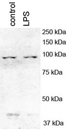
Application: Western BlotSample Tested: Cell lysate, Sample Amount: 50ug-100ug proteinSpecies: Mouse and HumanVerified Customer | Posted 10/30/2018After LPS stimulation, PGC1a expression at different time points.

-
Application: Western BlotSample Tested: 3 human cancer cell linesSpecies: HumanVerified Customer | Posted 12/14/2017PGC1ALPHA EXPRESSION IN HUMAN CANCER CELL LINES.AB DILUTIION: 1/1000
-
Application: Western BlotSample Tested: Primary mouse hepatocytesSpecies: MouseVerified Customer | Posted 12/08/2017mouse hepatocytes were treated with 100 ng/ml LPS for 4 hoursmouse hepatocytes were treated with 100 ng/ml LPS for 4 hours
-
Application: Western BlotSample Tested: Mouse skeletal muscle homogenateSpecies: MouseVerified Customer | Posted 09/26/2017Western Blot: PGC1 alpha Antibody [NBP1-04676]Western Blot: PGC1 alpha Antibody [NBP1-04676] - Total protein from murine skeletal muscle tissue, separated on a 4-12% gel by SDS-PAGE, transferred to nitrocellulose membrane and blocked in 5% non-fat milk for 1h at room temperature. The membrane was probed with anti-PGC1-alpha 1:4000 in non-fat milk.
-
Application: Chromatin ImmunoprecipitationSample Tested: Adult spinal cordSpecies: RatVerified Customer | Posted 04/20/2017
-
Application: Western BlotSample Tested: mouse liver lysateSpecies: mus musculus and MouseVerified Customer | Posted 04/18/2017
-
Application: Western BlotSample Tested: Mouse skeletal muscleSpecies: MouseVerified Customer | Posted 03/22/2017C57BL/6 Mice
-
Application: Western BlotSample Tested: Rat Liver Nuclear Protein ExtractSpecies: RatVerified Customer | Posted 11/05/2015PGC-1alpha Western tested in Rat Liver Nuclear Protein Extract
-
Application: Western BlotSample Tested: rat spinal cordSpecies: RatVerified Customer | Posted 09/16/2015rat spinal cord
-
Application: Western BlotSample Tested:Species: OtherVerified Customer | Posted 04/08/2015PGC1 alpha antibody and actin in Siberian hamster
-
Application: Western BlotSample Tested: rat liver homogenates and human fibroblasts lysateSpecies: HumanVerified Customer | Posted 04/23/2013
There are no reviews that match your criteria.
Protocols
View specific protocols for PGC1 alpha Antibody - BSA Free (NBP1-04676):
Immunocytochemistry Protocol
Culture cells to appropriate density in 35 mm culture dishes or 6-well plates.
1. Remove culture medium and wash the cells briefly in PBS. Add 10% formalin to the dish and fix at room temperature for 10 minutes.
2. Remove the formalin and wash the cells in PBS.
3. Permeablize the cells with 0.1% Triton X100 or other suitable detergent for 10 min.
4. Remove the permeablization buffer and wash three times for 10 minutes each in PBS. Be sure to not let the specimen dry out.
5. To block nonspecific antibody binding, incubate in 10% normal goat serum from 1 hour to overnight at room temperature.
6. Add primary antibody at appropriate dilution and incubate overnight at 4C.
7. Remove primary antibody and replace with PBS. Wash three times for 10 minutes each.
8. Add secondary antibody at appropriate dilution. Incubate for 1 hour at room temperature.
9. Remove secondary antibody and replace with PBS. Wash three times for 10 minutes each.
10. Counter stain DNA with DAPi if required.
Immunohistochemistry-Paraffin Embedded Sections
Antigen Unmasking:
Bring slides to a boil in 10 mM sodium citrate buffer (pH 6.0) then maintain at a sub-boiling temperature for 10 minutes. Cool slides on bench-top for 30 minutes.
Staining:
1. Wash sections in deionized water three times for 5 minutes each.
2. Wash sections in wash buffer for 5 minutes.
3. Block each section with 100-400 ul blocking solution for 1 hour at room temperature.
4. Remove blocking solution and add 100-400 ul diluted primary antibody. Incubate overnight at 4 C.
5. Remove antibody solution and wash sections in wash buffer three times for 5 minutes each.
6. Add 100-400 ul biotinylated diluted secondary antibody. Incubate 30 minutes at room temperature.
7. Remove secondary antibody solution and wash sections three times with wash buffer for 5 minutes each.
8. Add 100-400 ul Streptavidin-HRP reagent to each section and incubate for 30 minutes at room temperature.
9. Wash sections three times in wash buffer for 5 minutes each.
10. Add 100-400 ul DAB substrate to each section and monitor staining closely.
11. As soon as the sections develop, immerse slides in deionized water.
12. Counterstain sections in hematoxylin.
13. Wash sections in deionized water two times for 5 minutes each.
14. Dehydrate sections.
15. Mount coverslips.
*The above information is only intended as a guide. The researcher should determine what protocol best meets their needs. Please follow safe laboratory procedures.
Western Blot Protocol
1. Perform SDS-PAGE on samples to be analyzed, loading 40 ug of total protein per lane.
2. Transfer proteins to membrane according to the instructions provided by the manufacturer of the membrane and transfer apparatus.
3. Stain according to standard Ponceau S procedure (or similar product) to assess transfer success, and mark molecular weight standards where appropriate.
4. Rinse the blot.
5. Block the membrane using standard blocking buffer for at least 1 hour.
6. Wash the membrane in wash buffer three times for 10 minutes each.
7. Dilute primary antibody in blocking buffer and incubate 1 hour at room temperature.
8. Wash the membrane in wash buffer three times for 10 minutes each.
9. Apply the diluted HRP conjugated secondary antibody in blocking buffer (as per manufacturers instructions) and incubate 1 hour at room temperature.
10. Wash the blot in wash buffer three times for 10 minutes each (this step can be repeated as required to reduce background).
11. Apply the detection reagent of choice in accordance with the manufacturers instructions.
Note: Tween-20 can be added to the blocking or antibody dilution buffer at a final concentration of 0.05-0.2%.
Find general support by application which include: protocols, troubleshooting, illustrated assays, videos and webinars.
- 7-Amino Actinomycin D (7-AAD) Cell Viability Flow Cytometry Protocol
- Antigen Retrieval Protocol (PIER)
- Antigen Retrieval for Frozen Sections Protocol
- Appropriate Fixation of IHC/ICC Samples
- Cellular Response to Hypoxia Protocols
- ChIP Protocol Video
- Chromatin Immunoprecipitation (ChIP) Protocol
- Chromatin Immunoprecipitation Protocol
- Chromogenic IHC Staining of Formalin-Fixed Paraffin-Embedded (FFPE) Tissue Protocol
- Chromogenic Immunohistochemistry Staining of Frozen Tissue
- ClariTSA™ Fluorophore Kits
- Detection & Visualization of Antibody Binding
- Extracellular Membrane Flow Cytometry Protocol
- Flow Cytometry Protocol for Cell Surface Markers
- Flow Cytometry Protocol for Staining Membrane Associated Proteins
- Flow Cytometry Staining Protocols
- Flow Cytometry Troubleshooting Guide
- Fluorescent IHC Staining of Frozen Tissue Protocol
- Graphic Protocol for Heat-induced Epitope Retrieval
- Graphic Protocol for the Preparation and Fluorescent IHC Staining of Frozen Tissue Sections
- Graphic Protocol for the Preparation and Fluorescent IHC Staining of Paraffin-embedded Tissue Sections
- Graphic Protocol for the Preparation of Gelatin-coated Slides for Histological Tissue Sections
- ICC Cell Smear Protocol for Suspension Cells
- ICC Immunocytochemistry Protocol Videos
- ICC for Adherent Cells
- IHC Sample Preparation (Frozen sections vs Paraffin)
- Immunocytochemistry (ICC) Protocol
- Immunocytochemistry Troubleshooting
- Immunofluorescence of Organoids Embedded in Cultrex Basement Membrane Extract
- Immunofluorescent IHC Staining of Formalin-Fixed Paraffin-Embedded (FFPE) Tissue Protocol
- Immunohistochemistry (IHC) and Immunocytochemistry (ICC) Protocols
- Immunohistochemistry Frozen Troubleshooting
- Immunohistochemistry Paraffin Troubleshooting
- Immunoprecipitation Protocol
- Intracellular Flow Cytometry Protocol Using Alcohol (Methanol)
- Intracellular Flow Cytometry Protocol Using Detergents
- Intracellular Nuclear Staining Flow Cytometry Protocol Using Detergents
- Intracellular Staining Flow Cytometry Protocol Using Alcohol Permeabilization
- Intracellular Staining Flow Cytometry Protocol Using Detergents to Permeabilize Cells
- Preparing Samples for IHC/ICC Experiments
- Preventing Non-Specific Staining (Non-Specific Binding)
- Primary Antibody Selection & Optimization
- Propidium Iodide Cell Viability Flow Cytometry Protocol
- Protocol for Heat-Induced Epitope Retrieval (HIER)
- Protocol for Liperfluo
- Protocol for Making a 4% Formaldehyde Solution in PBS
- Protocol for VisUCyte™ HRP Polymer Detection Reagent
- Protocol for the Characterization of Human Th22 Cells
- Protocol for the Characterization of Human Th9 Cells
- Protocol for the Fluorescent ICC Staining of Cell Smears - Graphic
- Protocol for the Fluorescent ICC Staining of Cultured Cells on Coverslips - Graphic
- Protocol for the Preparation & Fixation of Cells on Coverslips
- Protocol for the Preparation and Chromogenic IHC Staining of Frozen Tissue Sections
- Protocol for the Preparation and Chromogenic IHC Staining of Frozen Tissue Sections - Graphic
- Protocol for the Preparation and Chromogenic IHC Staining of Paraffin-embedded Tissue Sections
- Protocol for the Preparation and Chromogenic IHC Staining of Paraffin-embedded Tissue Sections - Graphic
- Protocol for the Preparation and Fluorescent ICC Staining of Cells on Coverslips
- Protocol for the Preparation and Fluorescent ICC Staining of Non-adherent Cells
- Protocol for the Preparation and Fluorescent ICC Staining of Stem Cells on Coverslips
- Protocol for the Preparation and Fluorescent IHC Staining of Frozen Tissue Sections
- Protocol for the Preparation and Fluorescent IHC Staining of Paraffin-embedded Tissue Sections
- Protocol for the Preparation of Gelatin-coated Slides for Histological Tissue Sections
- Protocol for the Preparation of a Cell Smear for Non-adherent Cell ICC - Graphic
- Protocol: Annexin V and PI Staining by Flow Cytometry
- Protocol: Annexin V and PI Staining for Apoptosis by Flow Cytometry
- R&D Systems Quality Control Western Blot Protocol
- TUNEL and Active Caspase-3 Detection by IHC/ICC Protocol
- The Importance of IHC/ICC Controls
- Troubleshooting Guide: Fluorokine Flow Cytometry Kits
- Troubleshooting Guide: Immunohistochemistry
- Troubleshooting Guide: Western Blot Figures
- Western Blot Conditions
- Western Blot Protocol
- Western Blot Protocol for Cell Lysates
- Western Blot Troubleshooting
- Western Blot Troubleshooting Guide
- View all Protocols, Troubleshooting, Illustrated assays and Webinars
FAQs for PGC1 alpha Antibody - BSA Free
Showing
1
-
4 of
4 FAQs
Showing All
-
Q: Do you have any information about the specificity of NBP1-04676 with the methylated form of PGC1 alpha?
A: According to UniProt, there are no methylated a.a. reported for this protein target. However there are 13 N6-acetyllysine reported, and a.a. 413, 442, and 451 are included in the immunogen. Since this is a polyclonal antibody, it's anticipated that this antibody will detect both unacetylated and acetylated PGC1 alpha, regardless of where or how many of a.a. (lysine) residues are modified.
-
Q: I am looking for a primary PGC1 alpha antibody against human to be used in fluorescence. However, it cannot be conjugated with FITC (Green). It has to be conjugated with something else (Gold, red, orange, blue or aqua). Do you have such a product?
A: NBP1-04676R is conjugated to DyLight 550 which is an equivalent of TRITC. NBP1-04676C is conjugated to DyLight 650 which is an equivalent of APC. One of these two should fit your needs.
-
Q: Please differentiate to me between PPAR and PGC clearly. I am confused with the difference between these two
A:
Thank you very much for contacting Novus Biologicals technical support team and sharing your query on the differences between PGC-1 alpha and PPAR. These are two different proteins encoded by their respective genes and serves different functions. PGC-1 alpha (PGC1A or PPAR gamma coactivator 1-alpha) is a transcriptional co-activator for steroid receptors and nuclear receptors, and it regulates diverse aspects of cellular physiology. It up-regulates the transcriptional activity of PPAR-gamma /thyroid hormone receptor on the uncoupling protein promoter; regulates the key mitochondrial genes involved in adaptive thermogenesis; implicates in the metabolic reprogramming in response to nutrients availability through the coordination of the expression of a wide array of genes involved in the regulation of glucose and fatty acid metabolism. Among our PGC-1 alpha antibodies, NBP1-04676 is our best selling product with nice customer feedback and citations in at least 13 research publications. PPAR (PPAR alpha) on the other hand is a ligand-activated transcription factor which gets activated by the endogenous ligand 1-palmitoyl-2-oleoyl-sn-glycerol-3-phosphocholine, and oleylethanolamide (a naturally occurring lipid that regulates satiety), and acts as a key regulator of lipid metabolism. It also acts as a receptor for peroxisome proliferators such as hypolipidemic drugs and fatty acids. It regulates the peroxisomal beta-oxidation pathway of fatty acids, and also functions as transcription activator for the ACOX1 and P450 genes. We have a variety of PPAR alpha antibodies. I hope you will find this information helpful but please let me know if I can support you with anything else from my end. Thank you very much for choosing Novus Biologicals as your quality reagent supplier and we wish you the best with your research projects.
-
Q: What is the second band in the image from verified customer review: Western Blot: PGC1 alpha Antibody [NBP1-04676] - PGC1 alpha expression in lysates from Siberian hamster using anti-PGC1 alpha antibody?
A: The second band in the customer review image on our datasheet is from their actin loading control. Our band just below 50 kDa in human kidney lysate has not yet been identified. It is possible that it was heavy chain IgG detection from the secondary
-
Q: Do you have any information about the specificity of NBP1-04676 with the methylated form of PGC1 alpha?
A: According to UniProt, there are no methylated a.a. reported for this protein target. However there are 13 N6-acetyllysine reported, and a.a. 413, 442, and 451 are included in the immunogen. Since this is a polyclonal antibody, it's anticipated that this antibody will detect both unacetylated and acetylated PGC1 alpha, regardless of where or how many of a.a. (lysine) residues are modified.
-
Q: I am looking for a primary PGC1 alpha antibody against human to be used in fluorescence. However, it cannot be conjugated with FITC (Green). It has to be conjugated with something else (Gold, red, orange, blue or aqua). Do you have such a product?
A: NBP1-04676R is conjugated to DyLight 550 which is an equivalent of TRITC. NBP1-04676C is conjugated to DyLight 650 which is an equivalent of APC. One of these two should fit your needs.
-
Q: Please differentiate to me between PPAR and PGC clearly. I am confused with the difference between these two
A:
Thank you very much for contacting Novus Biologicals technical support team and sharing your query on the differences between PGC-1 alpha and PPAR. These are two different proteins encoded by their respective genes and serves different functions. PGC-1 alpha (PGC1A or PPAR gamma coactivator 1-alpha) is a transcriptional co-activator for steroid receptors and nuclear receptors, and it regulates diverse aspects of cellular physiology. It up-regulates the transcriptional activity of PPAR-gamma /thyroid hormone receptor on the uncoupling protein promoter; regulates the key mitochondrial genes involved in adaptive thermogenesis; implicates in the metabolic reprogramming in response to nutrients availability through the coordination of the expression of a wide array of genes involved in the regulation of glucose and fatty acid metabolism. Among our PGC-1 alpha antibodies, NBP1-04676 is our best selling product with nice customer feedback and citations in at least 13 research publications. PPAR (PPAR alpha) on the other hand is a ligand-activated transcription factor which gets activated by the endogenous ligand 1-palmitoyl-2-oleoyl-sn-glycerol-3-phosphocholine, and oleylethanolamide (a naturally occurring lipid that regulates satiety), and acts as a key regulator of lipid metabolism. It also acts as a receptor for peroxisome proliferators such as hypolipidemic drugs and fatty acids. It regulates the peroxisomal beta-oxidation pathway of fatty acids, and also functions as transcription activator for the ACOX1 and P450 genes. We have a variety of PPAR alpha antibodies. I hope you will find this information helpful but please let me know if I can support you with anything else from my end. Thank you very much for choosing Novus Biologicals as your quality reagent supplier and we wish you the best with your research projects.
-
Q: What is the second band in the image from verified customer review: Western Blot: PGC1 alpha Antibody [NBP1-04676] - PGC1 alpha expression in lysates from Siberian hamster using anti-PGC1 alpha antibody?
A: The second band in the customer review image on our datasheet is from their actin loading control. Our band just below 50 kDa in human kidney lysate has not yet been identified. It is possible that it was heavy chain IgG detection from the secondary
Loading...
Associated Pathways
