Podoplanin Antibody (8.1.1) - BSA Free
Novus Biologicals | Catalog # NB600-1015
Key Product Details
Validated by
Biological Validation
Species Reactivity
Validated:
Mouse, Human (Negative)
Cited:
Mouse
Applications
Validated:
Immunohistochemistry, Immunohistochemistry-Paraffin, Immunohistochemistry-Frozen, Western Blot, Flow Cytometry, Immunocytochemistry/ Immunofluorescence, Immunoprecipitation, Electron Microscopy, CyTOF-ready
Cited:
Immunohistochemistry, Immunohistochemistry-Paraffin, Immunohistochemistry-Frozen, Western Blot, Flow Cytometry, Immunocytochemistry/ Immunofluorescence, IF/IHC, Electron Microscopy
Label
Unconjugated
Antibody Source
Monoclonal Golden Syrian Hamster IgG Clone # 8.1.1
Format
BSA Free
Loading...
Product Specifications
Immunogen
Murine thymic stromal cell lines
Reactivity Notes
Does not cross-react with human.
Localization
Membrane; Single-pass type I membrane protein.
Marker
Lymphatic Endothelium Marker
Clonality
Monoclonal
Host
Golden Syrian Hamster
Isotype
IgG
Theoretical MW
40 kDa.
Disclaimer note: The observed molecular weight of the protein may vary from the listed predicted molecular weight due to post translational modifications, post translation cleavages, relative charges, and other experimental factors.
Disclaimer note: The observed molecular weight of the protein may vary from the listed predicted molecular weight due to post translational modifications, post translation cleavages, relative charges, and other experimental factors.
Scientific Data Images for Podoplanin Antibody (8.1.1) - BSA Free
![Western Blot: Podoplanin Antibody (8.1.1)BSA Free [NB600-1015]](https://resources.rndsystems.com/images/products/Podoplanin-Antibody-8-1-1-BSA-Free-Western-Blot-NB600-1015-img0012.jpg "Western Blot: Podoplanin Antibody (8.1.1)BSA Free [NB600-1015]")
Western Blot: Podoplanin Antibody (8.1.1)BSA Free [NB600-1015]
Podoplanin-Antibody-8-1-1-BSA-Free-Western-Blot-NB600-1015-img0012.jpg![Immunocytochemistry/ Immunofluorescence: Podoplanin Antibody (8.1.1) - BSA Free [NB600-1015]](https://resources.rndsystems.com/images/products/Podoplanin-Antibody-8-1-1-BSA-Free-Immunocytochemistry-Immunofluorescence-NB600-1015-img0009.jpg "Immunocytochemistry/ Immunofluorescence: Podoplanin Antibody (8.1.1) - BSA Free [NB600-1015]")
Immunocytochemistry/ Immunofluorescence: Podoplanin Antibody (8.1.1) - BSA Free [NB600-1015]
Immunocytochemistry/Immunofluorescence: Podoplanin Antibody (8.1.1) - BSA Free [NB600-1015] - Neuro2a cells were fixed for 10 minutes using 10% formalin and then permeabilized for 5 minutes using 1X PBS + 0.05% Triton X-100. The cells were incubated with anti- at 5 ug/ml overnight at 4C and detected with an anti-Golden Syrian hamster DyLight 488 (Green) at a 1:500 dilution. Nuclei were counterstained with DAPI (Blue). Cells were imaged using a 40X objective.![Immunohistochemistry-Paraffin: Podoplanin Antibody (8.1.1) - BSA Free [NB600-1015]](https://resources.rndsystems.com/images/products/Podoplanin-Antibody-8-1-1-BSA-Free-Immunohistochemistry-Paraffin-NB600-1015-img0010.jpg "Immunohistochemistry-Paraffin: Podoplanin Antibody (8.1.1) - BSA Free [NB600-1015]")
Immunohistochemistry-Paraffin: Podoplanin Antibody (8.1.1) - BSA Free [NB600-1015]
Immunohistochemistry-Paraffin: Podoplanin Antibody (8.1.1) - BSA Free [NB600-1015] - Staining for podoplanin (red) and cytokeratins (green) in malignant serosal tumors morphologically consistent with sarcomatous mesothelioma. In A, the lining mesothelium (solid arrows) stained positively for podoplanin in this double label immunofluorescent image of diaphragm. The cells beneath the mesothelial lining are also red due to expression of podoplanin and these are cells of a malignant serosal tumor. Promotion of lung adenocarcinoma following inhalation exposure to multi-walled carbon nanotubes. Part Fibre Toxicol (2014)![Western Blot: Podoplanin Antibody (8.1.1)BSA Free [NB600-1015]](https://resources.rndsystems.com/images/products/Podoplanin-Antibody-8-1-1-BSA-Free-Western-Blot-NB600-1015-img0001.jpg "Western Blot: Podoplanin Antibody (8.1.1)BSA Free [NB600-1015]")
Western Blot: Podoplanin Antibody (8.1.1)BSA Free [NB600-1015]
Western Blot: Podoplanin Antibody (8.1.1) - BSA Free [NB600-1015] - Analysis of Podoplanin in mouse kidney tissue extract.![Immunocytochemistry/ Immunofluorescence: Podoplanin Antibody (8.1.1) - BSA Free [NB600-1015]](https://resources.rndsystems.com/images/products/Podoplanin-Antibody-8-1-1-BSA-Free-Immunocytochemistry-Immunofluorescence-NB600-1015-img0013.jpg "Immunocytochemistry/ Immunofluorescence: Podoplanin Antibody (8.1.1) - BSA Free [NB600-1015]")
![Immunohistochemistry-Paraffin: Podoplanin Antibody (8.1.1) - BSA Free [NB600-1015]](https://resources.rndsystems.com/images/products/Podoplanin-Antibody-8-1-1-BSA-Free-Immunohistochemistry-Paraffin-NB600-1015-img0004.jpg "Immunohistochemistry-Paraffin: Podoplanin Antibody (8.1.1) - BSA Free [NB600-1015]")
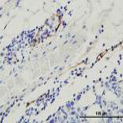
Immunohistochemistry-Paraffin: Podoplanin Antibody (8.1.1) - BSA Free [NB600-1015]
Immunohistochemistry-Paraffin: Podoplanin Antibody (8.1.1) - BSA Free [NB600-1015] - Analysis of Podoplanin in mouse mammary gland after MDA-MB-231 orthotopic transplantation. Image courtesy of product review by Luana Schito.![Immunohistochemistry: Podoplanin Antibody (8.1.1) - BSA Free [NB600-1015]](https://resources.rndsystems.com/images/products/Podoplanin-Antibody-8-1-1-BSA-Free-Immunohistochemistry-NB600-1015-img0008.jpg "Immunohistochemistry: Podoplanin Antibody (8.1.1) - BSA Free [NB600-1015]")
Immunohistochemistry: Podoplanin Antibody (8.1.1) - BSA Free [NB600-1015]
Immunohistochemistry: Podoplanin Antibody (8.1.1) - BSA Free [NB600-1015] - Podoplanin (8.1.1) antibody labeling (Green) of glomeruli from mouse kidney. Nuclei were counterstained with DAPI (Blue).![Immunohistochemistry-Paraffin: Podoplanin Antibody (8.1.1) - BSA Free [NB600-1015]](https://resources.rndsystems.com/images/products/Podoplanin-Antibody-8-1-1-BSA-Free-Immunohistochemistry-Paraffin-NB600-1015-img0011.jpg "Immunohistochemistry-Paraffin: Podoplanin Antibody (8.1.1) - BSA Free [NB600-1015]")
Immunohistochemistry-Paraffin: Podoplanin Antibody (8.1.1) - BSA Free [NB600-1015]
Immunohistochemistry-Paraffin: Podoplanin Antibody (8.1.1) - BSA Free [NB600-1015] - Staining for podoplanin (red) and cytokeratins (green) in malignant serosal tumors morphologically consistent with sarcomatous mesothelioma. In C, a double label immunofluorescent image demonstrates a malignant serosal tumor between the liver and gall bladder that is lined by reactive mesothelium (solid arrow) which stains red for podoplanin as well as green for cytokeratins. The cells of the subjacent malignant serosal tumor stain weakly red for podoplanin. Promotion of lung adenocarcinoma following inhalation exposure to multi-walled carbon nanotubes. Part Fibre Toxicol (2014) - BSA Free [NB600-1015] -")
Immunofluorescence: Podoplanin Antibody (8.1.1) - BSA Free [NB600-1015] -
Podolanin expressing on Mɸs mediates interactions with platelets.Mɸs were isolated from wild-type (WT) mice treated with acetaminophen (APAP) for 3 hr. The cells were treated in vitro with either control IgG (Ctrl IgG) or an anti-podoplanin antibody ( alpha -podoplanin Ab) before incubation with platelets. Immunofluorescence (IF) staining was performed to detect podoplanin on Mɸs and C-type lectin-like receptor 2 (Clec-2) on platelets. Scale bar, 25 μm. - BSA Free [NB600-1015] -")
Immunocytochemistry/ Immunofluorescence: Podoplanin Antibody (8.1.1) - BSA Free [NB600-1015] -
Immunocytochemistry/ Immunofluorescence: Podoplanin Antibody (8.1.1) - BSA Free [NB600-1015] - Immunofluorescence staining for podoplanin (red) & cytokeratins (green) in malignant serosal tumors morphologically consistent with sarcomatous mesothelioma. In A, the lining mesothelium (solid arrows) stained positively for podoplanin in this double label immunofluorescent image of diaphragm. The cells beneath the mesothelial lining are also red due to expression of podoplanin & these are cells of a malignant serosal tumor. In B, only the single label green fluorescence is shown to demonstrate weak expression of cytokeratins in the lining mesothelium (solid arrow), while staining of the subjacent tumor for cytokeratins is equivocal. In C, a double label immunofluorescent image demonstrates a malignant serosal tumor between the liver & gall bladder that is lined by reactive mesothelium (solid arrow) which stains red for podoplanin as well as green for cytokeratins. The cells of the subjacent malignant serosal tumor stain weakly red for podoplanin. In D, the photomicrograph shows this same tumor but only the green fluorescence for cytokeratins. The reactive mesothelium lining the malignant serosal tumor (solid arrow) stains green for cytokeratins while the serosal tumor has no evidence of cytokeratin expression. The normal mesothelium lining the liver is weakly positive for cytokeratins (dashed arrow). The epithelium lining the gall bladder (open arrow) strongly expresses cytokeratins. Magnification bar is 50 μm. Image collected & cropped by CiteAb from the following publication (http://particleandfibretoxicology.biomedcentral.com/articles/10.1186/17…), licensed under a CC-BY license. Not internally tested by Novus Biologicals. - BSA Free [NB600-1015] -")
Immunocytochemistry/ Immunofluorescence: Podoplanin Antibody (8.1.1) - BSA Free [NB600-1015] -
Immunocytochemistry/ Immunofluorescence: Podoplanin Antibody (8.1.1) - BSA Free [NB600-1015] - Immunofluorescence staining for podoplanin (red) & cytokeratins (green) in malignant serosal tumors morphologically consistent with sarcomatous mesothelioma. In A, the lining mesothelium (solid arrows) stained positively for podoplanin in this double label immunofluorescent image of diaphragm. The cells beneath the mesothelial lining are also red due to expression of podoplanin & these are cells of a malignant serosal tumor. In B, only the single label green fluorescence is shown to demonstrate weak expression of cytokeratins in the lining mesothelium (solid arrow), while staining of the subjacent tumor for cytokeratins is equivocal. In C, a double label immunofluorescent image demonstrates a malignant serosal tumor between the liver & gall bladder that is lined by reactive mesothelium (solid arrow) which stains red for podoplanin as well as green for cytokeratins. The cells of the subjacent malignant serosal tumor stain weakly red for podoplanin. In D, the photomicrograph shows this same tumor but only the green fluorescence for cytokeratins. The reactive mesothelium lining the malignant serosal tumor (solid arrow) stains green for cytokeratins while the serosal tumor has no evidence of cytokeratin expression. The normal mesothelium lining the liver is weakly positive for cytokeratins (dashed arrow). The epithelium lining the gall bladder (open arrow) strongly expresses cytokeratins. Magnification bar is 50 μm. Image collected & cropped by CiteAb from the following publication (http://particleandfibretoxicology.biomedcentral.com/articles/10.1186/17…), licensed under a CC-BY license. Not internally tested by Novus Biologicals.
Immunofluorescent Staining of Adult stem cell-derived Lung Organoids.
Adult stem cells isolated from human lung biopsy tissue were cultured following the steps detailed in the human lung organoid culture protocol. Lung organoids were stained with a (A) Goat Anti-Human p63/TP73L Polyclonal Antibody (Catalog # AF1916; red) and a rabbit anti-human cytokeratin 5 (KRT5) monoclonal antibody (green) to visualize basal cells; a (B) Hamster Anti-Mouse Podoplanin (PDPN) Monoclonal Antibody (Novus Biologicals, Catalog # NB600-1015; green) to visualize alveolar type I cells and a Goat Anti-Human p63/TP73L Polyclonal Antibody (Catalog # AF1916; red) to visualize basal cells; and a (C, D) Mouse Anti-MUC5AC Monoclonal Antibody (Novus Biologicals, Catalog # NBP2-15196; green) to visualize goblet cells and a Mouse Anti-Human/Mouse/Rat SOX2 Monoclonal Antibody (Catalog # MAB2018; red). All samples were counterstained with DAPI (Catalog # 5748; blue).Applications for Podoplanin Antibody (8.1.1) - BSA Free
Application
Recommended Usage
Flow Cytometry
1:400
Immunocytochemistry/ Immunofluorescence
1-5ug/ml
Immunohistochemistry
1:100-1:500
Immunohistochemistry-Paraffin
1:100-1:500
Immunoprecipitation
1:10-1:500
Western Blot
1:1000-1:2000
Application Notes
Optimal dilutions/concentrations should be determined by the end user. This antibody is CyTOF ready.
Reviewed Applications
Read 1 review rated 5 using NB600-1015 in the following applications:
Flow Cytometry Panel Builder
Bio-Techne Knows Flow Cytometry
Save time and reduce costly mistakes by quickly finding compatible reagents using the Panel Builder Tool.
Advanced Features
- Spectra Viewer - Custom analysis of spectra from multiple fluorochromes
- Spillover Popups - Visualize the spectra of individual fluorochromes
- Antigen Density Selector - Match fluorochrome brightness with antigen density

Formulation, Preparation, and Storage
Purification
Protein G purified
Formulation
PBS
Format
BSA Free
Preservative
0.02% Sodium Azide
Concentration
1 mg/ml
Shipping
The product is shipped with polar packs. Upon receipt, store it immediately at the temperature recommended below.
Stability & Storage
Store at 4C short term. Aliquot and store at -20C long term. Avoid freeze-thaw cycles.
Background: Podoplanin
Long Name
Lung Type-I Cell Membrane-associated Glycoprotein, Isoform A
Alternate Names
Aggrus, Gp38, PDPN, RANDAM-2, T1A-2
Entrez Gene IDs
14726 (Mouse)
Gene Symbol
PDPN
UniProt
Additional Podoplanin Products
Product Documents for Podoplanin Antibody (8.1.1) - BSA Free
Certificate of Analysis
To download a Certificate of Analysis, please enter a lot or batch number in the search box below.
Product Specific Notices for Podoplanin Antibody (8.1.1) - BSA Free
This product is for research use only and is not approved for use in humans or in clinical diagnosis. Primary Antibodies are guaranteed for 1 year from date of receipt.
Related Research Areas
Citations for Podoplanin Antibody (8.1.1) - BSA Free
 Powered by Bioz
Customer Reviews for Podoplanin Antibody (8.1.1) - BSA Free (1)
5 out of 5
1 Customer Rating
Have you used Podoplanin Antibody (8.1.1) - BSA Free?
Submit a review and receive an Amazon gift card!
$25/€18/£15/$25CAN/¥2500 Yen for a review with an image
$10/€7/£6/$10CAN/¥1110 Yen for a review without an image
Submit a review
Customer Images
Showing
1
-
1 of
1 review
Showing All
Filter By:
-
Application: Immunohistochemistry-ParaffinSample Tested: Mouse mammary gland after MDA-MB-231 orthotopic transplantationSpecies: MouseVerified Customer | Posted 05/17/2011

There are no reviews that match your criteria.
Protocols
Find general support by application which include: protocols, troubleshooting, illustrated assays, videos and webinars.
- 7-Amino Actinomycin D (7-AAD) Cell Viability Flow Cytometry Protocol
- Antigen Retrieval Protocol (PIER)
- Antigen Retrieval for Frozen Sections Protocol
- Appropriate Fixation of IHC/ICC Samples
- Cellular Response to Hypoxia Protocols
- Chromogenic IHC Staining of Formalin-Fixed Paraffin-Embedded (FFPE) Tissue Protocol
- Chromogenic Immunohistochemistry Staining of Frozen Tissue
- ClariTSA™ Fluorophore Kits
- Detection & Visualization of Antibody Binding
- Extracellular Membrane Flow Cytometry Protocol
- Flow Cytometry Protocol for Cell Surface Markers
- Flow Cytometry Protocol for Staining Membrane Associated Proteins
- Flow Cytometry Staining Protocols
- Flow Cytometry Troubleshooting Guide
- Fluorescent IHC Staining of Frozen Tissue Protocol
- Graphic Protocol for Heat-induced Epitope Retrieval
- Graphic Protocol for the Preparation and Fluorescent IHC Staining of Frozen Tissue Sections
- Graphic Protocol for the Preparation and Fluorescent IHC Staining of Paraffin-embedded Tissue Sections
- Graphic Protocol for the Preparation of Gelatin-coated Slides for Histological Tissue Sections
- ICC Cell Smear Protocol for Suspension Cells
- ICC Immunocytochemistry Protocol Videos
- ICC for Adherent Cells
- IHC Sample Preparation (Frozen sections vs Paraffin)
- Immunocytochemistry (ICC) Protocol
- Immunocytochemistry Troubleshooting
- Immunofluorescence of Organoids Embedded in Cultrex Basement Membrane Extract
- Immunofluorescent IHC Staining of Formalin-Fixed Paraffin-Embedded (FFPE) Tissue Protocol
- Immunohistochemistry (IHC) and Immunocytochemistry (ICC) Protocols
- Immunohistochemistry Frozen Troubleshooting
- Immunohistochemistry Paraffin Troubleshooting
- Immunoprecipitation Protocol
- Intracellular Flow Cytometry Protocol Using Alcohol (Methanol)
- Intracellular Flow Cytometry Protocol Using Detergents
- Intracellular Nuclear Staining Flow Cytometry Protocol Using Detergents
- Intracellular Staining Flow Cytometry Protocol Using Alcohol Permeabilization
- Intracellular Staining Flow Cytometry Protocol Using Detergents to Permeabilize Cells
- Preparing Samples for IHC/ICC Experiments
- Preventing Non-Specific Staining (Non-Specific Binding)
- Primary Antibody Selection & Optimization
- Propidium Iodide Cell Viability Flow Cytometry Protocol
- Protocol for Heat-Induced Epitope Retrieval (HIER)
- Protocol for Liperfluo
- Protocol for Making a 4% Formaldehyde Solution in PBS
- Protocol for VisUCyte™ HRP Polymer Detection Reagent
- Protocol for the Characterization of Human Th22 Cells
- Protocol for the Characterization of Human Th9 Cells
- Protocol for the Fluorescent ICC Staining of Cell Smears - Graphic
- Protocol for the Fluorescent ICC Staining of Cultured Cells on Coverslips - Graphic
- Protocol for the Preparation & Fixation of Cells on Coverslips
- Protocol for the Preparation and Chromogenic IHC Staining of Frozen Tissue Sections
- Protocol for the Preparation and Chromogenic IHC Staining of Frozen Tissue Sections - Graphic
- Protocol for the Preparation and Chromogenic IHC Staining of Paraffin-embedded Tissue Sections
- Protocol for the Preparation and Chromogenic IHC Staining of Paraffin-embedded Tissue Sections - Graphic
- Protocol for the Preparation and Fluorescent ICC Staining of Cells on Coverslips
- Protocol for the Preparation and Fluorescent ICC Staining of Non-adherent Cells
- Protocol for the Preparation and Fluorescent ICC Staining of Stem Cells on Coverslips
- Protocol for the Preparation and Fluorescent IHC Staining of Frozen Tissue Sections
- Protocol for the Preparation and Fluorescent IHC Staining of Paraffin-embedded Tissue Sections
- Protocol for the Preparation of Gelatin-coated Slides for Histological Tissue Sections
- Protocol for the Preparation of a Cell Smear for Non-adherent Cell ICC - Graphic
- Protocol: Annexin V and PI Staining by Flow Cytometry
- Protocol: Annexin V and PI Staining for Apoptosis by Flow Cytometry
- R&D Systems Quality Control Western Blot Protocol
- TUNEL and Active Caspase-3 Detection by IHC/ICC Protocol
- The Importance of IHC/ICC Controls
- Troubleshooting Guide: Fluorokine Flow Cytometry Kits
- Troubleshooting Guide: Immunohistochemistry
- Troubleshooting Guide: Western Blot Figures
- Western Blot Conditions
- Western Blot Protocol
- Western Blot Protocol for Cell Lysates
- Western Blot Troubleshooting
- Western Blot Troubleshooting Guide
- View all Protocols, Troubleshooting, Illustrated assays and Webinars
Loading...