Presenilin-1 Antibody (APS 11)
Novus Biologicals | Catalog # NB300-749
![Western Blot: Presenilin-1 Antibody (APS 11) [NB300-749]](https://resources.rndsystems.com/images/products/Presenilin-1-Antibody-APS-11-Western-Blot-NB300-749-img0007.jpg "Western Blot: Presenilin-1 Antibody (APS 11) [NB300-749]")
Loading...
Key Product Details
Species Reactivity
Validated:
Human, Mouse, Rat, Primate
Cited:
Rat
Applications
Validated:
Immunohistochemistry, Immunohistochemistry-Paraffin, Western Blot, ELISA, Immunocytochemistry/ Immunofluorescence, Immunoprecipitation
Cited:
Western Blot
Label
Unconjugated
Antibody Source
Monoclonal Mouse IgG1 Clone # APS 11
Loading...
Product Specifications
Immunogen
Synthetic peptide corresponding to residues C H(21) L S N T V R S Q N D N R E(34) of human PS1.
Reactivity Notes
Please note that this antibody is reactive to Mouse and derived from the same host, Mouse. Additional Mouse on Mouse blocking steps may be required for IHC and ICC experiments. Please contact Technical Support for more information.
Localization
Integral membrane protein. Golgi and endoplasmic reticulum. Bound to NOTCH1 also at the cell surface.
Specificity
No cross-reactivity is seen with presenilin 2.
Clonality
Monoclonal
Host
Mouse
Isotype
IgG1
Scientific Data Images for Presenilin-1 Antibody (APS 11)
Western Blot: Presenilin-1 Antibody (APS 11) [NB300-749]
Western Blot: Presenilin-1 Antibody (APS 11) [NB300-749] - Analysis of 25 ug of SH-SY5Y (lane 1), human brain (lane 2) and mouse brain (lane 3).![Immunocytochemistry/ Immunofluorescence: Presenilin-1 Antibody (APS 11) [NB300-749]](https://resources.rndsystems.com/images/products/Presenilin-1-Antibody-APS-11-Immunocytochemistry-Immunofluorescence-NB300-749-img0004.jpg "Immunocytochemistry/ Immunofluorescence: Presenilin-1 Antibody (APS 11) [NB300-749]")
Immunocytochemistry/ Immunofluorescence: Presenilin-1 Antibody (APS 11) [NB300-749]
Immunocytochemistry/Immunofluorescence: Presenilin-1 Antibody (APS 11) [NB300-749] - Mouse fibroblasts.![Immunohistochemistry-Paraffin: Presenilin-1 Antibody (APS 11) [NB300-749]](https://resources.rndsystems.com/images/products/Presenilin-1-Antibody-APS-11-Immunohistochemistry-Paraffin-NB300-749-img0006.jpg "Immunohistochemistry-Paraffin: Presenilin-1 Antibody (APS 11) [NB300-749]")
Immunohistochemistry-Paraffin: Presenilin-1 Antibody (APS 11) [NB300-749]
Immunohistochemistry-Paraffin: Presenilin-1 Antibody (APS 11) [NB300-749] - Immunohistochemistry was performed on normal biopsies of deparaffinized Human liver tissue. To expose target proteins, heat induced antigen retrieval was performed using 10mM sodium citrate (pH6.0) buffer, microwaved for 8-15 minutes. Following antigen retrieval tissues were blocked in 3% BSA-PBS for 30 minutes at room temperature. Tissues were then probed at a dilution of 1:200 with a mouse monoclonal antibody recognizing Presenilin 1 or without primary antibody (negative control) overnight at 4C in a humidified chamber. Tissues were washed extensively with PBST and endogenous peroxidase activity was quenched with a peroxidase suppressor. Tissues were counterstained with hematoxylin and prepped for mounting.![Immunohistochemistry-Paraffin: Presenilin-1 Antibody (APS 11) [NB300-749]](https://resources.rndsystems.com/images/products/Presenilin-1-Antibody-APS-11-Immunohistochemistry-Paraffin-NB300-749-img0005.jpg "Immunohistochemistry-Paraffin: Presenilin-1 Antibody (APS 11) [NB300-749]")
Immunohistochemistry-Paraffin: Presenilin-1 Antibody (APS 11) [NB300-749]
Immunohistochemistry-Paraffin: Presenilin-1 Antibody (APS 11) [NB300-749] - Immunohistochemistry was performed on normal biopsies of deparaffinized Human brain tissue. To expose target proteins, heat induced antigen retrieval was performed using 10mM sodium citrate (pH6.0) buffer, microwaved for 8-15 minutes. Following antigen retrieval tissues were blocked in 3% BSA-PBS for 30 minutes at room temperature. Tissues were then probed at a dilution of 1:20 with a mouse monoclonal antibody recognizing Presenilin 1 or without primary antibody (negative control) overnight at 4C in a humidified chamber. Tissues were washed extensively with PBST and endogenous peroxidase activity was quenched with a peroxidase suppressor. Tissues were counterstained with hematoxylin and prepped for mounting.Applications for Presenilin-1 Antibody (APS 11)
Application
Recommended Usage
ELISA
4.5 ug/ml
Immunocytochemistry/ Immunofluorescence
0.75 ug/ml
Immunohistochemistry
1:10 - 1:500
Immunohistochemistry-Paraffin
0.75 ug/ml
Immunoprecipitation
1:10 - 1:500
Western Blot
1:100 - 1:1000
Application Notes
IHC-P: Staining of both the human brain plaque core and dystrophic neurites. WB: Detects an approx. 28 kDa protein representing PS1 N-terminus cleavage product.
Reviewed Applications
Read 1 review rated 4 using NB300-749 in the following applications:
Formulation, Preparation, and Storage
Purification
Protein G purified
Formulation
PBS with 1 mg/ml BSA
Preservative
0.05% Sodium Azide
Concentration
1 mg/ml
Shipping
The product is shipped with polar packs. Upon receipt, store it immediately at the temperature recommended below.
Stability & Storage
Store at -20C. Avoid freeze-thaw cycles.
Background: Presenilin-1
Alternate Names
Presenilin1, PSEN1, S182
Gene Symbol
PSEN1
UniProt
Additional Presenilin-1 Products
Product Documents for Presenilin-1 Antibody (APS 11)
Certificate of Analysis
To download a Certificate of Analysis, please enter a lot or batch number in the search box below.
Product Specific Notices for Presenilin-1 Antibody (APS 11)
This product is for research use only and is not approved for use in humans or in clinical diagnosis. Primary Antibodies are guaranteed for 1 year from date of receipt.
Related Research Areas
Citations for Presenilin-1 Antibody (APS 11)
 Powered by Bioz
Customer Reviews for Presenilin-1 Antibody (APS 11) (1)
4 out of 5
1 Customer Rating
Have you used Presenilin-1 Antibody (APS 11)?
Submit a review and receive an Amazon gift card!
$25/€18/£15/$25CAN/¥2500 Yen for a review with an image
$10/€7/£6/$10CAN/¥1110 Yen for a review without an image
Submit a review
Customer Images
Showing
1
-
1 of
1 review
Showing All
Filter By:
-
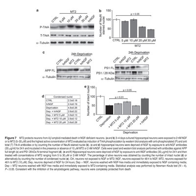
Application: Western BlotVerified Customer | Posted 12/07/2012MT2 protects neurons from Ab amyloid-mediated death in NGF-deficient neurons

There are no reviews that match your criteria.
Protocols
Find general support by application which include: protocols, troubleshooting, illustrated assays, videos and webinars.
- Antigen Retrieval Protocol (PIER)
- Antigen Retrieval for Frozen Sections Protocol
- Appropriate Fixation of IHC/ICC Samples
- Cellular Response to Hypoxia Protocols
- Chromogenic IHC Staining of Formalin-Fixed Paraffin-Embedded (FFPE) Tissue Protocol
- Chromogenic Immunohistochemistry Staining of Frozen Tissue
- ClariTSA™ Fluorophore Kits
- Detection & Visualization of Antibody Binding
- ELISA Sample Preparation & Collection Guide
- ELISA Troubleshooting Guide
- Fluorescent IHC Staining of Frozen Tissue Protocol
- Graphic Protocol for Heat-induced Epitope Retrieval
- Graphic Protocol for the Preparation and Fluorescent IHC Staining of Frozen Tissue Sections
- Graphic Protocol for the Preparation and Fluorescent IHC Staining of Paraffin-embedded Tissue Sections
- Graphic Protocol for the Preparation of Gelatin-coated Slides for Histological Tissue Sections
- How to Run an R&D Systems DuoSet ELISA
- How to Run an R&D Systems Quantikine ELISA
- How to Run an R&D Systems Quantikine™ QuicKit™ ELISA
- ICC Cell Smear Protocol for Suspension Cells
- ICC Immunocytochemistry Protocol Videos
- ICC for Adherent Cells
- IHC Sample Preparation (Frozen sections vs Paraffin)
- Immunocytochemistry (ICC) Protocol
- Immunocytochemistry Troubleshooting
- Immunofluorescence of Organoids Embedded in Cultrex Basement Membrane Extract
- Immunofluorescent IHC Staining of Formalin-Fixed Paraffin-Embedded (FFPE) Tissue Protocol
- Immunohistochemistry (IHC) and Immunocytochemistry (ICC) Protocols
- Immunohistochemistry Frozen Troubleshooting
- Immunohistochemistry Paraffin Troubleshooting
- Immunoprecipitation Protocol
- Preparing Samples for IHC/ICC Experiments
- Preventing Non-Specific Staining (Non-Specific Binding)
- Primary Antibody Selection & Optimization
- Protocol for Heat-Induced Epitope Retrieval (HIER)
- Protocol for Making a 4% Formaldehyde Solution in PBS
- Protocol for VisUCyte™ HRP Polymer Detection Reagent
- Protocol for the Fluorescent ICC Staining of Cell Smears - Graphic
- Protocol for the Fluorescent ICC Staining of Cultured Cells on Coverslips - Graphic
- Protocol for the Preparation & Fixation of Cells on Coverslips
- Protocol for the Preparation and Chromogenic IHC Staining of Frozen Tissue Sections
- Protocol for the Preparation and Chromogenic IHC Staining of Frozen Tissue Sections - Graphic
- Protocol for the Preparation and Chromogenic IHC Staining of Paraffin-embedded Tissue Sections
- Protocol for the Preparation and Chromogenic IHC Staining of Paraffin-embedded Tissue Sections - Graphic
- Protocol for the Preparation and Fluorescent ICC Staining of Cells on Coverslips
- Protocol for the Preparation and Fluorescent ICC Staining of Non-adherent Cells
- Protocol for the Preparation and Fluorescent ICC Staining of Stem Cells on Coverslips
- Protocol for the Preparation and Fluorescent IHC Staining of Frozen Tissue Sections
- Protocol for the Preparation and Fluorescent IHC Staining of Paraffin-embedded Tissue Sections
- Protocol for the Preparation of Gelatin-coated Slides for Histological Tissue Sections
- Protocol for the Preparation of a Cell Smear for Non-adherent Cell ICC - Graphic
- Quantikine HS ELISA Kit Assay Principle, Alkaline Phosphatase
- Quantikine HS ELISA Kit Principle, Streptavidin-HRP Polymer
- R&D Systems Quality Control Western Blot Protocol
- Sandwich ELISA (Colorimetric) – Biotin/Streptavidin Detection Protocol
- Sandwich ELISA (Colorimetric) – Direct Detection Protocol
- TUNEL and Active Caspase-3 Detection by IHC/ICC Protocol
- The Importance of IHC/ICC Controls
- Troubleshooting Guide: ELISA
- Troubleshooting Guide: Immunohistochemistry
- Troubleshooting Guide: Western Blot Figures
- Western Blot Conditions
- Western Blot Protocol
- Western Blot Protocol for Cell Lysates
- Western Blot Troubleshooting
- Western Blot Troubleshooting Guide
- View all Protocols, Troubleshooting, Illustrated assays and Webinars
FAQs for Presenilin-1 Antibody (APS 11)
Showing
1
-
1 of
1 FAQ
Showing All
-
Q: What is the exact immuogen?
A: The immunogen range for this antibody falls between amino acids 340-370.
Loading...
Associated Pathways

