
Key Product Details
Species Reactivity
Validated:
Rat
Cited:
Human, Mouse, Rat, Primate - Macaca fascicularis (Crab-eating Monkey or Cynomolgus Macaque), Rabbit, Transgenic Mouse
Applications
Validated:
Immunohistochemistry, Western Blot, Immunocytochemistry, Immunoprecipitation, Immunoaffinity Purification
Cited:
Immunohistochemistry, Immunohistochemistry-Paraffin, Western Blot, Immunocytochemistry, Immunoprecipitation
Label
Unconjugated
Antibody Source
Monoclonal Mouse IgG1 Clone # 1F8
Loading...
Product Specifications
Immunogen
Partially purified vesicles containing Glut4
Specificity
Recognizes an epitope in the cytoplasmic portion of rat Glut4 and has been shown to bind only to the muscle-adipose isoform of the glucose transporter, which is antigenically unique from the other transport proteins (1-3). Recognizes Glut4 in human, monkey, rat, mouse, and rabbit systems. It does not recognize this protein in canine systems. Its ability to bind to glucose transporter from other species has not been tested.
Clonality
Monoclonal
Host
Mouse
Isotype
IgG1
Scientific Data Images for Rat Glut4 Antibody (1F8)
Glut4 in L6 Rat Cell Line.
Glut4 was detected in immersion fixed L6 rat myoblast cell line (differentiated to muscle) using 10 µg/mL Rat Glut4 Monoclonal Antibody (Catalog # MAB1262) for 3 hours at room temperature. Cells were stained with the NorthernLights™ 557-conjugated Anti-Mouse IgG Secondary Antibody (red; Catalog # NL007) and counterstained with DAPI (blue). View our protocol for Fluorescent ICC Staining of Cells on Coverslips.
Glut4 in Rat Heart Tissue.
Glut4 was detected in immersion fixed paraffin-embedded sections of rat heart tissue using Mouse Anti-Rat Glut4 Monoclonal Antibody (Catalog # MAB1262) at 5 µg/mL overnight at 4 °C. Before incubation with the primary antibody, tissue was subjected to heat-induced epitope retrieval using Antigen Retrieval Reagent-Basic (Catalog # CTS013). Tissue was stained using the Anti-Mouse IgG VisUCyte™ HRP Polymer Antibody (brown; Catalog # VC001) and counterstained with hematoxylin (blue). Specific staining was localized to cytoplasm in cardiomyocytes. View our protocol for IHC Staining with VisUCyte HRP Polymer Detection Reagents.
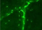
Detection of Human Glut4 by Immunocytochemistry/Immunofluorescence
Primary podocytes can be cultured from kidney organoid glomeruli. a Isolated organoid glomeruli show evidence of podocyte cell migration (OrgPods) displaying thin arborized projections (TAPs) (inset). Inverted image shown to provide maximum contrast, scale bar 100 µm. b TAPS from newly emerged podocytes are composed of F-actin shown by phallodin immunofluorescent staining. Inverted image, scale bar 50 µm. c Immunostaining at 36 h post-plating shows a strong positively stained 3D OrgGlom with a migrating 2D OrgPod population. Left panel 2D images, right panel 3D reconstruction of Z-stack. Scale bars 50 µm. d At 48 h post-plating OrgPods display a flattened, arborized morphology with processes connecting adjacent cells (arrow), scale bar 50 µm. e Immunostaining of ciPods for SYNAPTOPODIN showed expression is absent in undifferentiated cells (ciPod: Un), only becoming evident following 14 days induced differentiation at 37 °C (ciPod: Diff). OrgPods also display strong SYNAPTOPODIN protein expression, aligned with F-actin stress fibres. Scale bars 100 µm. f OrgPods express the neonatal Fc receptor (FcRN) and actively endocytose fluorescein isothiocyanate (FITC)-labelled albumin at 37 °C resulting in FITC-accumulation in endosomes on the cell surface. This is process halted when performed at 4 °C. Scale bars 50 µm. g OrgPods stimulated with insulin (10 mg/ml) for 10 min showed cortical reorganisation of their actin cytoskeleton with GLUT4 translocation from a vesicular to plasma membrane localisation. Scale bars 50 µm. All representative images reflect a minimum of three biological replicates. For immunofluorescence, images are shown in greyscale for single channels, and merged images in colour Image collected and cropped by CiteAb from the following publication (https://pubmed.ncbi.nlm.nih.gov/30514835), licensed under a CC-BY license. Not internally tested by R&D Systems.Applications for Rat Glut4 Antibody (1F8)
Application
Recommended Usage
Immunoaffinity Purification
Membrane vesicles from fat and muscle were immunoadsorbed using this antibody linked to sepharose (3, 4).
Immunocytochemistry
8-25 µg/mL
Sample: Immersion fixed L6 rat myoblast cell line (differentiated to muscle)
Sample: Immersion fixed L6 rat myoblast cell line (differentiated to muscle)
Immunohistochemistry
5-25 µg/mL
Sample: Immersion fixed paraffin-embedded sections of rat heart tissue
Sample: Immersion fixed paraffin-embedded sections of rat heart tissue
Immunoprecipitation
Fischer, et al. (1997) J. Biol. Chem. 272:7085.
Western Blot
Zorzano, et al. (1989) J. Biol. Chem. 264:12358.
Reviewed Applications
Read 1 review rated 5 using MAB1262 in the following applications:
Formulation, Preparation, and Storage
Purification
Protein A or G purified from hybridoma culture supernatant
Reconstitution
For liquid material, refer to CoA for concentration.
Formulation
Supplied as a 0.2 um filtered solution in PBS. *Small pack size (SP) is supplied either lyophilized or as a 0.2 µm filtered solution in PBS.
Shipping
Lyophilized product is shipped at ambient temperature. Liquid small pack size (-SP) is shipped with polar packs. Upon receipt, store immediately at the temperature recommended below.
Stability & Storage
Use a manual defrost freezer and avoid repeated freeze-thaw cycles.
- 12 months from date of receipt, -20 to -70 °C, as supplied.
- 1 month, 2 to 8 °C under sterile conditions after opening.
- 6 months, -20 to -70 °C under sterile conditions after opening.
Calculators
Background: Glut4
References
- James, et al. (1988) Nature 333:183.
- Fukumoto, et al. (1989) J. Biol. Chem. 264:7776.
- Zorzano, et al. (1989) J. Biol. Chem. 264:12358.
- Rodnick, et al. (1992) J. Biol. Chem. 267:6278.
Alternate Names
SLC2A4
Gene Symbol
SLC2A4
Additional Glut4 Products
Product Documents for Rat Glut4 Antibody (1F8)
Certificate of Analysis
To download a Certificate of Analysis, please enter a lot or batch number in the search box below.
Note: Certificate of Analysis not available for kit components.
Product Specific Notices for Rat Glut4 Antibody (1F8)
For research use only
Related Research Areas
Citations for Rat Glut4 Antibody (1F8)
 Powered by Bioz
Customer Reviews for Rat Glut4 Antibody (1F8) (1)
5 out of 5
1 Customer Rating
Have you used Rat Glut4 Antibody (1F8)?
Submit a review and receive an Amazon gift card!
$25/€18/£15/$25CAN/¥2500 Yen for a review with an image
$10/€7/£6/$10CAN/¥1110 Yen for a review without an image
Submit a review
Customer Images
Showing
1
-
1 of
1 review
Showing All
Filter By:
-
Application: ImmunohistochemistrySample Tested: Skeletal muscleSpecies: RatVerified Customer | Posted 09/01/2021

There are no reviews that match your criteria.
Protocols
Find general support by application which include: protocols, troubleshooting, illustrated assays, videos and webinars.
- Antigen Retrieval Protocol (PIER)
- Antigen Retrieval for Frozen Sections Protocol
- Appropriate Fixation of IHC/ICC Samples
- Cellular Response to Hypoxia Protocols
- Chromogenic IHC Staining of Formalin-Fixed Paraffin-Embedded (FFPE) Tissue Protocol
- Chromogenic Immunohistochemistry Staining of Frozen Tissue
- ClariTSA™ Fluorophore Kits
- Detection & Visualization of Antibody Binding
- Fluorescent IHC Staining of Frozen Tissue Protocol
- Graphic Protocol for Heat-induced Epitope Retrieval
- Graphic Protocol for the Preparation and Fluorescent IHC Staining of Frozen Tissue Sections
- Graphic Protocol for the Preparation and Fluorescent IHC Staining of Paraffin-embedded Tissue Sections
- Graphic Protocol for the Preparation of Gelatin-coated Slides for Histological Tissue Sections
- ICC Cell Smear Protocol for Suspension Cells
- ICC Immunocytochemistry Protocol Videos
- ICC for Adherent Cells
- IHC Sample Preparation (Frozen sections vs Paraffin)
- Immunocytochemistry (ICC) Protocol
- Immunocytochemistry Troubleshooting
- Immunofluorescence of Organoids Embedded in Cultrex Basement Membrane Extract
- Immunofluorescent IHC Staining of Formalin-Fixed Paraffin-Embedded (FFPE) Tissue Protocol
- Immunohistochemistry (IHC) and Immunocytochemistry (ICC) Protocols
- Immunohistochemistry Frozen Troubleshooting
- Immunohistochemistry Paraffin Troubleshooting
- Immunoprecipitation Protocol
- Preparing Samples for IHC/ICC Experiments
- Preventing Non-Specific Staining (Non-Specific Binding)
- Primary Antibody Selection & Optimization
- Protocol for Heat-Induced Epitope Retrieval (HIER)
- Protocol for Making a 4% Formaldehyde Solution in PBS
- Protocol for VisUCyte™ HRP Polymer Detection Reagent
- Protocol for the Fluorescent ICC Staining of Cell Smears - Graphic
- Protocol for the Fluorescent ICC Staining of Cultured Cells on Coverslips - Graphic
- Protocol for the Preparation & Fixation of Cells on Coverslips
- Protocol for the Preparation and Chromogenic IHC Staining of Frozen Tissue Sections
- Protocol for the Preparation and Chromogenic IHC Staining of Frozen Tissue Sections - Graphic
- Protocol for the Preparation and Chromogenic IHC Staining of Paraffin-embedded Tissue Sections
- Protocol for the Preparation and Chromogenic IHC Staining of Paraffin-embedded Tissue Sections - Graphic
- Protocol for the Preparation and Fluorescent ICC Staining of Cells on Coverslips
- Protocol for the Preparation and Fluorescent ICC Staining of Non-adherent Cells
- Protocol for the Preparation and Fluorescent ICC Staining of Stem Cells on Coverslips
- Protocol for the Preparation and Fluorescent IHC Staining of Frozen Tissue Sections
- Protocol for the Preparation and Fluorescent IHC Staining of Paraffin-embedded Tissue Sections
- Protocol for the Preparation of Gelatin-coated Slides for Histological Tissue Sections
- Protocol for the Preparation of a Cell Smear for Non-adherent Cell ICC - Graphic
- R&D Systems Quality Control Western Blot Protocol
- TUNEL and Active Caspase-3 Detection by IHC/ICC Protocol
- The Importance of IHC/ICC Controls
- Troubleshooting Guide: Immunohistochemistry
- Troubleshooting Guide: Western Blot Figures
- Western Blot Conditions
- Western Blot Protocol
- Western Blot Protocol for Cell Lysates
- Western Blot Troubleshooting
- Western Blot Troubleshooting Guide
- View all Protocols, Troubleshooting, Illustrated assays and Webinars
Loading...
Associated Pathways

