RHAMM/CD168 Antibody - BSA Free
Novus Biologicals | Catalog # NBP1-76538

Key Product Details
Validated by
Knockout/Knockdown
Species Reactivity
Validated:
Human, Mouse, Rat, Porcine
Cited:
Human, Mouse, Porcine
Applications
Validated:
Immunohistochemistry, Immunohistochemistry-Paraffin, Western Blot, ELISA, Flow Cytometry, Immunocytochemistry/ Immunofluorescence
Cited:
Immunohistochemistry, Flow Cytometry, IF/IHC
Label
Unconjugated
Antibody Source
Polyclonal Rabbit IgG
Format
BSA Free
Loading...
Product Specifications
Immunogen
Antibody was raised against a 18 amino acid synthetic peptide near the amino terminus of human RHAMM. The immunogen is located within amino acids 80 - 130 of RHAMM.
Reactivity Notes
Porcine reactivity reported in scientific literature (PMID: 27856308).
Clonality
Polyclonal
Host
Rabbit
Isotype
IgG
Theoretical MW
35 kDa.
Disclaimer note: The observed molecular weight of the protein may vary from the listed predicted molecular weight due to post translational modifications, post translation cleavages, relative charges, and other experimental factors.
Disclaimer note: The observed molecular weight of the protein may vary from the listed predicted molecular weight due to post translational modifications, post translation cleavages, relative charges, and other experimental factors.
Scientific Data Images for RHAMM/CD168 Antibody - BSA Free
Western Blot: RHAMM/CD168 Antibody - BSA Free [NBP1-76538] -
Western Blot: RHAMM/CD168 Antibody - BSA Free [NBP1-76538] - Loading: 10 μg of lysate Antibodies: RHAMM/CD168, 1 ug/mL and beta-actin, 1ug/mL, 1 h incubation at RT in 5% NFDM/TBST. Secondary: Goat Anti-Rabbit IgG HRP conjugate at 1:10000 dilution.![Western Blot: RHAMM/CD168 AntibodyBSA Free [NBP1-76538]](https://resources.rndsystems.com/images/products/RHAMM-CD168-Antibody-Western-Blot-NBP1-76538-img0004.jpg "Western Blot: RHAMM/CD168 AntibodyBSA Free [NBP1-76538]")
Western Blot: RHAMM/CD168 AntibodyBSA Free [NBP1-76538]
Western Blot: RHAMM/CD168 Antibody [NBP1-76538] - Analysis in rat stomach tissue lysate with antibody at 0.5 ug/mL.![Immunohistochemistry: RHAMM/CD168 Antibody - BSA Free [NBP1-76538]](https://resources.rndsystems.com/images/products/RHAMM-CD168-Antibody-Immunohistochemistry-NBP1-76538-img0003.jpg "Immunohistochemistry: RHAMM/CD168 Antibody - BSA Free [NBP1-76538]")
Immunohistochemistry: RHAMM/CD168 Antibody - BSA Free [NBP1-76538]
Immunohistochemistry: RHAMM/CD168 Antibody [NBP1-76538] - Human stomach tissue.![Flow Cytometry: RHAMM/CD168 Antibody - BSA Free [NBP1-76538]](https://resources.rndsystems.com/images/products/RHAMM-CD168-Antibody-Flow-Cytometry-NBP1-76538-img0005.jpg "Flow Cytometry: RHAMM/CD168 Antibody - BSA Free [NBP1-76538]")
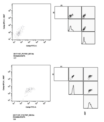
Flow Cytometry: RHAMM/CD168 Antibody - BSA Free [NBP1-76538]
Flow Cytometry: RHAMM/CD168 Antibody [NBP1-76538] - Human megakaryocytes. 47,000 CD41+ CD42+ human iPS-derived megakaryocytes [Moreau et al., 2016] were stained for 20 min at room temperature. Cells were then washed in 500 uL PBS/BSA/EDTA and viable DAPI cells were analysed using a Galios flow cytometer. Flow cytometry image submitted by a verified customer review. Image from the Alexa Fluor 700 version of this antibody.
Immunocytochemistry/ Immunofluorescence: RHAMM/CD168 Antibody - BSA Free [NBP1-76538] -
Immunocytochemistry/ Immunofluorescence: RHAMM/CD168 Antibody - BSA Free [NBP1-76538] - Immunofluorescence of RHAMM/CD168 in human stomach tissue with RHAM antibody at 20 ug/mL.Applications for RHAMM/CD168 Antibody - BSA Free
Application
Recommended Usage
ELISA
1:100-1:2000
Immunocytochemistry/ Immunofluorescence
5 ug/ml
Immunohistochemistry
2.5 ug/ml
Immunohistochemistry-Paraffin
2.5 ug/ml
Western Blot
0.5 ug/ml
Reviewed Applications
Read 1 review rated 5 using NBP1-76538 in the following applications:
Flow Cytometry Panel Builder
Bio-Techne Knows Flow Cytometry
Save time and reduce costly mistakes by quickly finding compatible reagents using the Panel Builder Tool.
Advanced Features
- Spectra Viewer - Custom analysis of spectra from multiple fluorochromes
- Spillover Popups - Visualize the spectra of individual fluorochromes
- Antigen Density Selector - Match fluorochrome brightness with antigen density

Formulation, Preparation, and Storage
Purification
Peptide affinity purified
Formulation
PBS
Format
BSA Free
Preservative
0.02% Sodium Azide
Concentration
1 mg/ml
Shipping
The product is shipped with polar packs. Upon receipt, store it immediately at the temperature recommended below.
Stability & Storage
Store at 4C short term. Aliquot and store at -20C long term. Avoid freeze-thaw cycles.
Background: RHAMM/CD168
Long Name
Receptor For Hyaluronan-Mediated Motility
Alternate Names
CD168, HMMR, IHABP
Gene Symbol
HMMR
UniProt
Additional RHAMM/CD168 Products
Product Documents for RHAMM/CD168 Antibody - BSA Free
Certificate of Analysis
To download a Certificate of Analysis, please enter a lot or batch number in the search box below.
Product Specific Notices for RHAMM/CD168 Antibody - BSA Free
This product is for research use only and is not approved for use in humans or in clinical diagnosis. Primary Antibodies are guaranteed for 1 year from date of receipt.
Related Research Areas
Citations for RHAMM/CD168 Antibody - BSA Free
 Powered by Bioz
Customer Reviews for RHAMM/CD168 Antibody - BSA Free (1)
5 out of 5
1 Customer Rating
Have you used RHAMM/CD168 Antibody - BSA Free?
Submit a review and receive an Amazon gift card!
$25/€18/£15/$25CAN/¥2500 Yen for a review with an image
$10/€7/£6/$10CAN/¥1110 Yen for a review without an image
Submit a review
Customer Images
Showing
1
-
1 of
1 review
Showing All
Filter By:
-
Application: Flow CytometrySample Tested: Hippocampal Cell SuspensionSpecies: MouseVerified Customer | Posted 01/25/2018See experimental details.Used antibody for FACS. 5 x 10^6 Mouse primary hippocampal cells suspended in complete neurosphere supplement media at 4' C. Cells are not permeabilized. Primary antibody used at 1:200 dilution for an hour on ice. Secondary goat antibody diluted to 1:2000 used. BD FACS Aria Fusion Sorter was used to analyze the sample. Excitation wavelength 488nm. Emission Filter 530/30. Events acquired ~70,000. Controls used were as follows: 1) No staining without Propidium Iodide. 2)No staining with Propidium Iodide. 3)Secondary only with Propidium Iodide. Rhamm only with Propidium Iodide.

There are no reviews that match your criteria.
Protocols
Find general support by application which include: protocols, troubleshooting, illustrated assays, videos and webinars.
- 7-Amino Actinomycin D (7-AAD) Cell Viability Flow Cytometry Protocol
- Antigen Retrieval Protocol (PIER)
- Antigen Retrieval for Frozen Sections Protocol
- Appropriate Fixation of IHC/ICC Samples
- Cellular Response to Hypoxia Protocols
- Chromogenic IHC Staining of Formalin-Fixed Paraffin-Embedded (FFPE) Tissue Protocol
- Chromogenic Immunohistochemistry Staining of Frozen Tissue
- ClariTSA™ Fluorophore Kits
- Detection & Visualization of Antibody Binding
- ELISA Sample Preparation & Collection Guide
- ELISA Troubleshooting Guide
- Extracellular Membrane Flow Cytometry Protocol
- Flow Cytometry Protocol for Cell Surface Markers
- Flow Cytometry Protocol for Staining Membrane Associated Proteins
- Flow Cytometry Staining Protocols
- Flow Cytometry Troubleshooting Guide
- Fluorescent IHC Staining of Frozen Tissue Protocol
- Graphic Protocol for Heat-induced Epitope Retrieval
- Graphic Protocol for the Preparation and Fluorescent IHC Staining of Frozen Tissue Sections
- Graphic Protocol for the Preparation and Fluorescent IHC Staining of Paraffin-embedded Tissue Sections
- Graphic Protocol for the Preparation of Gelatin-coated Slides for Histological Tissue Sections
- How to Run an R&D Systems DuoSet ELISA
- How to Run an R&D Systems Quantikine ELISA
- How to Run an R&D Systems Quantikine™ QuicKit™ ELISA
- ICC Cell Smear Protocol for Suspension Cells
- ICC Immunocytochemistry Protocol Videos
- ICC for Adherent Cells
- IHC Sample Preparation (Frozen sections vs Paraffin)
- Immunocytochemistry (ICC) Protocol
- Immunocytochemistry Troubleshooting
- Immunofluorescence of Organoids Embedded in Cultrex Basement Membrane Extract
- Immunofluorescent IHC Staining of Formalin-Fixed Paraffin-Embedded (FFPE) Tissue Protocol
- Immunohistochemistry (IHC) and Immunocytochemistry (ICC) Protocols
- Immunohistochemistry Frozen Troubleshooting
- Immunohistochemistry Paraffin Troubleshooting
- Intracellular Flow Cytometry Protocol Using Alcohol (Methanol)
- Intracellular Flow Cytometry Protocol Using Detergents
- Intracellular Nuclear Staining Flow Cytometry Protocol Using Detergents
- Intracellular Staining Flow Cytometry Protocol Using Alcohol Permeabilization
- Intracellular Staining Flow Cytometry Protocol Using Detergents to Permeabilize Cells
- Preparing Samples for IHC/ICC Experiments
- Preventing Non-Specific Staining (Non-Specific Binding)
- Primary Antibody Selection & Optimization
- Propidium Iodide Cell Viability Flow Cytometry Protocol
- Protocol for Heat-Induced Epitope Retrieval (HIER)
- Protocol for Liperfluo
- Protocol for Making a 4% Formaldehyde Solution in PBS
- Protocol for VisUCyte™ HRP Polymer Detection Reagent
- Protocol for the Characterization of Human Th22 Cells
- Protocol for the Characterization of Human Th9 Cells
- Protocol for the Fluorescent ICC Staining of Cell Smears - Graphic
- Protocol for the Fluorescent ICC Staining of Cultured Cells on Coverslips - Graphic
- Protocol for the Preparation & Fixation of Cells on Coverslips
- Protocol for the Preparation and Chromogenic IHC Staining of Frozen Tissue Sections
- Protocol for the Preparation and Chromogenic IHC Staining of Frozen Tissue Sections - Graphic
- Protocol for the Preparation and Chromogenic IHC Staining of Paraffin-embedded Tissue Sections
- Protocol for the Preparation and Chromogenic IHC Staining of Paraffin-embedded Tissue Sections - Graphic
- Protocol for the Preparation and Fluorescent ICC Staining of Cells on Coverslips
- Protocol for the Preparation and Fluorescent ICC Staining of Non-adherent Cells
- Protocol for the Preparation and Fluorescent ICC Staining of Stem Cells on Coverslips
- Protocol for the Preparation and Fluorescent IHC Staining of Frozen Tissue Sections
- Protocol for the Preparation and Fluorescent IHC Staining of Paraffin-embedded Tissue Sections
- Protocol for the Preparation of Gelatin-coated Slides for Histological Tissue Sections
- Protocol for the Preparation of a Cell Smear for Non-adherent Cell ICC - Graphic
- Protocol: Annexin V and PI Staining by Flow Cytometry
- Protocol: Annexin V and PI Staining for Apoptosis by Flow Cytometry
- Quantikine HS ELISA Kit Assay Principle, Alkaline Phosphatase
- Quantikine HS ELISA Kit Principle, Streptavidin-HRP Polymer
- R&D Systems Quality Control Western Blot Protocol
- Sandwich ELISA (Colorimetric) – Biotin/Streptavidin Detection Protocol
- Sandwich ELISA (Colorimetric) – Direct Detection Protocol
- TUNEL and Active Caspase-3 Detection by IHC/ICC Protocol
- The Importance of IHC/ICC Controls
- Troubleshooting Guide: ELISA
- Troubleshooting Guide: Fluorokine Flow Cytometry Kits
- Troubleshooting Guide: Immunohistochemistry
- Troubleshooting Guide: Western Blot Figures
- Western Blot Conditions
- Western Blot Protocol
- Western Blot Protocol for Cell Lysates
- Western Blot Troubleshooting
- Western Blot Troubleshooting Guide
- View all Protocols, Troubleshooting, Illustrated assays and Webinars
Loading...