Serpin E1/PAI-1 Antibody - BSA Free
Novus Biologicals | Catalog # NBP1-19773
![Western Blot: Serpin E1/PAI-1 AntibodyBSA Free [NBP1-19773]](https://resources.rndsystems.com/images/products/Serpin-E1-PAI-1-Antibody-Western-Blot-NBP1-19773-img0005.jpg "Western Blot: Serpin E1/PAI-1 AntibodyBSA Free [NBP1-19773]")
Key Product Details
Validated by
Biological Validation
Species Reactivity
Validated:
Human, Mouse, Rat
Cited:
Human, Mouse, Rat
Applications
Validated:
Immunohistochemistry, Immunohistochemistry-Paraffin, Immunohistochemistry-Frozen, Western Blot, ELISA, Immunocytochemistry/ Immunofluorescence
Cited:
Immunohistochemistry, Immunohistochemistry-Paraffin, Immunohistochemistry-Frozen, Western Blot, ELISA, Immunocytochemistry/ Immunofluorescence, IF/IHC
Label
Unconjugated
Antibody Source
Polyclonal Rabbit IgG
Format
BSA Free
Loading...
Product Specifications
Immunogen
A synthetic peptide made to an internal portion of the human PAI1/Serpine 1 protein (between residues 300-400) [UniProt P05121]
Reactivity Notes
Use in Rat reported in scientific literature (PMID:35386330).
Localization
Secreted.
Specificity
PAI-1 (N315) pAb detects endogenous levels of PAI-1 protein.
Clonality
Polyclonal
Host
Rabbit
Isotype
IgG
Theoretical MW
45 kDa.
Disclaimer note: The observed molecular weight of the protein may vary from the listed predicted molecular weight due to post translational modifications, post translation cleavages, relative charges, and other experimental factors.
Disclaimer note: The observed molecular weight of the protein may vary from the listed predicted molecular weight due to post translational modifications, post translation cleavages, relative charges, and other experimental factors.
Scientific Data Images for Serpin E1/PAI-1 Antibody - BSA Free
Western Blot: Serpin E1/PAI-1 AntibodyBSA Free [NBP1-19773]
Western Blot: Serpin E1/PAI-1 Antibody [NBP1-19773] - Extracts from Jurkat cells.![Immunohistochemistry-Paraffin: Serpin E1/PAI-1 Antibody - BSA Free [NBP1-19773]](https://resources.rndsystems.com/images/products/Serpin-E1-PAI-1-Antibody-Immunohistochemistry-Paraffin-NBP1-19773-img0007.jpg "Immunohistochemistry-Paraffin: Serpin E1/PAI-1 Antibody - BSA Free [NBP1-19773]")
Immunohistochemistry-Paraffin: Serpin E1/PAI-1 Antibody - BSA Free [NBP1-19773]
Immunohistochemistry-Paraffin: Serpin E1/PAI-1 Antibody [NBP1-19773] - Staining of PAI1/Serpine 1 in mouse pancreas.![Western Blot: Serpin E1/PAI-1 AntibodyBSA Free [NBP1-19773]](https://resources.rndsystems.com/images/products/Serpin-E1-PAI-1-Antibody-Western-Blot-NBP1-19773-img0008.jpg "Western Blot: Serpin E1/PAI-1 AntibodyBSA Free [NBP1-19773]")
![Immunocytochemistry/ Immunofluorescence: Serpin E1/PAI-1 Antibody - BSA Free [NBP1-19773]](https://resources.rndsystems.com/images/products/Serpin-E1-PAI-1-Antibody-Immunocytochemistry-Immunofluorescence-NBP1-19773-img0006.jpg "Immunocytochemistry/ Immunofluorescence: Serpin E1/PAI-1 Antibody - BSA Free [NBP1-19773]")
Immunocytochemistry/ Immunofluorescence: Serpin E1/PAI-1 Antibody - BSA Free [NBP1-19773]
Immunocytochemistry/Immunofluorescence: Serpin E1/PAI-1 Antibody [NBP1-19773] - PAI1/Serpine1 antibody was tested in Hela cells with DyLight 488 (green). Nuclei and alpha-tubulin were counterstained with DAPI (blue) and Dylight 550 (red).![Western Blot: Serpin E1/PAI-1 AntibodyBSA Free [NBP1-19773]](https://resources.rndsystems.com/images/products/Serpin-E1-PAI-1-Antibody-Western-Blot-NBP1-19773-img0009.jpg "Western Blot: Serpin E1/PAI-1 AntibodyBSA Free [NBP1-19773]")

Western Blot: Serpin E1/PAI-1 Antibody - BSA Free [NBP1-19773] -
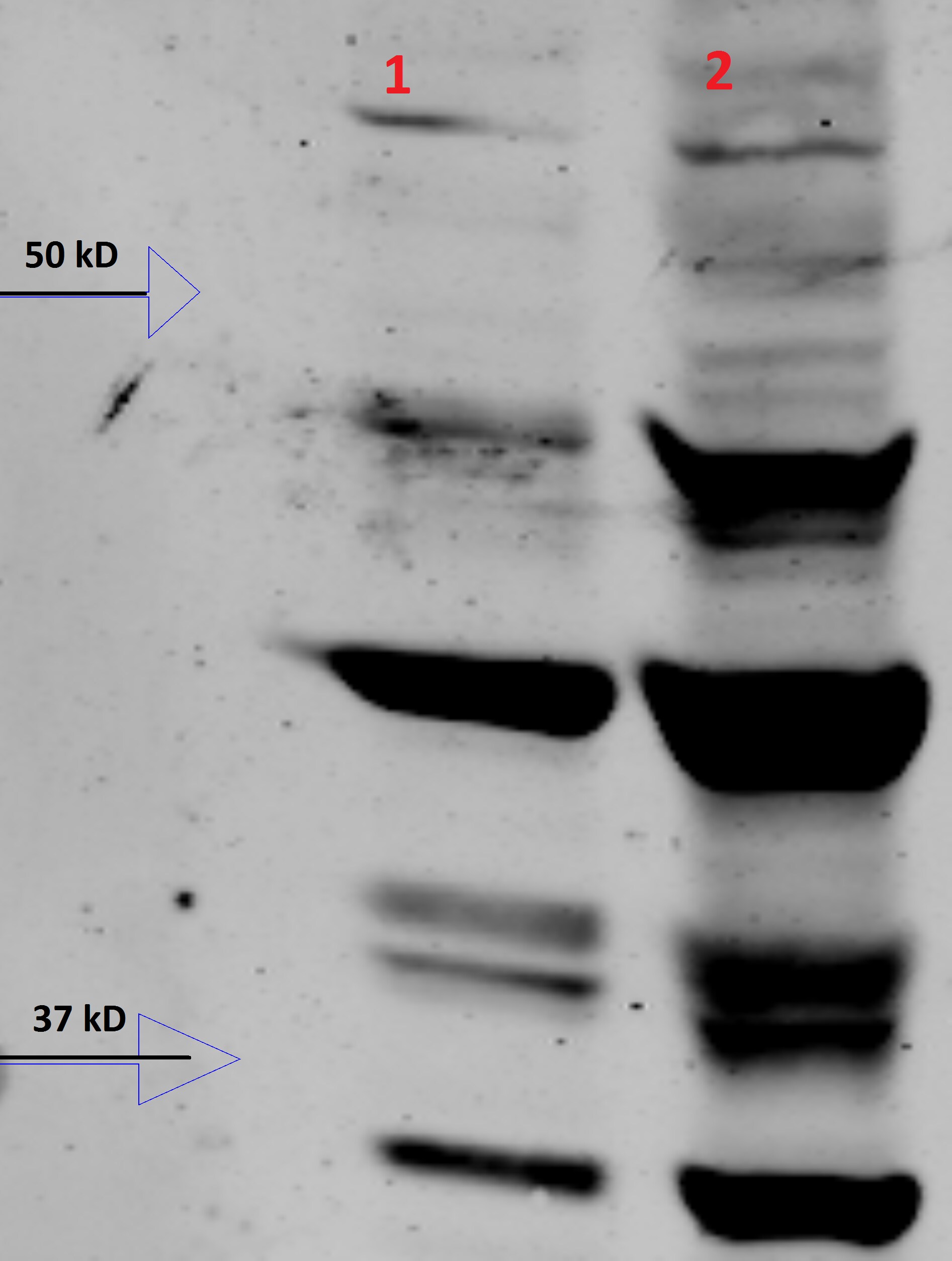
Western Blot: Serpin E1/PAI-1 Antibody - BSA Free [NBP1-19773] - (A) Immunoblots showing protein expression of ALK5, pSMAD2/3, PAI-1, pVHL, & beta -actin in A498 cells after treatment with TGF-beta at given point of time; (B) Immunoblots showing protein expression of ALK5, pSMAD2/3, PAI-1, pVHL, & beta -actin in ACHN cells after treatment with TGF-beta at given point of time; (C) Immunoblots showing protein expression of pVHL, ALK5, pSMAD2/3, PAI-1, & beta -actin after transfection of indicated vectors in A498 cells followed by TGF-beta treatment for 6 hours; (D) Immunoblots showing protein expression of pVHL, HA, pSMAD2/3, PAI-1, & beta -actin after transfection of indicated vectors in ACHN cells (48h) followed by TGF-beta treatment for 6 hours; (E) Invasion assay showing the invasiveness induced by TGF-beta in A498 cells after re-introduction of VHL (n=3 independent experiments *P< 0. 05); (F) Invasion assay showing invasiveness by TGF-beta in ACHN cells after siRNA VHL knockdown (n=3 independent experiments *P< 0.05). Image collected & cropped by CiteAb from the following publication (https://www.oncotarget.com/lookup/doi/10.18632/oncotarget.24631), licensed under a CC-BY license. Not internally tested by Novus Biologicals.
Western Blot: Serpin E1/PAI-1 Antibody - BSA Free [NBP1-19773] -
Western Blot: Serpin E1/PAI-1 Antibody - BSA Free [NBP1-19773] - (A) Immunoblots showing protein expression of ALK5, pSMAD2/3, PAI-1, pVHL, & beta -actin in A498 cells after treatment with TGF-beta at given point of time; (B) Immunoblots showing protein expression of ALK5, pSMAD2/3, PAI-1, pVHL, & beta -actin in ACHN cells after treatment with TGF-beta at given point of time; (C) Immunoblots showing protein expression of pVHL, ALK5, pSMAD2/3, PAI-1, & beta -actin after transfection of indicated vectors in A498 cells followed by TGF-beta treatment for 6 hours; (D) Immunoblots showing protein expression of pVHL, HA, pSMAD2/3, PAI-1, & beta -actin after transfection of indicated vectors in ACHN cells (48h) followed by TGF-beta treatment for 6 hours; (E) Invasion assay showing the invasiveness induced by TGF-beta in A498 cells after re-introduction of VHL (n=3 independent experiments *P< 0. 05); (F) Invasion assay showing invasiveness by TGF-beta in ACHN cells after siRNA VHL knockdown (n=3 independent experiments *P< 0.05). Image collected & cropped by CiteAb from the following publication (https://www.oncotarget.com/lookup/doi/10.18632/oncotarget.24631), licensed under a CC-BY license. Not internally tested by Novus Biologicals.
Immunocytochemistry/ Immunofluorescence: Serpin E1/PAI-1 Antibody - BSA Free [NBP1-19773] -
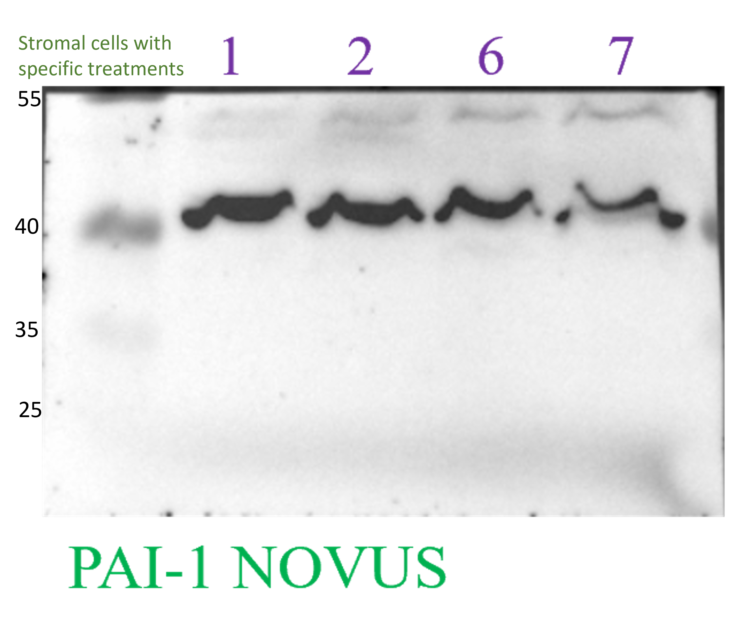
Potential effect on MMT of the H-HPMA hydrogel by regulating the TGF-beta 1/Smad signal pathway. (A) Relative TGF-beta 1 mRNA expression in the peritoneal tissues; (B) the expression of TGF-beta 1, Smad2/3 and Smad7 proteins in relative to the GAPDH in the peritoneal tissue analyzed with western blotting. e,f) Representative immunofluorescence images in the rat peritoneal membrane on day 10 post-surgery. Sections were stained with DAPI (blue), PAI-1-Fluor 594 (red) and t-PA-Alexa Fluor488 (green). Magnification: 200 × ; Scale bars: 100 μm. All data are presented as mean +/- SD (n = 3 per group); the ns means no significant difference; *p < 0.05; **p < 0.01; ***p < 0.001; ****p < 0.0001. Image collected and cropped by CiteAb from the following open publication (https://pubmed.ncbi.nlm.nih.gov/35386330), licensed under a CC-BY license. Not internally tested by Novus Biologicals.Applications for Serpin E1/PAI-1 Antibody - BSA Free
Application
Recommended Usage
Immunocytochemistry/ Immunofluorescence
5 - 10 ug/ml
Immunohistochemistry
1:50 - 1:100
Immunohistochemistry-Frozen
1:50 - 1:100
Immunohistochemistry-Paraffin
1:50 - 1:100
Western Blot
1.0 ug/ml
Reviewed Applications
Read 3 reviews rated 4.3 using NBP1-19773 in the following applications:
Formulation, Preparation, and Storage
Purification
Immunogen affinity purified
Formulation
PBS
Format
BSA Free
Preservative
0.02% Sodium Azide
Concentration
1.0 mg/ml
Shipping
The product is shipped with polar packs. Upon receipt, store it immediately at the temperature recommended below.
Stability & Storage
Store at 4C short term. Aliquot and store at -20C long term. Avoid freeze-thaw cycles.
Background: Serpin E1/PAI-1
Long Name
Plasminogen Activator Inhibitor
Alternate Names
Nexin, PAI-1, PAI1, PLANH1
Gene Symbol
SERPINE1
UniProt
Additional Serpin E1/PAI-1 Products
Product Documents for Serpin E1/PAI-1 Antibody - BSA Free
Certificate of Analysis
To download a Certificate of Analysis, please enter a lot or batch number in the search box below.
Product Specific Notices for Serpin E1/PAI-1 Antibody - BSA Free
This product is for research use only and is not approved for use in humans or in clinical diagnosis. Primary Antibodies are guaranteed for 1 year from date of receipt.
Citations for Serpin E1/PAI-1 Antibody - BSA Free
 Powered by Bioz
Customer Reviews for Serpin E1/PAI-1 Antibody - BSA Free (3)
4.3 out of 5
3 Customer Ratings
Have you used Serpin E1/PAI-1 Antibody - BSA Free?
Submit a review and receive an Amazon gift card!
$25/€18/£15/$25CAN/¥2500 Yen for a review with an image
$10/€7/£6/$10CAN/¥1110 Yen for a review without an image
Submit a review
Customer Images
-(01-mg)_NBP1-19773_8421.jpg)
Showing
1
-
3 of
3 reviews
Showing All
Filter By:
-
Application: Western BlotSample Tested: fibroblastsSpecies: HumanVerified Customer | Posted 04/25/2024We investigated the effects of our newly designed therapeutic strategy on the cultured tumor microenvironment. The antibody was used at a 1:1000 dilution, and the bands on the Western blot were clearly visible for identification.

-
Application: Western BlotSample Tested: HEK 293 cell line vs. HEK 293 cell line treated with TGF-betaSpecies: HumanVerified Customer | Posted 11/26/2015HEK 293 cell line vs. HEK 293 cell line treated with TGF-beta
-
Application: Western BlotSample Tested: Renal Cell Carcinoma tissuesSpecies: HumanVerified Customer | Posted 06/24/2014PAI-1 protein expression in Renal Cell Carcinoma samples.
There are no reviews that match your criteria.
Protocols
View specific protocols for Serpin E1/PAI-1 Antibody - BSA Free (NBP1-19773):
Immunocytochemistry Protocol
Culture cells to appropriate density in 35 mm culture dishes or 6-well plates.
1. Remove culture medium and add 10% formalin to the dish. Fix at room temperature for 30 minutes.
2. Remove the formalin and add ice cold methanol. Incubate for 5-10 minutes.
3. Remove methanol and add washing solution (i.e. PBS). Be sure to not let the specimen dry out. Wash three times for 10 minutes.
4. To block nonspecific antibody binding incubate in 10% normal goat serum from 1 hour to overnight at room temperature.
5. Add primary antibody at appropriate dilution and incubate at room temperature from 2 hours to overnight at room temperature.
6. Remove primary antibody and replace with washing solution. Wash three times for 10 minutes.
7. Add secondary antibody at appropriate dilution. Incubate for 1 hour at room temperature.
8. Remove antibody and replace with wash solution, then wash for 10 minutes. Add Hoechst 33258 to wash solution at 1:25,0000 and incubate for 10 minutes. Wash a third time for 10 minutes.
9. Cells can be viewed directly after washing. The plates can also be stored in PBS containing Azide covered in Parafilm (TM). Cells can also be cover-slipped using Fluoromount, with appropriate sealing.
*The above information is only intended as a guide. The researcher should determine what protocol best meets their needs. Please follow safe laboratory procedures.
Immunohistochemistry-Paraffin Embedded Sections
Antigen Unmasking:
Bring slides to a boil in 10 mM sodium citrate buffer (pH 6.0) then maintain at a sub-boiling temperature for 10 minutes. Cool slides on bench-top for 30 minutes.
Staining:
1. Wash sections in deionized water three times for 5 minutes each.
2. Wash sections in wash buffer for 5 minutes.
3. Block each section with 100-400 ul blocking solution for 1 hour at room temperature.
4. Remove blocking solution and add 100-400 ul diluted primary antibody. Incubate overnight at 4 C.
5. Remove antibody solution and wash sections in wash buffer three times for 5 minutes each.
6. Add 100-400 ul biotinylated diluted secondary antibody. Incubate 30 minutes at room temperature.
7. Remove secondary antibody solution and wash sections three times with wash buffer for 5 minutes each.
8. Add 100-400 ul Streptavidin-HRP reagent to each section and incubate for 30 minutes at room temperature.
9. Wash sections three times in wash buffer for 5 minutes each.
10. Add 100-400 ul DAB substrate to each section and monitor staining closely.
11. As soon as the sections develop, immerse slides in deionized water.
12. Counterstain sections in hematoxylin.
13. Wash sections in deionized water two times for 5 minutes each.
14. Dehydrate sections.
15. Mount coverslips.
Western Blot Protocol
1. Perform SDS-PAGE on samples to be analyzed, loading 40 ug of total protein per lane.
2. Transfer proteins to membrane according to the instructions provided by the manufacturer of the membrane and transfer apparatus.
3. Stain according to standard Ponceau S procedure (or similar product) to assess transfer success, and mark molecular weight standards where appropriate.
4. Rinse the blot.
5. Block the membrane using standard blocking buffer for at least 1 hour.
6. Wash the membrane in wash buffer three times for 10 minutes each.
7. Dilute primary antibody in blocking buffer and incubate 1 hour at room temperature.
8. Wash the membrane in wash buffer three times for 10 minutes each.
9. Apply the diluted HRP conjugated secondary antibody in blocking buffer (as per manufacturers instructions) and incubate 1 hour at room temperature.
10. Wash the blot in wash buffer three times for 10 minutes each (this step can be repeated as required to reduce background).
11. Apply the detection reagent of choice in accordance with the manufacturers instructions.
Note: Tween-20 can be added to the blocking or antibody dilution buffer at a final concentration of 0.05-0.2%.
Find general support by application which include: protocols, troubleshooting, illustrated assays, videos and webinars.
- Antigen Retrieval Protocol (PIER)
- Antigen Retrieval for Frozen Sections Protocol
- Appropriate Fixation of IHC/ICC Samples
- Cellular Response to Hypoxia Protocols
- Chromogenic IHC Staining of Formalin-Fixed Paraffin-Embedded (FFPE) Tissue Protocol
- Chromogenic Immunohistochemistry Staining of Frozen Tissue
- ClariTSA™ Fluorophore Kits
- Detection & Visualization of Antibody Binding
- ELISA Sample Preparation & Collection Guide
- ELISA Troubleshooting Guide
- Fluorescent IHC Staining of Frozen Tissue Protocol
- Graphic Protocol for Heat-induced Epitope Retrieval
- Graphic Protocol for the Preparation and Fluorescent IHC Staining of Frozen Tissue Sections
- Graphic Protocol for the Preparation and Fluorescent IHC Staining of Paraffin-embedded Tissue Sections
- Graphic Protocol for the Preparation of Gelatin-coated Slides for Histological Tissue Sections
- How to Run an R&D Systems DuoSet ELISA
- How to Run an R&D Systems Quantikine ELISA
- How to Run an R&D Systems Quantikine™ QuicKit™ ELISA
- ICC Cell Smear Protocol for Suspension Cells
- ICC Immunocytochemistry Protocol Videos
- ICC for Adherent Cells
- IHC Sample Preparation (Frozen sections vs Paraffin)
- Immunocytochemistry (ICC) Protocol
- Immunocytochemistry Troubleshooting
- Immunofluorescence of Organoids Embedded in Cultrex Basement Membrane Extract
- Immunofluorescent IHC Staining of Formalin-Fixed Paraffin-Embedded (FFPE) Tissue Protocol
- Immunohistochemistry (IHC) and Immunocytochemistry (ICC) Protocols
- Immunohistochemistry Frozen Troubleshooting
- Immunohistochemistry Paraffin Troubleshooting
- Preparing Samples for IHC/ICC Experiments
- Preventing Non-Specific Staining (Non-Specific Binding)
- Primary Antibody Selection & Optimization
- Protocol for Heat-Induced Epitope Retrieval (HIER)
- Protocol for Making a 4% Formaldehyde Solution in PBS
- Protocol for VisUCyte™ HRP Polymer Detection Reagent
- Protocol for the Fluorescent ICC Staining of Cell Smears - Graphic
- Protocol for the Fluorescent ICC Staining of Cultured Cells on Coverslips - Graphic
- Protocol for the Preparation & Fixation of Cells on Coverslips
- Protocol for the Preparation and Chromogenic IHC Staining of Frozen Tissue Sections
- Protocol for the Preparation and Chromogenic IHC Staining of Frozen Tissue Sections - Graphic
- Protocol for the Preparation and Chromogenic IHC Staining of Paraffin-embedded Tissue Sections
- Protocol for the Preparation and Chromogenic IHC Staining of Paraffin-embedded Tissue Sections - Graphic
- Protocol for the Preparation and Fluorescent ICC Staining of Cells on Coverslips
- Protocol for the Preparation and Fluorescent ICC Staining of Non-adherent Cells
- Protocol for the Preparation and Fluorescent ICC Staining of Stem Cells on Coverslips
- Protocol for the Preparation and Fluorescent IHC Staining of Frozen Tissue Sections
- Protocol for the Preparation and Fluorescent IHC Staining of Paraffin-embedded Tissue Sections
- Protocol for the Preparation of Gelatin-coated Slides for Histological Tissue Sections
- Protocol for the Preparation of a Cell Smear for Non-adherent Cell ICC - Graphic
- Quantikine HS ELISA Kit Assay Principle, Alkaline Phosphatase
- Quantikine HS ELISA Kit Principle, Streptavidin-HRP Polymer
- R&D Systems Quality Control Western Blot Protocol
- Sandwich ELISA (Colorimetric) – Biotin/Streptavidin Detection Protocol
- Sandwich ELISA (Colorimetric) – Direct Detection Protocol
- TUNEL and Active Caspase-3 Detection by IHC/ICC Protocol
- The Importance of IHC/ICC Controls
- Troubleshooting Guide: ELISA
- Troubleshooting Guide: Immunohistochemistry
- Troubleshooting Guide: Western Blot Figures
- Western Blot Conditions
- Western Blot Protocol
- Western Blot Protocol for Cell Lysates
- Western Blot Troubleshooting
- Western Blot Troubleshooting Guide
- View all Protocols, Troubleshooting, Illustrated assays and Webinars
Loading...
Associated Pathways
