Survivin Antibody (60.11) - BSA Free
Novus Biologicals | Catalog # NB500-238
Key Product Details
Validated by
Knockout/Knockdown, Independent Antibodies
Species Reactivity
Validated:
Human, Mouse, Rat
Cited:
Human, Mouse, Rat
Applications
Validated:
Immunohistochemistry, Immunohistochemistry-Paraffin, Immunohistochemistry-Frozen, Western Blot, ELISA, Flow Cytometry, Flow (Intracellular), Immunocytochemistry/ Immunofluorescence, Proximity Ligation Assay, Knockdown Validated
Cited:
Immunohistochemistry, Immunohistochemistry-Paraffin, Immunohistochemistry-Frozen, Western Blot, ELISA, Flow Cytometry, Immunocytochemistry/ Immunofluorescence, Proximity Ligation Assay, IF/IHC
Label
Unconjugated
Antibody Source
Monoclonal Mouse IgG2a Kappa Clone # 60.11
Format
BSA Free
Loading...
Product Specifications
Immunogen
This Survivin Antibody (60.11) [Alexa Fluor 350] was developed against full length recombinant human Survivin [UniProt# O15392]
Epitope
The epitope recognized is between amino acids 27-67
Localization
Cytoplasmic
Specificity
Survivin Antibody (60.11) [NB500-238] is specific for the cytoplasmic form of Survivin.
Clonality
Monoclonal
Host
Mouse
Isotype
IgG2a Kappa
Theoretical MW
16 kDa.
Disclaimer note: The observed molecular weight of the protein may vary from the listed predicted molecular weight due to post translational modifications, post translation cleavages, relative charges, and other experimental factors.
Disclaimer note: The observed molecular weight of the protein may vary from the listed predicted molecular weight due to post translational modifications, post translation cleavages, relative charges, and other experimental factors.
Scientific Data Images for Survivin Antibody (60.11) - BSA Free
![Immunohistochemistry: Survivin Antibody (60.11) - BSA Free [NB500-238]](https://resources.rndsystems.com/images/products/Survivin-Antibody-60-11-Immunohistochemistry-NB500-238-img0019.jpg "Immunohistochemistry: Survivin Antibody (60.11) - BSA Free [NB500-238]")
![Flow (Intracellular): Survivin Antibody (60.11) - BSA Free [NB500-238]](https://resources.rndsystems.com/images/products/Survivin-Antibody-60-11-Flow-Intracellular-NB500-238-img0015.jpg "Flow (Intracellular): Survivin Antibody (60.11) - BSA Free [NB500-238]")
Flow (Intracellular): Survivin Antibody (60.11) - BSA Free [NB500-238]
Flow (Intracellular): Survivin Antibody (60.11) [NB500-238] - An intracellular stain was performed on A549 cells with Survivin Antibody (60.11) [NB500-238] and a matched isotype control. Cells were fixed with 4% PFA and then permeablized with 0.1% saponin. Cells were incubated in an antibody dilution of 1 ug/mL for 30 minutes at room temperature, followed by mouse F(ab)2 IgG (H+L) APC-conjugated secondary antibody (F0101B, R&D Systems). Image using the Unpurified form of this antibody.![Western Blot: Survivin Antibody (60.11)BSA Free [NB500-238]](https://resources.rndsystems.com/images/products/Survivin-Antibody-60-11-Western-Blot-NB500-238-img0006.jpg "Western Blot: Survivin Antibody (60.11)BSA Free [NB500-238]")
Western Blot: Survivin Antibody (60.11)BSA Free [NB500-238]
Western Blot: Survivin Antibody (60.11) [NB500-238] - Survivin detection in 30ug of HeLa whole cell extract using [NB500-238] at 1ug/ml. Detected at predicted molecular weight of 16 kDa.![Knockdown Validated: Survivin Antibody (60.11) - BSA Free [NB500-238]](https://resources.rndsystems.com/images/products/Survivin-Antibody-60-11-Knockdown-Validated-NB500-238-img0020.jpg "Western Blot: Survivin Antibody (60.11) - BSA Free [NB500-238]")
![Immunocytochemistry/ Immunofluorescence: Survivin Antibody (60.11) - BSA Free [NB500-238]](https://resources.rndsystems.com/images/products/Survivin-Antibody-60-11-Immunocytochemistry-Immunofluorescence-NB500-238-img0021.jpg "Immunocytochemistry/ Immunofluorescence: Survivin Antibody (60.11) - BSA Free [NB500-238]")
Immunocytochemistry/ Immunofluorescence: Survivin Antibody (60.11) - BSA Free [NB500-238]
Immunocytochemistry/Immunofluorescence: Survivin Antibody (60.11) [NB500-238] - HeLa cells stained NB500-205 (Green) detected with DyLight Fluor 488 conjugated anti-mouse IgG secondary antibody. Nuclei are counterstained with Hoechst 33258 (Blue). Image using the Unpurified format of this antibody.![Immunohistochemistry-Paraffin: Survivin Antibody (60.11) - BSA Free [NB500-238]](https://resources.rndsystems.com/images/products/Survivin-Antibody-60-11-Immunohistochemistry-Paraffin-NB500-238-img0022.jpg "Immunohistochemistry-Paraffin: Survivin Antibody (60.11) - BSA Free [NB500-238]")
Immunohistochemistry-Paraffin: Survivin Antibody (60.11) - BSA Free [NB500-238]
Immunohistochemistry-Paraffin: Survivin Antibody (60.11) [NB500-238] - Staining of ovary cancer. Image using the Unpurified format of this antibody.![Flow Cytometry: Survivin Antibody (60.11) - BSA Free [NB500-238]](https://resources.rndsystems.com/images/products/Survivin-Antibody-60-11-Flow-Cytometry-NB500-238-img0017.jpg "Flow Cytometry: Survivin Antibody (60.11) - BSA Free [NB500-238]")
Flow Cytometry: Survivin Antibody (60.11) - BSA Free [NB500-238]
Flow Cytometry: Survivin Antibody (60.11) [NB500-238] - An intracellular stain was performed on U2OS cells with Survivin Antibody (60.11) [NB500-238AF700] (blue) and a matched isotype control (orange). Cells were fixed with 4% PFA and then permeabilized with 0.1% saponin. Cells were incubated in an antibody dilution of 5 ug/mL for 30 minutes at room temperature. Both antibodies were conjugated to Alexa Fluor 700.![Flow (Intracellular): Survivin Antibody (60.11) - BSA Free [NB500-238]](https://resources.rndsystems.com/images/products/Survivin-Antibody-60-11-Flow-Intracellular-NB500-238-img0014.jpg "Flow (Intracellular): Survivin Antibody (60.11) - BSA Free [NB500-238]")
Flow (Intracellular): Survivin Antibody (60.11) - BSA Free [NB500-238]
Flow (Intracellular): Survivin Antibody (60.11) [NB500-238] - An intracellular stain was performed on Daudi cells with Survivin Antibody (60.11)[NB500-238] (blue) and a matched isotype control, Mouse IgG2a Kappa Light Chain Isotype Control (MG2a-53) [NB600-986] (orange). Cells were fixed with 4% PFA and then permeablized with 0.1% saponin. Cells were incubated in an antibody dilution of 1 ug/mL for 30 minutes at room temperature, followed by mouse F(ab)2 IgG (H+L) APC-conjugated secondary antibody (F0101B, R&D Systems). - BSA Free [NB500-238] -")
Western Blot: Survivin Antibody (60.11) - BSA Free [NB500-238] -
Binding of antibody (60.11) used in imaging flow cytometry to full-length survivin protein and to survivin vaccine peptide aa53-67/M57 and wild type peptide aa53-67 in A431 Human Cell Line by Flow Cytometry.")
Detection of Survivin (60.11) in A431 Human Cell Line by Flow Cytometry.
A431 human skin carcinoma cell line was stained with Mouse anti-Survivin (60.11) Protein-G purified Monoclonal Antibody conjugated to Alexa Fluor® 647 (Catalog # NB500-238AF647, blue histogram) or matched control antibody (orange histogram). - BSA Free [NB500-238] -")
Western Blot: Survivin Antibody (60.11) - BSA Free [NB500-238] -
Western Blot: Survivin Antibody (60.11) - BSA Free [NB500-238] - Binding of antibody (60.11) used in imaging flow cytometry to full-length survivin protein & to survivin vaccine peptide aa53-67/M57 & wild type peptide aa53-67 Image collected & cropped by CiteAb from the following publication (https://www.oncotarget.com/lookup/doi/10.18632/oncotarget.21773), licensed under a CC-BY license. Not internally tested by Novus Biologicals. - BSA Free [NB500-238] -")
Western Blot: Survivin Antibody (60.11) - BSA Free [NB500-238] -
Western Blot: Survivin Antibody (60.11) - BSA Free [NB500-238] - CD133+ cells are more resistant to chemotherapeutic agents. STA-ET-8.2 cells were FACS-sorted & CD133+ & CD133- fractions treated with increasing concentrations of (A) Doxorubicin (Doxo), (B) Etoposide (Etop), (C) Vincristine (Vinc) or (D) a combination of all three drugs at high (Doxo 10 μM; Etop 10 ug/ml; Vinc 100 ng/ml), medium (Doxo 2.5 μM; 20 Etop 2.5 ug/ml; Vinc 25 ng/ml) & low (Doxo 0.5 μM; Etop 0.5 ug/ml; Vinc5 ng/ml) concentrations. Viability was assessed by MTS assay after 96 hrs (*p < 0.05) & CD133+ cells displayed increased drug resistance. (E): CD133+ & CD133- TC-71 cells are equally sensitive to Doxo, Etop, & Vinc. (F): Western blot reveals no difference in apoptosis & drug resistance protein expression between unsorted & CD133-sorted STA-ET-8.2 cell fractions. Image collected & cropped by CiteAb from the following publication (https://pubmed.ncbi.nlm.nih.gov/20346143), licensed under a CC-BY license. Not internally tested by Novus Biologicals.Applications for Survivin Antibody (60.11) - BSA Free
Application
Recommended Usage
ELISA
reported in scientific literature (PMID 11861764)
Flow Cytometry
1 ug/mL. Use reported in scientific literature (PMID 25050620)
Immunocytochemistry/ Immunofluorescence
1:50-1:200
Immunohistochemistry
1:50-1:200
Immunohistochemistry-Paraffin
1:50-1:200
Proximity Ligation Assay
reported in scientific literature (PMID 28077791)
Western Blot
1:1000
Application Notes
By WB, this antibody recognizes a band at ~16.5 kDa representing Survivin.
Reviewed Applications
Read 3 reviews rated 3.7 using NB500-238 in the following applications:
Flow Cytometry Panel Builder
Bio-Techne Knows Flow Cytometry
Save time and reduce costly mistakes by quickly finding compatible reagents using the Panel Builder Tool.
Advanced Features
- Spectra Viewer - Custom analysis of spectra from multiple fluorochromes
- Spillover Popups - Visualize the spectra of individual fluorochromes
- Antigen Density Selector - Match fluorochrome brightness with antigen density

Formulation, Preparation, and Storage
Purification
Protein G purified
Formulation
PBS
Format
BSA Free
Preservative
0.1% Sodium Azide
Concentration
1.0 mg/ml
Shipping
The product is shipped with polar packs. Upon receipt, store it immediately at the temperature recommended below.
Stability & Storage
Aliquot and store at -20C or -80C. Avoid freeze-thaw cycles.
Background: Survivin
Besides being highly abundant in fetal development and expressed in proliferating adult cells such as activated T lymphocytes, erythroblasts, and self-renewing stem cells, survivin is generally absent in adult tissues. However, it is elevated in common cancers such as lung, colon, pancreas, breast and prostate where it drives proliferation, metastasis, poor prognosis, and decreased patient survival (2).
Survivin has been shown to be involved in multiple cellular processes including cell cycle progression, mitotic spindle assembly, kinetochore attachment, angiogenesis, migration, and its anti-apoptotic activity has been linked to both its monomeric and homodimeric forms. Survivin impacts the function of other IAP members, c-IAP1 and c-IAP-2, or modulates the inhibitory activity of XIAP against caspases by forming a stable complex with XIAP and HBXIP. During the intrinsic apoptotic pathway, survivin may prevent the release of mitochondrial APAF1 into the cytoplasm or hinder the association of SMAC with other IAPS, which results in prolonged cell survival (3).
References
1. Sah NK, Seniya C. (2015) Survivin splice variants and their diagnostic significance. Tumour Biol. 36(9):6623-31. PMID: 26245993
2. Lladser A, Sanhueza C, Kiessling R, Quest AF. (2011) Is survivin the potential Achilles' heel of cancer? Adv Cancer Res. 111:1-37. PMID: 21704829
3. Wheatley SP, Altieri DC. (2019) Survivin at a glance. J Cell Sci. 132(7). PMID: 30948431
Alternate Names
API4, BIRC5
Gene Symbol
BIRC5
Additional Survivin Products
Product Documents for Survivin Antibody (60.11) - BSA Free
Certificate of Analysis
To download a Certificate of Analysis, please enter a lot or batch number in the search box below.
Product Specific Notices for Survivin Antibody (60.11) - BSA Free
This product is for research use only and is not approved for use in humans or in clinical diagnosis. Primary Antibodies are guaranteed for 1 year from date of receipt.
Related Research Areas
Citations for Survivin Antibody (60.11) - BSA Free
 Powered by Bioz
Customer Reviews for Survivin Antibody (60.11) - BSA Free (3)
3.7 out of 5
3 Customer Ratings
Have you used Survivin Antibody (60.11) - BSA Free?
Submit a review and receive an Amazon gift card!
$25/€18/£15/$25CAN/¥2500 Yen for a review with an image
$10/€7/£6/$10CAN/¥1110 Yen for a review without an image
Submit a review
Customer Images
Showing
1
-
3 of
3 reviews
Showing All
Filter By:
-
Application: MicroarraySample Tested: EDTA PlasmaSpecies: HumanVerified Customer | Posted 12/04/2018
-
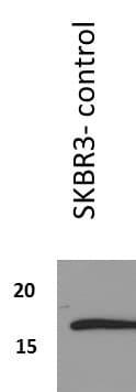
Application: Western BlotSample Tested: Human Breast Cancer Cell line SKBR3 cell lysatesSpecies: HumanVerified Customer | Posted 08/24/2017Band corresponds to survivin expression in untreated SKBR3 cell lysates used as a positive control for an experiment. Survivin antibody was diluted 1:1000 dilution in 3% BSA and incubated overnight at 4 degree C.

-
Application: Western BlotSample Tested: Mouse heartSpecies: MouseVerified Customer | Posted 12/08/2011
There are no reviews that match your criteria.
Protocols
View specific protocols for Survivin Antibody (60.11) - BSA Free (NB500-238):
Immunohistochemistry - FFPE sections
I. Deparaffinization:
A. Treat slides with Xylene: 3 changes for 5 minutes each. Drain slides for 10 seconds between changes.
B. Treat slides with 100% Reagent Alcohol: 3 changes for 5 minutes each. Drain slides for 10 seconds between changes.
II. Quench Endogenous Peroxidase:
A. Place slides in peroxidase quenching solution: 15-30 minutes.
To Prepare 200 ml of Quenching Solution:
Add 3 ml of 30% Hydrogen Peroxide to 200 ml of Methanol.
Use within 4 hours of preparation
B. Place slides in distilled water: 2 changes for 2 minutes each.
III. Retrieve Epitopes:
A. Preheat Citrate Buffer. Place 200 ml of Citrate Buffer Working Solution into container, cover and place into steamer. Heat to 90-96 degrees Celcius.
B. Place rack of slides into hot Citrate Buffer for 20 minutes. Cover.
C. Carefully remove container with slides from steamer and cool on bench, uncovered, for 20 minutes.
D. Slowly add distilled water to further cool for 5 minutes.E. Rinse slides with distilled water. 2 changes for 2 minutes each.
IV. Immunostaining Procedure:
A. Remove each slide from rack and circle tissue section with a hydrophobic barrier pen (e.g. Liquid Blocker-Super Pap Pen).
B. Flood slide with Wash Solution. Do not allow tissue sections to dry for the rest of the procedure.
C. Drain wash solution and apply 4 drops of Blocking Reagent to each slide and incubate for 15 minutes.
D. Drain Blocking Reagent (do not wash off the Blocking Reagent), apply 200 ul of primary antibody solution to each slide, and incubate for 1 hour.
E. Wash slides with Wash Solution: 3 changes for 5 minutes each.
F. Drain wash solution, apply 4 drops of Secondary antibody to each slide and incubate for 1 hour.
G. Wash slides with Wash Solution: 3 changes for 5 minutes each.
H. Drain wash solution, apply 4 drops of DAB Substrate to each slide and develop for 5-10 minutes. Check development with microscope.
I. Wash slides with Wash Solution: 3 changes for 5 minutes each.
J. Drain wash solution, apply 4 drops of Hematoxylin to each slide and stain for 1-3 minutes. Increase time if darker counterstaining is desired.
K. Wash slides with Wash Solution: 2-3 changes for 2 minutes each.
L. Drain wash solution and apply 4 drops of Bluing Solution to each slide for 1-2 minutes.
M. Rinse slides in distilled water.
N. Soak slides in 70% reagent alcohol: 3 minutes with intermittent agitation.
O. Soak slides in 95% reagent alcohol: 2 changes for 3 minutes each with intermittent agitation.
P. Soak slides in 100% reagent alcohol: 3 changes for 3 minutes each with intermittent agitation. Drain slides for 10 seconds between each change.
Q. Soak slides in Xylene: 3 changes for 3 minutes each with intermittent agitation. Drain slides for 10 seconds between each change.
R. Apply 2-3 drops of non-aqueous mounting media to each slide and mount coverslip.
S. Lay slides on a flat surface to dry prior to viewing under microscope.
NOTES:
Use treated slides (e.g. HistoBond) to assure adherence of FFPE sections to slide.
Prior to deparaffinization, heat slides overnight in a 60 degrees Celcius oven.
All steps in which Xylene is used should be performed in a fume hood.
For Epitope Retrieval, a microwave or pressure cooker may be substituted for the steamer method. Adjust times as necessary depending on conditions.
For the initial IHC run with a new primary antibody, test tissues with and without Epitope Retrieval. In some
instances, Epitope Retrieval may not be necessary.
200 ul is the recommended maximum volume to apply to a slide for full coverage. Using more than 200 ul may allow solutions to wick off the slide and create drying artifacts. For small tissue sections less than 200 ul may be used.
5 minutes of development with DAB Substrate should be sufficient. Do not develop for more than 10 minutes. If 5 minutes of development causes background staining, further dilution of the primary antibody may be necessary.
Hematoxylin should produce a light nuclear counterstain so as not to obscure the DAB staining. Counterstain for 1-1 1/2 minutes for nuclear antigens. Counterstain for 2-3 minutes for cytoplasmic and membranous antigens. If darker counterstaining is desired increase time (up to 10 minutes).
Find general support by application which include: protocols, troubleshooting, illustrated assays, videos and webinars.
- 7-Amino Actinomycin D (7-AAD) Cell Viability Flow Cytometry Protocol
- Antigen Retrieval Protocol (PIER)
- Antigen Retrieval for Frozen Sections Protocol
- Appropriate Fixation of IHC/ICC Samples
- Cellular Response to Hypoxia Protocols
- Chromogenic IHC Staining of Formalin-Fixed Paraffin-Embedded (FFPE) Tissue Protocol
- Chromogenic Immunohistochemistry Staining of Frozen Tissue
- ClariTSA™ Fluorophore Kits
- Detection & Visualization of Antibody Binding
- ELISA Sample Preparation & Collection Guide
- ELISA Troubleshooting Guide
- Extracellular Membrane Flow Cytometry Protocol
- Flow Cytometry Protocol for Cell Surface Markers
- Flow Cytometry Protocol for Staining Membrane Associated Proteins
- Flow Cytometry Staining Protocols
- Flow Cytometry Troubleshooting Guide
- Fluorescent IHC Staining of Frozen Tissue Protocol
- Graphic Protocol for Heat-induced Epitope Retrieval
- Graphic Protocol for the Preparation and Fluorescent IHC Staining of Frozen Tissue Sections
- Graphic Protocol for the Preparation and Fluorescent IHC Staining of Paraffin-embedded Tissue Sections
- Graphic Protocol for the Preparation of Gelatin-coated Slides for Histological Tissue Sections
- How to Run an R&D Systems DuoSet ELISA
- How to Run an R&D Systems Quantikine ELISA
- How to Run an R&D Systems Quantikine™ QuicKit™ ELISA
- ICC Cell Smear Protocol for Suspension Cells
- ICC Immunocytochemistry Protocol Videos
- ICC for Adherent Cells
- IHC Sample Preparation (Frozen sections vs Paraffin)
- Immunocytochemistry (ICC) Protocol
- Immunocytochemistry Troubleshooting
- Immunofluorescence of Organoids Embedded in Cultrex Basement Membrane Extract
- Immunofluorescent IHC Staining of Formalin-Fixed Paraffin-Embedded (FFPE) Tissue Protocol
- Immunohistochemistry (IHC) and Immunocytochemistry (ICC) Protocols
- Immunohistochemistry Frozen Troubleshooting
- Immunohistochemistry Paraffin Troubleshooting
- Intracellular Flow Cytometry Protocol Using Alcohol (Methanol)
- Intracellular Flow Cytometry Protocol Using Detergents
- Intracellular Nuclear Staining Flow Cytometry Protocol Using Detergents
- Intracellular Staining Flow Cytometry Protocol Using Alcohol Permeabilization
- Intracellular Staining Flow Cytometry Protocol Using Detergents to Permeabilize Cells
- Preparing Samples for IHC/ICC Experiments
- Preventing Non-Specific Staining (Non-Specific Binding)
- Primary Antibody Selection & Optimization
- Propidium Iodide Cell Viability Flow Cytometry Protocol
- Protocol for Heat-Induced Epitope Retrieval (HIER)
- Protocol for Liperfluo
- Protocol for Making a 4% Formaldehyde Solution in PBS
- Protocol for VisUCyte™ HRP Polymer Detection Reagent
- Protocol for the Characterization of Human Th22 Cells
- Protocol for the Characterization of Human Th9 Cells
- Protocol for the Fluorescent ICC Staining of Cell Smears - Graphic
- Protocol for the Fluorescent ICC Staining of Cultured Cells on Coverslips - Graphic
- Protocol for the Preparation & Fixation of Cells on Coverslips
- Protocol for the Preparation and Chromogenic IHC Staining of Frozen Tissue Sections
- Protocol for the Preparation and Chromogenic IHC Staining of Frozen Tissue Sections - Graphic
- Protocol for the Preparation and Chromogenic IHC Staining of Paraffin-embedded Tissue Sections
- Protocol for the Preparation and Chromogenic IHC Staining of Paraffin-embedded Tissue Sections - Graphic
- Protocol for the Preparation and Fluorescent ICC Staining of Cells on Coverslips
- Protocol for the Preparation and Fluorescent ICC Staining of Non-adherent Cells
- Protocol for the Preparation and Fluorescent ICC Staining of Stem Cells on Coverslips
- Protocol for the Preparation and Fluorescent IHC Staining of Frozen Tissue Sections
- Protocol for the Preparation and Fluorescent IHC Staining of Paraffin-embedded Tissue Sections
- Protocol for the Preparation of Gelatin-coated Slides for Histological Tissue Sections
- Protocol for the Preparation of a Cell Smear for Non-adherent Cell ICC - Graphic
- Protocol: Annexin V and PI Staining by Flow Cytometry
- Protocol: Annexin V and PI Staining for Apoptosis by Flow Cytometry
- Quantikine HS ELISA Kit Assay Principle, Alkaline Phosphatase
- Quantikine HS ELISA Kit Principle, Streptavidin-HRP Polymer
- R&D Systems Quality Control Western Blot Protocol
- Sandwich ELISA (Colorimetric) – Biotin/Streptavidin Detection Protocol
- Sandwich ELISA (Colorimetric) – Direct Detection Protocol
- TUNEL and Active Caspase-3 Detection by IHC/ICC Protocol
- The Importance of IHC/ICC Controls
- Troubleshooting Guide: ELISA
- Troubleshooting Guide: Fluorokine Flow Cytometry Kits
- Troubleshooting Guide: Immunohistochemistry
- Troubleshooting Guide: Western Blot Figures
- Western Blot Conditions
- Western Blot Protocol
- Western Blot Protocol for Cell Lysates
- Western Blot Troubleshooting
- Western Blot Troubleshooting Guide
- View all Protocols, Troubleshooting, Illustrated assays and Webinars
FAQs for Survivin Antibody (60.11) - BSA Free
Showing
1
-
1 of
1 FAQ
Showing All
-
Q: Can I use this antibody with species other than those listed?
A: The species we have listed are validated and therefore have a 100% guarantee to work with this antibody. We cannot guarantee that this will work with other species.
Loading...
Associated Pathways
