Survivin Antibody - BSA Free
Novus Biologicals | Catalog # NB500-201
Key Product Details
Validated by
Knockout/Knockdown, Orthogonal Validation, Biological Validation
Species Reactivity
Validated:
Human, Mouse, Rat, Canine, Feline, Guinea Pig, Hamster
Cited:
Human, Mouse, Rat, Canine, Guinea Pig, Hamster
Applications
Validated:
Immunohistochemistry, Immunohistochemistry-Paraffin, Immunohistochemistry-Frozen, Western Blot, ELISA, Flow Cytometry, Dual RNAscope ISH-IHC, Immunocytochemistry/ Immunofluorescence, Simple Western, Immunoprecipitation, Chromatin Immunoprecipitation (ChIP), Knockdown Validated
Cited:
Immunohistochemistry, Immunohistochemistry-Paraffin, Immunohistochemistry-Frozen, Western Blot, ELISA, Flow Cytometry, Immunocytochemistry/ Immunofluorescence, Simple Western, Immunoprecipitation, Proximity Ligation Assay, IF/IHC, Knockdown Validated
Label
Unconjugated
Antibody Source
Polyclonal Rabbit IgG
Format
BSA Free
Loading...
Product Specifications
Immunogen
This Survivin Antibody was developed against full length recombinant human Survivin [UniProt# O15392]
Reactivity Notes
Hamster reactivity reported in scientific literature (PMID: 23405201). Guinea Pig reactivity reported in scientific literature (PMID: 21364656).
Localization
Nuclear
Clonality
Polyclonal
Host
Rabbit
Isotype
IgG
Theoretical MW
16 kDa.
Disclaimer note: The observed molecular weight of the protein may vary from the listed predicted molecular weight due to post translational modifications, post translation cleavages, relative charges, and other experimental factors.
Disclaimer note: The observed molecular weight of the protein may vary from the listed predicted molecular weight due to post translational modifications, post translation cleavages, relative charges, and other experimental factors.
Scientific Data Images for Survivin Antibody - BSA Free
Dual RNAscope ISH-IHC: Survivin Antibody [NB500-201] - Formalin-fixed paraffin-embedded tissue sections of human esophagus squamous cell carcinoma were probed for Survivin mRNA (ACD RNAScope Probe, [465361]; Fast Red chromogen, ACD [322360]). Adjacent tissue section was processed for immunohistochemistry using rabbit polyclonal [NB500-201] at 1.5ug/mL with overnight incubation at 4 degrees Celsius followed by incubation with anti-rabbit IgG VisUCyte HRP Polymer Antibody [VC003] and DAB chromogen (yellow-brown). Tissue was counterstained with hematoxylin (blue). Specific staining was localized to tumor cells.
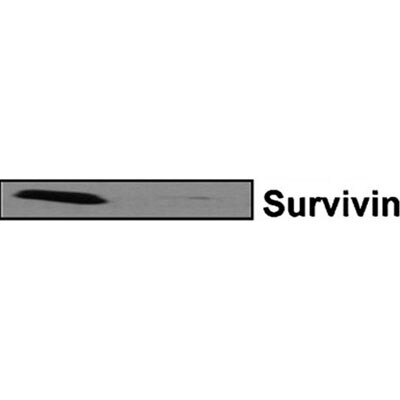
![Knockdown Validated: Survivin Antibody - BSA Free [NB500-201]](https://resources.rndsystems.com/images/products/Survivin-Antibody-Knockdown-Validated-NB500-201-img0037.jpg "Western Blot: Survivin Antibody - BSA Free [NB500-201]")
![Immunohistochemistry: Survivin Antibody - BSA Free [NB500-201]](https://resources.rndsystems.com/images/products/Survivin-Antibody-Immunohistochemistry-NB500-201-img0038.jpg "Immunohistochemistry: Survivin Antibody - BSA Free [NB500-201]")
Immunohistochemistry: Survivin Antibody - BSA Free [NB500-201]
Survivin-Antibody-Immunohistochemistry-NB500-201-img0038.jpg![Knockdown Validated: Survivin Antibody - BSA Free [NB500-201]](https://resources.rndsystems.com/images/products/Survivin-Antibody-Knockdown-Validated-NB500-201-img0033.jpg "Knockdown Validated: Survivin Antibody - BSA Free [NB500-201]")
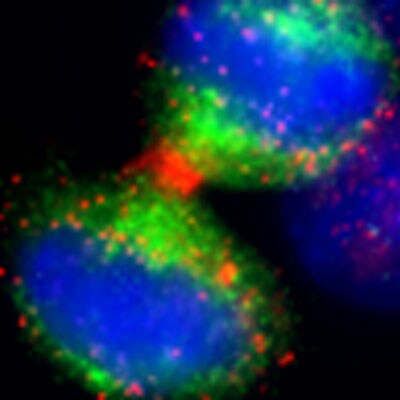
![Immunocytochemistry/ Immunofluorescence: Survivin Antibody - BSA Free [NB500-201]](https://resources.rndsystems.com/images/products/Survivin-Antibody-Immunocytochemistry-Immunofluorescence-NB500-201-img0018.jpg "Immunocytochemistry/ Immunofluorescence: Survivin Antibody - BSA Free [NB500-201]")
Immunocytochemistry/ Immunofluorescence: Survivin Antibody - BSA Free [NB500-201]
Immunocytochemistry/Immunofluorescence: Survivin Antibody [NB500-201] - Analysis using the HRP conjugate of [NB500-201]. Staining of Telophase with accumulation of survivin in the midbodies of two daughter cells. Survivin detection using [NB500-201].![Western Blot: Survivin AntibodyBSA Free [NB500-201]](https://resources.rndsystems.com/images/products/Survivin-Antibody-Western-Blot-NB500-201-img0017.jpg "Western Blot: Survivin AntibodyBSA Free [NB500-201]")
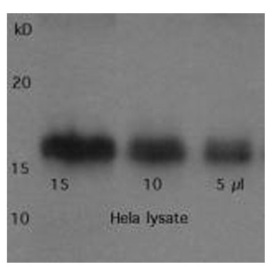
Western Blot: Survivin AntibodyBSA Free [NB500-201]
Western Blot: Survivin Antibody [NB500-201] - Analysis of 30ug of HeLa whole cell lysate [NB800-PC1] using rabbit polyclonal [NB500-201] at 1ug/ml. Detection was performed using ECL method with 1 minute exposure. Band detected at higher molecular weight than the predicted MW (16 kDa).![Immunohistochemistry-Paraffin: Survivin Antibody - BSA Free [NB500-201]](https://resources.rndsystems.com/images/products/Survivin-Antibody-Immunohistochemistry-Paraffin-NB500-201-img0008.jpg "Immunohistochemistry-Paraffin: Survivin Antibody - BSA Free [NB500-201]")
Immunohistochemistry-Paraffin: Survivin Antibody - BSA Free [NB500-201]
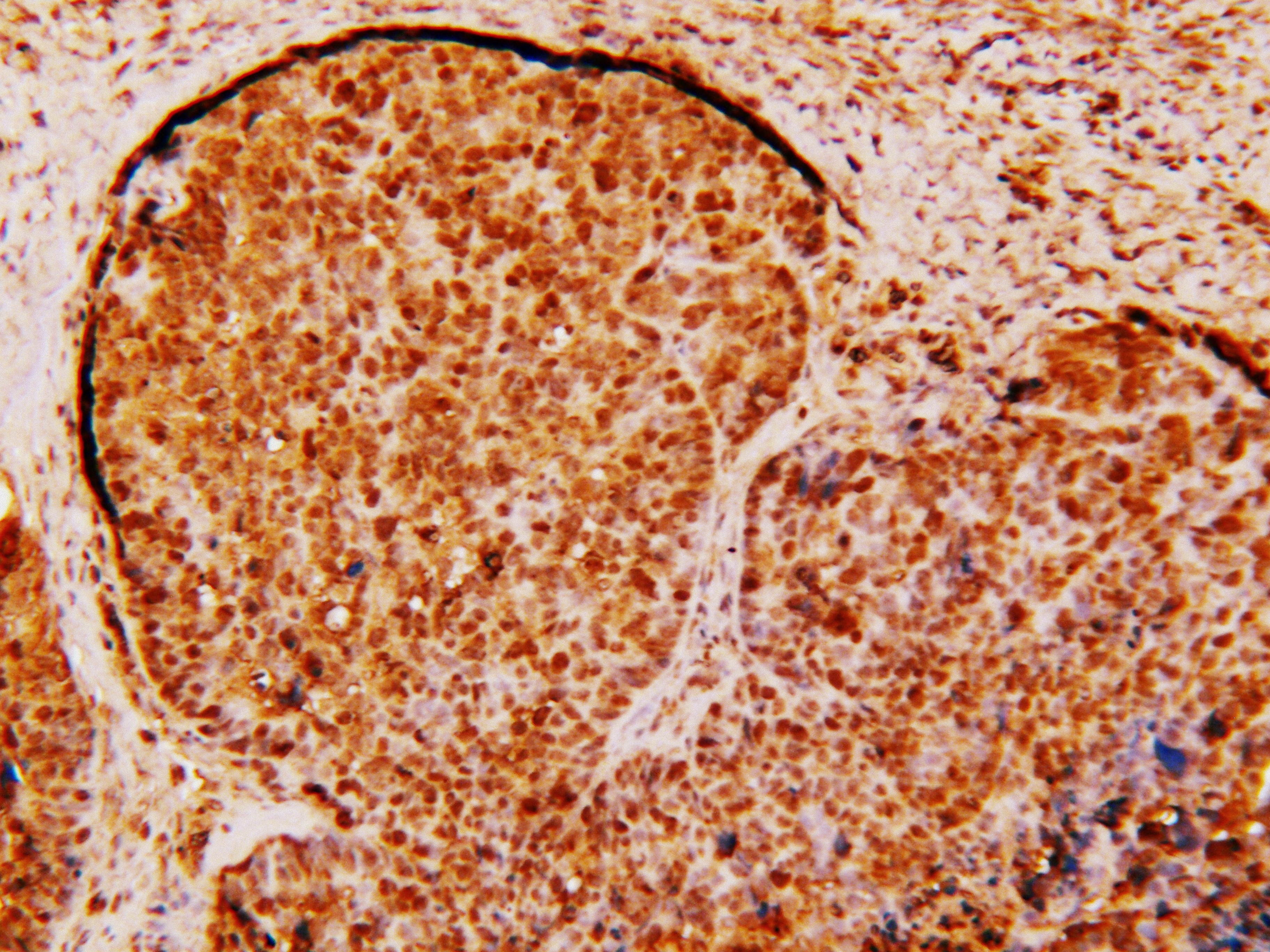
Immunohistochemistry-Paraffin: Survivin Antibody [NB500-201] - Immunohistochemical staining of Survivin in human rectal cancer using [NB500-201] and DAB with hematoxylin counterstain.![Flow Cytometry: Survivin Antibody - BSA Free [NB500-201]](https://resources.rndsystems.com/images/products/Survivin-Antibody-Flow-Cytometry-NB500-201-img0025.jpg "Flow Cytometry: Survivin Antibody - BSA Free [NB500-201]")
Flow Cytometry: Survivin Antibody - BSA Free [NB500-201]
Flow Cytometry: Survivin Antibody [NB500-201] - An intracellular stain was performed on HeLa cells with [NB500-201] and a matched isotype control. Cells were fixed with 4% PFA and then permeablized with 0.1% saponin. Cells were incubated in an antibody dilution of 2.5 ug/mL for 30 minutes at room temperature, followed by Rabbit IgG (H+L) Cross-Adsorbed Secondary Antibody, Dylight 550.![Western Blot: Survivin AntibodyBSA Free [NB500-201]](https://resources.rndsystems.com/images/products/Survivin-Antibody-Western-Blot-NB500-201-img0032.jpg "Western Blot: Survivin AntibodyBSA Free [NB500-201]")
Western Blot: Survivin AntibodyBSA Free [NB500-201]
Survivin-Antibody-Western-Blot-NB500-201-img0032.jpg![Western Blot: Survivin AntibodyBSA Free [NB500-201]](https://resources.rndsystems.com/images/products/Survivin-Antibody-Western-Blot-NB500-201-img0013.jpg "Western Blot: Survivin AntibodyBSA Free [NB500-201]")
Western Blot: Survivin AntibodyBSA Free [NB500-201]
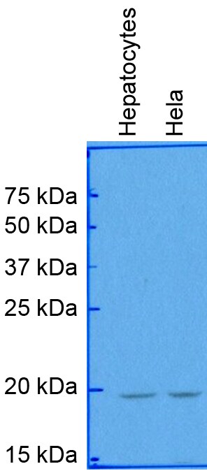
Western Blot: Survivin Antibody [NB500-201] - Analysis of Survivin in human hepatocytes from cancer patient (left) and HeLa cell lysate (right) using [NB500-201]. Image from verified customer review. Note: bands detected at higher molecular weight than predicted (16 kDa)![Western Blot: Survivin AntibodyBSA Free [NB500-201]](https://resources.rndsystems.com/images/products/Survivin-Antibody-Western-Blot-NB500-201-img0028.jpg "Western Blot: Survivin AntibodyBSA Free [NB500-201]")
![Immunocytochemistry/ Immunofluorescence: Survivin Antibody - BSA Free [NB500-201]](https://resources.rndsystems.com/images/products/Survivin-Antibody-Immunocytochemistry-Immunofluorescence-NB500-201-img0009.jpg "Immunocytochemistry/ Immunofluorescence: Survivin Antibody - BSA Free [NB500-201]")
Immunocytochemistry/ Immunofluorescence: Survivin Antibody - BSA Free [NB500-201]
Immunocytochemistry/Immunofluorescence: Survivin Antibody [NB500-201] - Analysis of HeLa cells using Survivin Antibody ([NB500-201], 1:10). An Alexa Fluor 488-conjugated Goat to rabbit IgG was used as secondary antibody (green). Actin filaments were labeled with Alexa Fluor 568 phalloidin (red). DAPI was used to stain the cell nuclei (blue).![Immunohistochemistry-Paraffin: Survivin Antibody - BSA Free [NB500-201]](https://resources.rndsystems.com/images/products/Survivin-Antibody-Immunohistochemistry-Paraffin-NB500-201-img0010.jpg "Immunohistochemistry-Paraffin: Survivin Antibody - BSA Free [NB500-201]")
Immunohistochemistry-Paraffin: Survivin Antibody - BSA Free [NB500-201]
Immunohistochemistry-Paraffin: Survivin Antibody [NB500-201] - Analysis of Survivin in ovarian cancer tissue using [NB500-201]. Image from verified customer review.![Immunohistochemistry: Survivin Antibody - BSA Free [NB500-201]](https://resources.rndsystems.com/images/products/Survivin-Antibody-Immunohistochemistry-NB500-201-img0015.jpg "Immunohistochemistry: Survivin Antibody - BSA Free [NB500-201]")
Immunohistochemistry: Survivin Antibody - BSA Free [NB500-201]
Immunohistochemistry: Survivin Antibody [NB500-201] - HRP conjugated Survivin expression in BIRC5 transfected 293T cells using Survivin Antibody [NB500-201]. Image from verified customer review.![Immunohistochemistry: Survivin Antibody - BSA Free [NB500-201]](https://resources.rndsystems.com/images/products/Survivin-Antibody-Immunohistochemistry-NB500-201-img0016.jpg "Immunohistochemistry: Survivin Antibody - BSA Free [NB500-201]")
Immunohistochemistry: Survivin Antibody - BSA Free [NB500-201]
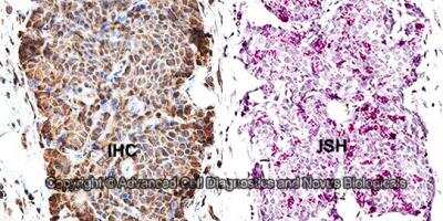
Immunohistochemistry: Survivin Antibody [NB500-201] - Immunohistochemical analysis using [NB500-201]. The top photo is a control stain and the bottom photo is anti-survivin staining of melanoma. Photo courtesy of Dr. Dario Altieri, Yale University.![Immunohistochemistry-Paraffin: Survivin Antibody - BSA Free [NB500-201]](https://resources.rndsystems.com/images/products/Survivin-Antibody-Immunohistochemistry-Paraffin-NB500-201-img0021.jpg "Immunohistochemistry-Paraffin: Survivin Antibody - BSA Free [NB500-201]")
Immunohistochemistry-Paraffin: Survivin Antibody - BSA Free [NB500-201]
Immunohistochemistry-Paraffin: Survivin Antibody [NB500-201] - Survivin shows lysates of human neuroblastoma cell line. Polyvinylidene fluoride (PVDF) membrane was probed with 1:200 dilution of 0.5 ug/mL of rabbit polyclonal [NB500-201], followed by 1:2000 dilution of goat anti-rabbit IgG.![Simple Western: Survivin AntibodyBSA Free [NB500-201]](https://resources.rndsystems.com/images/products/Survivin-Antibody-Simple-Western-NB500-201-img0011.jpg "Simple Western: Survivin AntibodyBSA Free [NB500-201]")
Simple Western: Survivin AntibodyBSA Free [NB500-201]
Simple Western: Survivin Antibody [NB500-201] - Simple Western analysis using [NB500-201]. Lane view shows a specific band for Survivin in 1.0 mg/ml of HeLa lysate. This experiment was performed under reducing conditions using the 12-230 kDa separation system. Theoretical molecular weight: 16 kDa.
Western Blot: Survivin Antibody - BSA Free [NB500-201] -
Radiation-resistant Caveolin-1 (CAV1)-silenced fibroblasts differentially express and secrete the apoptosis inhibiting protein TP53-regulated inhibitor of apoptosis 1 (TRIAP1).
Western Blot: Survivin Antibody - BSA Free [NB500-201] -
Expression of survivin protein in cell lines after transfection with siRNA.Survivin protein expression in CHS cell lines was evaluated by western blotting at 48 h after transfection with scrambled and survivin siRNA.

Western Blot: Survivin Antibody - BSA Free [NB500-201] -
(A) Levels of suppression of Survivin and expression of p53 for 2 h, 4 h and 8 h for MBA-MD 231 cells. (B) Light microscopy images of the three cell types using nanoconstruct with the AS1411 aptamer. Cell population had been observed to reduce for the MBA-MD 231 cells and AGS while the non-tumorigenic cells MCF-10a did not exhibit any appreciable loss in cell number as well as cell morphology.
Western Blot: Survivin Antibody - BSA Free [NB500-201] -
Detection of BCL2, Bcl-xL, XIAP and survivin protein content by western blotting 48 h after transfection with a total of 40 nM siRNA in EJ28 bladder cancer cells. Beta-actin was used for loading control.
Western Blot: Survivin Antibody - BSA Free [NB500-201] -
Western Blot: Survivin Antibody - BSA Free [NB500-201] - Deficiency in CCAR2 or Hsp60 reduces expression of survivin. SH-SY5Y cells were transfected with Universal (siU), CCAR2 (siC), or Hsp60 (siH) siRNA. Forty-eight hours later, expression of survivin protein was examined by western blotting. (A) Survivin expression was detected in whole cell lysates. The relative level of survivin protein is presented as the mean ± standard error of the mean (SEM) (n = 3). Asterisks (*) denote statistically significant differences (p < 0.05, one-way ANOVA). (B) Cytosolic & mitochondrial fractions were isolated to determine localization & expression of survivin. (C) Two different siRNAs specific for CCAR2 & Hsp60 were used to knock down their expressions. Image collected & cropped by CiteAb from the following publication (https://pubmed.ncbi.nlm.nih.gov/30609639), licensed under a CC-BY license. Not internally tested by Novus Biologicals.
Western Blot: Survivin Antibody - BSA Free [NB500-201] -
Western Blot: Survivin Antibody - BSA Free [NB500-201] - Survivin & BIM expression in response to lapatinibWestern analysis of Survivin & BIM protein levels after 24 hour exposure to 0.1% DMSO (−) or 2 μM lapatinib (+) in lapatinib-resistant cells (A), cells overexpressing t-Darpp (B), or SK/HerR cells transiently transfected with siRNA targeted to GFP (siCtrl) or Darpp-32/t-Darpp (siDp) for 72 hours (C). alpha -Tubulin was used as a loading control. Protein expression was quantified using ImageJ software. Data was normalized to alpha -Tubulin levels & expressed as the fold change in protein level after lapatinib treatment, relative to the DMSO control, for each cell line. Image collected & cropped by CiteAb from the following publication (https://www.oncotarget.com/lookup/doi/10.18632/oncotarget.5311), licensed under a CC-BY license. Not internally tested by Novus Biologicals.
Western Blot: Survivin Antibody - BSA Free [NB500-201] -
Western Blot: Survivin Antibody - BSA Free [NB500-201] - Expression of stem cell & differentiation markers in RAD, NRAD & TOT cells from cSCC. (A) Cells were analyzed immediately after separation, & levels of markers were determined by Western blot analysis. beta -actin was used as loading control; (B) Bar graphs show the average densitometry values normalized to actin. *p < 0.05; **p < 0.01. Image collected & cropped by CiteAb from the following publication (http://www.mdpi.com/1422-0067/14/10/19540), licensed under a CC-BY license. Not internally tested by Novus Biologicals.
Western Blot: Survivin Antibody - BSA Free [NB500-201] -
Western Blot: Survivin Antibody - BSA Free [NB500-201] - Expression of survivin protein in cell lines after transfection with siRNA.Survivin protein expression in CHS cell lines was evaluated by western blotting at 48 h after transfection with scrambled & survivin siRNA. Image collected & cropped by CiteAb from the following publication (https://pubmed.ncbi.nlm.nih.gov/24260303), licensed under a CC-BY license. Not internally tested by Novus Biologicals.
Western Blot: Survivin Antibody - BSA Free [NB500-201] -
Western Blot: Survivin Antibody - BSA Free [NB500-201] - Survivin protein stability is increased in the presence of autophagy inhibitors in serum-free media, but not in serum-containing media. Western blots & protein quantitation graphs showing survivin protein levels over time after addition of CHX (100 μM) to media. One representative western blot out of three repeat experiments was shown for each treatment & condition. The prominent band shown is survivin while the smaller bands are nonspecific, as determined via control experiments utilizing a neutralizing peptide for the survivin antibody. *Indicates the time at which survivin protein is half of the amount at 0 hours (half-life), P < 0.02. Image collected & cropped by CiteAb from the following publication (https://pubmed.ncbi.nlm.nih.gov/23431290), licensed under a CC-BY license. Not internally tested by Novus Biologicals.
Western Blot: Survivin Antibody - BSA Free [NB500-201] -
Western Blot: Survivin Antibody - BSA Free [NB500-201] - (A) Levels of suppression of Survivin & expression of p53 for 2 h, 4 h & 8 h for MBA-MD 231 cells. (B) Light microscopy images of the three cell types using nanoconstruct with the AS1411 aptamer. Cell population had been observed to reduce for the MBA-MD 231 cells & AGS while the non-tumorigenic cells MCF-10a did not exhibit any appreciable loss in cell number as well as cell morphology. Image collected & cropped by CiteAb from the following publication (https://www.nature.com/articles/s41598-017-00912-3), licensed under a CC-BY license. Not internally tested by Novus Biologicals.
Western Blot: Survivin Antibody - BSA Free [NB500-201] -
Western Blot: Survivin Antibody - BSA Free [NB500-201] - Deficiency in CCAR2 or Hsp60 reduces expression of survivin. SH-SY5Y cells were transfected with Universal (siU), CCAR2 (siC), or Hsp60 (siH) siRNA. Forty-eight hours later, expression of survivin protein was examined by western blotting. (A) Survivin expression was detected in whole cell lysates. The relative level of survivin protein is presented as the mean ± standard error of the mean (SEM) (n = 3). Asterisks (*) denote statistically significant differences (p < 0.05, one-way ANOVA). (B) Cytosolic & mitochondrial fractions were isolated to determine localization & expression of survivin. (C) Two different siRNAs specific for CCAR2 & Hsp60 were used to knock down their expressions. Image collected & cropped by CiteAb from the following publication (https://pubmed.ncbi.nlm.nih.gov/30609639), licensed under a CC-BY license. Not internally tested by Novus Biologicals.
Immunohistochemistry: Survivin Antibody - BSA Free [NB500-201] -
Immunohistochemistry: Survivin Antibody - BSA Free [NB500-201] - RAD & NRAD-derived tumor characterization. (A) Mitotic Index representing the number of cells undergoing mitosis over total cells were counted in RAD & NRAD-derived tumors **p < 0.01; (B) Survivin & Ki67 expression in RAD & NRAD-derived tumors by immunohistochemistry. Scale bar = 200 μm; (C) K10, E-FABP & involucrin expression in RAD & NRAD-derived tumors by immunohistochemistry. Scale bar = 200 μm. Image collected & cropped by CiteAb from the following publication (http://www.mdpi.com/1422-0067/14/10/19540), licensed under a CC-BY license. Not internally tested by Novus Biologicals.
Western Blot: Survivin Antibody - BSA Free [NB500-201] -
Western Blot: Survivin Antibody - BSA Free [NB500-201] - Gene-silencing of Bcl-2, Survivin & XIAP. Western blot showed efficient silencing in transfected MiaPaCa-2 (A) & AsPC-1 cells (B). 50,000 cells/well were single-transfected with carrier solution (lane 1) & siRNA against Luciferase (lane 2) as control. SGS, lowST & ST all effectively silenced the three target genes (lane 3-5). Efficient knock-down was also shown in the lowST group by RT-PCR in MiaPaCa-2 (C) & AsPC-1 cells (D). White bars show controls, grey bars signify transfected cells. All samples were normalized to beta -Actin as a house-keeping gene. SGS = Simultaneous gene silencing; lowST = Low dose siRNA transfection; ST = Standard dose siRNA transfection. Image collected & cropped by CiteAb from the following publication (https://pubmed.ncbi.nlm.nih.gov/20646298), licensed under a CC-BY license. Not internally tested by Novus Biologicals.
Western Blot: Survivin Antibody - BSA Free [NB500-201] -
Western Blot: Survivin Antibody - BSA Free [NB500-201] - CCAR2 binds Hsp60 & survivin. Interaction between CCAR2 & survivin was examined in SH-SY5Y or HEK293 cells by co-immunoprecipitation with either an anti-CCAR2 or an anti-survivin antibody, followed by western blotting. (A) Interaction between CCAR2 & survivin in whole cell lysates from SH-SY5Y cells was examined. (B) Interaction between CCAR2, survivin, & Hsp60 in cytosolic & mitochondrial fractions isolated from HEK293 cells was examined. (C) SH-SY5Y cells were depleted of Hsp60 & the interaction between CCAR2 & survivin was examined. siU, universal siRNA; siH, Hsp60 siRNA. Image collected & cropped by CiteAb from the following publication (https://pubmed.ncbi.nlm.nih.gov/30609639), licensed under a CC-BY license. Not internally tested by Novus Biologicals.
Western Blot: Survivin Antibody - BSA Free [NB500-201] -
Western Blot: Survivin Antibody - BSA Free [NB500-201] - Radiation-resistant Caveolin-1 (CAV1)-silenced fibroblasts differentially express & secrete the apoptosis inhibiting protein TP53-regulated inhibitor of apoptosis 1 (TRIAP1). Image collected & cropped by CiteAb from the following publication (https://pubmed.ncbi.nlm.nih.gov/30871022), licensed under a CC-BY license. Not internally tested by Novus Biologicals.
Western Blot: Survivin Antibody - BSA Free [NB500-201] -
Western Blot: Survivin Antibody - BSA Free [NB500-201] - Western blotting analysis in MDA-MB-231 cells & SUM 149 cells.The cells were treated for 24 h with C. juttae essential oil (EO) (46 & 64 μg/ml, respectively). The data shown are the results of a representative experiment. Image collected & cropped by CiteAb from the following publication (https://pubmed.ncbi.nlm.nih.gov/30921428), licensed under a CC-BY license. Not internally tested by Novus Biologicals.
Western Blot: Survivin Antibody - BSA Free [NB500-201] -
Western Blot: Survivin Antibody - BSA Free [NB500-201] - Overexpression of Survivin predominantly protects anoikis. B. Caspase-3 activation in EGFP- & EGFP-Survivin-expressing cells after serum starvation under attached or detached culture conditions. Experimental protocol was illustrated in Figure S1. Transfection frequencies checked by using fluorescence microscopy & confirmed to be 80–90%. Cells kept in serum-free medium for 24–72 h, harvested, & lysed in Laemmli SDS-sample buffer for immunoblot analysis with anti-GFP, anti-Survivin, anti-activated caspase-3 antibody, anti-LC3B, & anti-alpha -tubulin.Image collected & cropped by CiteAb from the following publication (https://dx.plos.org/10.1371/journal.pone.0055710), licensed under a CC-BY license. Not internally tested by Novus Biologicals.
Western Blot: Survivin Antibody - BSA Free [NB500-201] -
Western Blot: Survivin Antibody - BSA Free [NB500-201] - Representative Western blots showing the IAP levels in EV derived from individual European American (EA) (N1-N5) & (N7-N9), *N6 & African American (AA) (N1-N9) patients with prostate cancer (PCa).Specific antibodies against Survivin, XIAP, cIAP-1, cIAP-2, & Lamp1 were used for the Western blotting analysis of total exosomal proteins. The blots from both patient groups were processed under identical conditions; Lamp 1 was used as loading control. (*N6, Hispanic.) (Both blots were done side by side in the same gel running & transferring apparatus, blocking, washing buffers, & antibody incubations were done in the same time, in the same incubating trays under the identical exposure to keep the consistencies.) Image collected & cropped by CiteAb from the following publication (https://dx.plos.org/10.1371/journal.pone.0183122), licensed under a CC-BY license. Not internally tested by Novus Biologicals.
Western Blot: Survivin Antibody - BSA Free [NB500-201] -
Western Blot: Survivin Antibody - BSA Free [NB500-201] - CCAR2 binds Hsp60 & survivin. Interaction between CCAR2 & survivin was examined in SH-SY5Y or HEK293 cells by co-immunoprecipitation with either an anti-CCAR2 or an anti-survivin antibody, followed by western blotting. (A) Interaction between CCAR2 & survivin in whole cell lysates from SH-SY5Y cells was examined. (B) Interaction between CCAR2, survivin, & Hsp60 in cytosolic & mitochondrial fractions isolated from HEK293 cells was examined. (C) SH-SY5Y cells were depleted of Hsp60 & the interaction between CCAR2 & survivin was examined. siU, universal siRNA; siH, Hsp60 siRNA. Image collected & cropped by CiteAb from the following publication (https://pubmed.ncbi.nlm.nih.gov/30609639), licensed under a CC-BY license. Not internally tested by Novus Biologicals.
Western Blot: Survivin Antibody - BSA Free [NB500-201] -
Western Blot: Survivin Antibody - BSA Free [NB500-201] - Induction of cell death & suppression of survivin after L-OHP depends on p53(A) HCT116 wild typeand p53-/- cells were treated with 5 μM L-OHP or 10 μM CPT-11 for 24 hours. Protein levels of survivin, p53 & p21 were detected by Western blot analysis; vinculin serves as loading control. (B) Quantitative real-time PCR was performed to quantify BIRC5 mRNA levels in HCT116 wild type & p53-deficient cells after 24 hours treatment (** p < 0.01, n = 3). (C) Flow cytometric analysis of DNA content was done in HCT116 wild type & p53-/- cells after 24 hours treatment with L-OHP (n = 4). (D) SubG1-populations were detected in both cell lines after 48 hours treatment (*** p < 0.001, n = 4). Image collected & cropped by CiteAb from the following publication (https://pubmed.ncbi.nlm.nih.gov/29963241), licensed under a CC-BY license. Not internally tested by Novus Biologicals.
Western Blot: Survivin Antibody - BSA Free [NB500-201] -
Western Blot: Survivin Antibody - BSA Free [NB500-201] - Detection of BCL2, Bcl-xL, XIAP & survivin protein content by western blotting 48 h after transfection with a total of 40 nM siRNA in EJ28 bladder cancer cells. Beta-actin was used for loading control. Image collected & cropped by CiteAb from the following publication (http://www.spandidos-publications.com/10.3892/ijo.2012.1549), licensed under a CC-BY license. Not internally tested by Novus Biologicals.
Western Blot: Survivin Antibody - BSA Free [NB500-201] -
Western Blot: Survivin Antibody - BSA Free [NB500-201] - PARP6 inhibits cell growth & colony formationA. A schematic illustration of the human PARP6 protein. PARP6 consists of 630 amino acid residues & has a PARP catalytic domain in C-terminal region. delta C-PARP6 lacks the PARP catalytic domain. B. Cell proliferation after FL-PARP6 & delta C-PARP6 overexpression in SW480 cells was measured by MTT assays. After transfection with the p-EGFP-empty, p-EGFP-FL-PARP6 or p-EGFP-delta C-PARP6 plasmids, transfectant cells (1500 cells / well) were replated in 96-well plates. The MTT assay was performed to test the cell viability at 24 h, 48 h & 72 h. Values indicate mean ± SD (n=6). C. Colony formation aof FL-PARP6 & delta C-PARP6 transfectant SW480 cells. Cells were plated in 6 cm dishes at a density of 500 cells per dish. After 2 weeks, number of colonies were counted. The data represent the means ± S.D. of three independent experiments **P < 0.01. D. Expression of cell cycle related proteins including Survivin, Cyclin B, Aurora-B, p21 & p27 in empty vector, FL-PARP6 & delta C-PARP6 transfectant SW480 cells was examined by Western blot analysis. beta -actin expression was used as a loading control. E. Expression of apoptosis related proteins including cleaved Caspase-3, Caspase-3, Bax, Bcl-2 & Bcl-XL in empty vector, FL-PARP6 & delta C-PARP6 transfectant SW480 cells was examined by Western blot analysis. beta -actin expression was used as a loading control. Image collected & cropped by CiteAb from the following publication (https://pubmed.ncbi.nlm.nih.gov/26934315), licensed under a CC-BY license. Not internally tested by Novus Biologicals.
Western Blot: Survivin Antibody - BSA Free [NB500-201] -
Western Blot: Survivin Antibody - BSA Free [NB500-201] - Screening of epigenetic drugs for upregulation of miRNAs & downregulation of ZEB1Heat map showing the relative expression levels after drug treatment for 48 h in Panc1. Values measured by qRT–PCR were depicted with the software GENE-E. Only mocetinostat upregulated the miRNAs & downregulated ZEB1.Relative expression of indicated genes in Panc1 measured by qRT–PCR after treatment with different HDAC inhibitors. Note the downregulation of ZEB1 & upregulation of miR-203, miR-200, & E-cadherin by mocetinostat. n = 3, mean ± SEM; unpaired Student's t-test. For significance, see Supplementary Table S1.Immunoblot & immunofluorescence showing that mocetinostat treatment (1 μM, 48 h) reduced ZEB1 expression & induced E-cadherin in Panc1. Expression of histone deacetylases was not altered by mocetinostat, but histone acetylation was induced. Scale bar 10 μm.Chromatin immunoprecipitation analysis validated mocetinostat-induced (1 μM, 48 h) enrichment of the active histone marks H3ac, H4ac, H3K9ac, & H3K4me3 at ZEB1 target gene loci in Panc1. n = 3, mean ± SEM; unpaired Student's t-test.Mocetinostat treatment reduced expression of the anti-apoptotic miR-203 target survivin & sphere-forming capacity in Panc1 when pre-treated with mocetinostat for 48 h. n = 3, mean ± SEM; Mann–Whitney U-test.Source data are available online for this figure. Image collected & cropped by CiteAb from the following publication (https://pubmed.ncbi.nlm.nih.gov/25872941), licensed under a CC-BY license. Not internally tested by Novus Biologicals.
Western Blot: Survivin Antibody - BSA Free [NB500-201] -
Western Blot: Survivin Antibody - BSA Free [NB500-201] - Survivin is highly enriched in exosomes from PTX-treated cancer cells. (A) Western blot analysis using Survivin, flotillin, I kappa B alpha, & CD-63 antibodies was performed on lysates of MDAMB231 cells treated with either DMSO or PTX (lanes labeled WCL), as well as the exosomes (lanes labeled Exos) & MVs (lanes labeled MVs) generated by the cells. (B) The relative amounts of Survivin detected in exosomes generated by DMSO- & PTX-treated MDAMB231 cells. (C) Western blot analysis using Survivin & actin antibodies was performed on lysates of MDAMB231 cells treated with PTX for increasing lengths of time. (D) Immunofluorescence using a Survivin antibody was performed on MDAMB231 cells treated with either DMSO or PTX (top images). The cells were also stained with DAPI to label nuclei (bottom images). Arrows indicate areas where Survivin is detected as puncta in the cytosol of cells treated with PTX. Scale bar = 5 µm. (E) Western blot analysis was performed using a Survivin antibody on lysates of MDAMB231 cells, U87 glioblastoma cells, & SKBR3 cells that had been treated with DMSO or PTX (lanes labeled WCL), & on the exosomes these cells generated (lanes labeled Exos). (F) Western blot analysis was performed on lysates of exosomes from MDAMB231 cells that had been treated with the indicated chemotherapeutic agents & inhibitors. The experiments in B were performed a minimum of three separate times, with each experiment yielding similar results. Student t-tests were performed. *** p < 0.001. Image collected & cropped by CiteAb from the following publication (http://www.mdpi.com/2072-6694/8/12/111), licensed under a CC-BY license. Not internally tested by Novus Biologicals.
Western Blot: Survivin Antibody - BSA Free [NB500-201] -
Western Blot: Survivin Antibody - BSA Free [NB500-201] - NPC-derived EBNA1 is compromised for interaction with SurvivinA. LC-MS analysis of proteins associated with FLAG-EBNA1 (F-EBNA1) from B95-8 or with the NPC DNA binding domain. Unique peptides & spectral counts are shown for USP7, BIRC5 (Survivin), & Tankyrase 1 (TNKS). B. Western blot analysis of FLAG IP from HeLa cells with stable expression of FLAG-vector, F-EBNA1 (B95-8) or F-EBNA1 (NPC) probed with antibody to USP7, Tankyrase 1, Survivin, or FLAG. C. Extracts from stable HeLa cells shown in panel B, were subject to IP with antibody to Survivin probed with antibody to FLAG or Survivin (top panel). Extracts from stable HeLa cells shown in panel B were subject to IP with antibody to Tankyrase 1 probed with antibody to FLAG or Tankyrase1 (lower panel). D. MutuI cell (top panels) or C666-1 cell (lower panels) extracts were subject to IP with antibody to EBNA1 or control IgG, & then assayed by Western blot with antibody to Survivin or EBNA1, as indicated. E. Same as in panel D, except IP with Suvivin or control IgG. F. In situ Proximity Ligation Assay (in situ PLA) is shown for interphase cells from either MUTU-I or C666-1 using mouse anti-EBNA1 & rabbit anti-Survivin. G. Quantification of in situ PLA for n > 100. ** p-value <.01 using student t-test. H. PFGE analysis of EBV episomes & linears at 7 days post-transduction with lentivirus shRNA for shControl or shSurvivin in MUTU I or RAJI cell lines. Human alpha satellite DNA is used for DNA loading control Western blot of Survivin & Actin are shown below for each cell at 7 days post-transduction. Image collected & cropped by CiteAb from the following publication (https://pubmed.ncbi.nlm.nih.gov/28077791), licensed under a CC-BY license. Not internally tested by Novus Biologicals.
Western Blot: Survivin Antibody - BSA Free [NB500-201] -
Western Blot: Survivin Antibody - BSA Free [NB500-201] - Anti-tumor effects & functional evidence of sCA-survivin-siRNA in HCT116 & HT29 solid tumor models.(A) For western blot analysis, HCT116 cells were seeded into 6-well plates & transfected with 100 pmol/well of either control or survivin siRNA by Lp (Lipofectamine 2000) or sCA. Actin blots served as loading controls. (B) For proliferation assays, cells were uniformly seeded into 96-well plates (1 × 10⁴ cells/well), & 5 pmol/well siRNA was used. Cell viability was examined at 48 & 72 h by WST-8 assay. Data represent mean ± SEM. *P = 0.0294, **P = 0.0304 (n = 4, Wilcoxon rank test). (C) In vivo tumor growth. Each vehicle, carrying 15 μg of control siRNA or survivin siRNA, was administered by intravenous injection to mice with HCT116 tumors. Data represent the mean ± SEM (n = 10 tumors, Wilcoxon rank test). (D) Immunostaining of survivin in the tumor tissues on day 19. Scale bar, 50 μm. (E) In vivo live imaging of sCA-siRNA (6-FAM labeled) in HT29 tumor by multiphoton microscopy at 90 min. (F) Fluorescent detection of naked-siRNA (6-FAM labeled) or sCA-siRNA (6-FAM labeled) in the HT29 tumor at 4 h. Green: 6-FAM labeled siRNA; Red: microvasculature; Blue: DAPI stained nuclei. Scale bar, 50 μm. (G) Mice were administered with 40 μg of naked-survivin-siRNA or sCA-survivin-siRNA on days 0, 1, & 2. Tumors were removed on day 3, & western blot analysis for survivin was performed. (H) Many tumor cells treated with sCA-survivin-siRNA (6-FAM labeled) had condensed nuclei & were positive for TUNEL assay. Scale bar, 50 μm. Image collected & cropped by CiteAb from the following publication (https://pubmed.ncbi.nlm.nih.gov/25738937), licensed under a CC-BY license. Not internally tested by Novus Biologicals.
Western Blot: Survivin Antibody - BSA Free [NB500-201] -
Western Blot: Survivin Antibody - BSA Free [NB500-201] - Survivin regulates autophagic flux. (A) U2OS cells stably expressing survivinWTGFP or GFP alone were treated with RAP (200 nM) & CQ (120 µM) for 2 h, then lysed & immunoblotted with anti-LC3, anti-survivin & anti-tubulin antibodies. Immunoblot shown is representative of four independent experiments. (B) ImageJ quantitation of LC3II signals in (A), normalised against tubulin control & expressed as band intensity relative to untreated GFP cells. (C) Data from (B) expressed as a percentage increase in LC3II between CQ treated & untreated cells to indicate autophagic flux. (D) The above cell lines were treated with CQ (50 µM) for 8 h & p62 levels assessed by immunoblotting at 2 h intervals. Blot shown is representative of three independent experiments. (E) ImageJ quantitation of p62 signals in (D) normalised against tubulin & expressed as band intensity relative to untreated GFP cells. Error bars indicate s.e.m., N=3. *P<0.05. Image collected & cropped by CiteAb from the following publication (https://pubmed.ncbi.nlm.nih.gov/30348810), licensed under a CC-BY license. Not internally tested by Novus Biologicals.
Western Blot: Survivin Antibody - BSA Free [NB500-201] -
Western Blot: Survivin Antibody - BSA Free [NB500-201] - Cdh1 depletion induces early DNA replication. (A) Fzr1+/+ & Fzr1−/− MEFs were synchronized at G1-S phase by double thymidine block (DTB, see diagram). After release from G1-S arrest, cells were collected at the indicated times. Cell extracts were immunoblotted for the indicated proteins. M phase is evaluated by detection of FoxM1 p-T600. beta -actin is shown as a loading control. Asterisk shows the position of Aurora A p-T288. (B) Subcellular colocalization of Aurora B & histone H3 p-S10 in Fzr1+/+ & Fzr1−/− MEFs. Cells were fixed & stained using anti-H3 p-S10 (green) & anti-Aurora B (red) antibodies. Nuclei of the cells were stained using DAPI (blue). Arrowheads show H3 p-S10 in mitotic cells, & arrows show H3 p-S10 in interphase cells. Scale bar: 20 µm. (C) Fzr1+/+ & Fzr1−/− MEFs were fixed & stained using anti-H3 p-S10 (green) & anti-cyclin A (red) antibodies. Nuclei of the cells were stained with DAPI (blue). Cyclin A expression was used as a marker of G2 cells. Scale bar: 10 µm. (D) Fzr1−/− MEFs were treated with the Aurora B specific inhibitor barasertib (10 nM) for 24 h. Cells were fixed & stained using an anti-H3 p-S10 antibody (green). Nuclei of the cells were stained using DAPI (blue). Scale bar: 10 µm. (E) Fzr1+/+ & Fzr1−/− MEFs were serum starved for 72 h, forced to enter the cell cycle after stimulation with 15% FBS & pulsed with EdU. The percentage of EdU-positive cells was scored at different time points. Three independent experiments were used to calculate the mean±s.d. Blots & images in A–D are representative of at least two experiments. Image collected & cropped by CiteAb from the following publication (https://pubmed.ncbi.nlm.nih.gov/32934012), licensed under a CC-BY license. Not internally tested by Novus Biologicals.
Western Blot: Survivin Antibody - BSA Free [NB500-201] -
Western Blot: Survivin Antibody - BSA Free [NB500-201] - Borealin is degraded at G1 phase via APC/CCdh1-mediated polyubiquitylation. (A) 293T cells co-transfected w/ FLAG–Borealin & empty vector (EV), HA–Cdh1, HA–Cdc20, Myc–CUL1, Myc–CUL2, Myc–CUL3, Myc–CUL4, or Myc–CUL5. Then, cells treated w/ 10 µM MG132 for 5 h. Cell extracts immunoprecipitated (IP) w/ an affinity-purified pAb against HA or Myc, & analyzed by immunoblotting as indicated. beta -actin expression used as a loading control. WCE: whole cell extracts. Asterisk indicates non-specific bands. (B) GST-tagged wild-type (WT) borealin bound to glutathione sepharose 4B resin incubated w/ in vitro translated (IVT) Cdh1 for 2 h at 4C. After washing, these proteins eluted & analyzed by immunoblotting using anti-Cdh1 antibody. GST-tagged borealin & GST visualized by Coomassie Blue (CBB) staining. IB, immunoblot. (C) HA-tagged Cdh1 (0, 0.5, 1.0, 1.5 & 2.0 μg) co-transfected w/ FLAG-tagged borealin (0.5 μg) in 293T cells. Cells then collected & lysed for immunoblotting as indicated. beta -actin expression used as a loading control. (D) HeLa cells transfected w/ control or Cdh1 siRNA & synchronized at prometaphase arrest using nocodazole (Noc). After mitotic shake-off, cells released & collected at indicated times. Cell extracts immunoblotted for the indicated proteins. beta -actin expression used as a loading control. (E) An in vivo ubiquitylation assay performed. FLAG-tagged borealin & HA-tagged ubiquitin co-transfected w/ either control, Cdc20 or Cdh1 siRNA in 293T cells. Cell extracts immunoprecipitated using an anti-FLAG antibody, & the precipitates blotted w/ anti-HA & anti-FLAG antibodies. Expression of endogenous Cdc20 & Cdh1 also examined. beta -actin expression used as a loading control. Blots shown representative of two experiments. Image collected & cropped by CiteAb from the following publication (https://pubmed.ncbi.nlm.nih.gov/32934012), licensed under a CC-BY license. Not internally tested by Novus Biologicals.
Western Blot: Survivin Antibody - BSA Free [NB500-201] -
Western Blot: Survivin Antibody - BSA Free [NB500-201] - Borealin is degraded at G1 phase via the ubiquitin–proteasome pathway. (A) HeLa cells were released from a prometaphase arrest with nocodazole (Noc) & collected at the indicated times (left panel). In addition, HeLa cells were synchronized at the G1-S border using a double thymidine block (DTB). After release, cells were collected at the indicated time points (right panel). Cells were then lysed for immunoblotting as indicated. beta -actin expression was used as a loading control. As; asynchronous. (B) The schematic graph shows protein expression level of borealin & APC/C activity during cell-cycle progression, based on the results shown in A. (C) HeLa cells were synchronized in M phase by mitotic shake-off with nocodazole (M). After 2 h release from mitotic arrest (G1), cells were treated with or without 10 μM MG132 or 10 μM lactacystin for 5 h. Asynchronous cells (As) were also treated with or without 10 μM MG132 or 10 μM lactacystin for 5 h. Cells were then collected & lysed for immunoblotting as indicated. beta -actin expression was used as a loading control. Blots shown in A & C are representative of two experiments. Image collected & cropped by CiteAb from the following publication (https://pubmed.ncbi.nlm.nih.gov/32934012), licensed under a CC-BY license. Not internally tested by Novus Biologicals.
Western Blot: Survivin Antibody - BSA Free [NB500-201] -
Western Blot: Survivin Antibody - BSA Free [NB500-201] - Pro-apoptotic activity of SAB-13 & SAB-14. (A–C,E,F), FACS analysis of the cells after 48 h treatment. Analysis of apoptosis induction in 22Rv1 (A) & PC-3 cells (B) using Annexin-V-FITC/propidium iodide (PI) double staining. PC-3 cells were pre-treated with 100 µM of pan-caspase inhibitor z-VAD(OMe)-fmk (zVAD) for 1 h & then treated with indicated concentrations of the drugs for 48 h (B). Viable cells (Annexin-V-FITC(–)/PI(–), LL quadrant) or early apoptotic cells (Annexin-V-FITC(+)/PI(–), LR quadrant) were quantified using the Cell Quest Pro software (C) (mean ± SEM; n = 3; * p < 0.05, Student′s t-test). (D) Western blotting analysis of the protein expression in 22Rv1 cells after 48 h of treatment. beta -actin was used as a loading control (mean ± SEM; n = 3; one-way ANOVA test). Anisomycin (Aniso; treatment with 10 µM for 48 h) was used as a positive control. (E,F) Cell cycle analysis of 22Rv1 cells using PI staining, apoptotic cells were detected as sub-G1 population (E) (mean ± SEM; n = 3; * p < 0.05, one-way ANOVA test). Image collected & cropped by CiteAb from the following publication (https://pubmed.ncbi.nlm.nih.gov/32403427), licensed under a CC-BY license. Not internally tested by Novus Biologicals.
Western Blot: Survivin Antibody - BSA Free [NB500-201] -
Western Blot: Survivin Antibody - BSA Free [NB500-201] - Borealin is degraded at G1 phase via the ubiquitin–proteasome pathway. (A) HeLa cells were released from a prometaphase arrest with nocodazole (Noc) & collected at the indicated times (left panel). In addition, HeLa cells were synchronized at the G1-S border using a double thymidine block (DTB). After release, cells were collected at the indicated time points (right panel). Cells were then lysed for immunoblotting as indicated. beta -actin expression was used as a loading control. As; asynchronous. (B) The schematic graph shows protein expression level of borealin & APC/C activity during cell-cycle progression, based on the results shown in A. (C) HeLa cells were synchronized in M phase by mitotic shake-off with nocodazole (M). After 2 h release from mitotic arrest (G1), cells were treated with or without 10 μM MG132 or 10 μM lactacystin for 5 h. Asynchronous cells (As) were also treated with or without 10 μM MG132 or 10 μM lactacystin for 5 h. Cells were then collected & lysed for immunoblotting as indicated. beta -actin expression was used as a loading control. Blots shown in A & C are representative of two experiments. Image collected & cropped by CiteAb from the following publication (https://pubmed.ncbi.nlm.nih.gov/32934012), licensed under a CC-BY license. Not internally tested by Novus Biologicals.
Immunohistochemistry: Survivin Antibody - BSA Free [NB500-201] -
Immunohistochemistry: Survivin Antibody - BSA Free [NB500-201] - Protein expression pattern of Survivin, XIAP & XAF1 in HCC tissues & non-neoplastic liver parenchyma. IAP members immunoreactivity was estimated by tissue microarray in a subset of HCC patients (n = 40). A-D, Representative Survivin cytoplasmatic immunostaining in a tumor core (A), in a tumor proximal to cirrhosis (C, N: cirrhosis, K: HCC), & in adjacent & long-distance non-neoplastic parenchyma (B & D, respectively). XIAP marked (score 12) & moderate (score 8) immunoreactivity is shown for HCC (E & I, respectively) as well as for cirrhosis (F & L, respectively). G & H, Nuclear XAF1 staining is shown for tumor & non-neoplastic liver whereas XAF1 cytoplasmatic expression in HCC & cirrhosis is shown in panels M & N, respectively. Original magnification ×50 & ×250, for tissue cores & insets, respectively. Image collected & cropped by CiteAb from the following publication (https://bmccancer.biomedcentral.com/articles/10.1186/1471-2407-9-125), licensed under a CC-BY license. Not internally tested by Novus Biologicals.
Western Blot: Survivin Antibody - BSA Free [NB500-201] -
Western Blot: Survivin Antibody - BSA Free [NB500-201] - Survivin is highly enriched in exosomes from PTX-treated cancer cells. (A) Western blot analysis using Survivin, flotillin, I kappa B alpha, & CD-63 antibodies was performed on lysates of MDAMB231 cells treated with either DMSO or PTX (lanes labeled WCL), as well as the exosomes (lanes labeled Exos) & MVs (lanes labeled MVs) generated by the cells. (B) The relative amounts of Survivin detected in exosomes generated by DMSO- & PTX-treated MDAMB231 cells. (C) Western blot analysis using Survivin & actin antibodies was performed on lysates of MDAMB231 cells treated with PTX for increasing lengths of time. (D) Immunofluorescence using a Survivin antibody was performed on MDAMB231 cells treated with either DMSO or PTX (top images). The cells were also stained with DAPI to label nuclei (bottom images). Arrows indicate areas where Survivin is detected as puncta in the cytosol of cells treated with PTX. Scale bar = 5 µm. (E) Western blot analysis was performed using a Survivin antibody on lysates of MDAMB231 cells, U87 glioblastoma cells, & SKBR3 cells that had been treated with DMSO or PTX (lanes labeled WCL), & on the exosomes these cells generated (lanes labeled Exos). (F) Western blot analysis was performed on lysates of exosomes from MDAMB231 cells that had been treated with the indicated chemotherapeutic agents & inhibitors. The experiments in B were performed a minimum of three separate times, with each experiment yielding similar results. Student t-tests were performed. *** p < 0.001. Image collected & cropped by CiteAb from the following publication (http://www.mdpi.com/2072-6694/8/12/111), licensed under a CC-BY license. Not internally tested by Novus Biologicals.
Western Blot: Survivin Antibody - BSA Free [NB500-201] -
Western Blot: Survivin Antibody - BSA Free [NB500-201] - Survivin affects cellular susceptibility to chemotherapeutic drugs(A) siRNA-mediated knockdown of survivin was performed in HCT116 cells for 24 hours (scrambled siRNA (siCtrl) transfection serves as control). Thereafter, cells were treated with 10 μM CPT-11 for 24 hours. Western blot analysis detected protein levels of survivin, p53, as well as cleavage products of caspase-3 & PARP1; vinculin serves as loading control. (B) HCT116 cells were transfected with 0.1 μg & 0.25 μg survivin-MYC plasmid for 24 hours & were treated 5 μM L-OHP for additional 24 & 48 hours. Western blot analysis detected MYC-tag, cleavage of caspase-3 & PARP1; vinculin serves as loading control (n = 2). (C) HCT116 cells were treated with 3 μM ETP-46464 for 1 hour, after which 10 μM CPT-11 were added for additional 24 hours. Western blot was carried out as indicated, with vinculin as loading control (n = 2). (D) HCT116 cells were treated as described in C, but for 48 hours total incubation time. Cells were harvested & analyzed for the occurrence of cells in the subG1 fraction (n=3). Image collected & cropped by CiteAb from the following publication (https://pubmed.ncbi.nlm.nih.gov/29963241), licensed under a CC-BY license. Not internally tested by Novus Biologicals.
Western Blot: Survivin Antibody - BSA Free [NB500-201] -
Western Blot: Survivin Antibody - BSA Free [NB500-201] - Deficiency of CCAR2 or Hsp60 reduces expression of survivin mRNA. SH-SY5Y (A–E) or BE(2)-M17 cells (E) were transfected with Universal (siU), CCAR2 (siC), or Hsp60 (siH) siRNA. (A,B) Forty-eight hours later, the level of survivin protein was examined by western blotting. Cells deficient in CCAR2 & Hsp60 were treated with 25 μM MG132 (A) or 100 μM chloroquine (B) 4 h or 24 h prior to cell lysis, respectively. (C,D) The level of survivin mRNA in each group of siRNA-transfected cells was measured by RT-PCR. (C) Levels were normalized against beta -actin & quantified using ImageJ software. The relative level of survivin mRNA is expressed as the mean ± standard error of the mean (SEM) (n = 3). Asterisks (*) denote statistically significant differences (p < 0.05, one-way ANOVA). (D) Two different siRNAs targeting CCAR2 & Hsp60 were used to knock down their expressions. (E) The level of each protein was examined by western blotting. The relative level is expressed as the mean ± standard error of the mean (SEM) (n = 3). Indicators (*, #) denote statistically significant differences from the corresponding control cells (p < 0.05, one-way ANOVA). Image collected & cropped by CiteAb from the following publication (https://pubmed.ncbi.nlm.nih.gov/30609639), licensed under a CC-BY license. Not internally tested by Novus Biologicals.
Western Blot: Survivin Antibody - BSA Free [NB500-201] -
Western Blot: Survivin Antibody - BSA Free [NB500-201] - Correlation between PARP6 & Survivin expression in CRCA. Expression of PAPR6 & Survivin in 238 CRC cases was examined by immunohistochemistry. Representative images of PARP6, Ki-67 & Survivin in normal colonic mucosa, well differentiated adenocarcinoma & poorly differentiated adenocarcinoma cases. B. Correlation between PARP6 & proliferation marker, Ki-67 in CRC cases by immunohistochemistry. Graph shows Ki-67 index in CRC cases with low or high expression of PARP6. C. Correlation between PARP6 & Survivin in CRC cases by immunohistochemistry. Graph shows Ki-67 index in CRC cases with low or high expression of Survivin. D. The expression of PAPR6 & Survivin were determined in 4 CRC tissues & normal adjacent colorectal tissues by Western blot analysis. beta -actin was used as a loading control. Image collected & cropped by CiteAb from the following publication (https://pubmed.ncbi.nlm.nih.gov/26934315), licensed under a CC-BY license. Not internally tested by Novus Biologicals.
Western Blot: Survivin Antibody - BSA Free [NB500-201] -
Western Blot: Survivin Antibody - BSA Free [NB500-201] - Survivin is highly enriched in exosomes from PTX-treated cancer cells. (A) Western blot analysis using Survivin, flotillin, I kappa B alpha, & CD-63 antibodies was performed on lysates of MDAMB231 cells treated with either DMSO or PTX (lanes labeled WCL), as well as the exosomes (lanes labeled Exos) & MVs (lanes labeled MVs) generated by the cells. (B) The relative amounts of Survivin detected in exosomes generated by DMSO- & PTX-treated MDAMB231 cells. (C) Western blot analysis using Survivin & actin antibodies was performed on lysates of MDAMB231 cells treated with PTX for increasing lengths of time. (D) Immunofluorescence using a Survivin antibody was performed on MDAMB231 cells treated with either DMSO or PTX (top images). The cells were also stained with DAPI to label nuclei (bottom images). Arrows indicate areas where Survivin is detected as puncta in the cytosol of cells treated with PTX. Scale bar = 5 µm. (E) Western blot analysis was performed using a Survivin antibody on lysates of MDAMB231 cells, U87 glioblastoma cells, & SKBR3 cells that had been treated with DMSO or PTX (lanes labeled WCL), & on the exosomes these cells generated (lanes labeled Exos). (F) Western blot analysis was performed on lysates of exosomes from MDAMB231 cells that had been treated with the indicated chemotherapeutic agents & inhibitors. The experiments in B were performed a minimum of three separate times, with each experiment yielding similar results. Student t-tests were performed. *** p < 0.001. Image collected & cropped by CiteAb from the following publication (http://www.mdpi.com/2072-6694/8/12/111), licensed under a CC-BY license. Not internally tested by Novus Biologicals.
Proximity Ligation Assay: Survivin Antibody - BSA Free [NB500-201] -
Proximity Ligation Assay: Survivin Antibody - BSA Free [NB500-201] - NPC-derived EBNA1 is compromised for interaction with SurvivinA. LC-MS analysis of proteins associated with FLAG-EBNA1 (F-EBNA1) from B95-8 or with the NPC DNA binding domain. Unique peptides & spectral counts are shown for USP7, BIRC5 (Survivin), & Tankyrase 1 (TNKS). B. Western blot analysis of FLAG IP from HeLa cells with stable expression of FLAG-vector, F-EBNA1 (B95-8) or F-EBNA1 (NPC) probed with antibody to USP7, Tankyrase 1, Survivin, or FLAG. C. Extracts from stable HeLa cells shown in panel B, were subject to IP with antibody to Survivin probed with antibody to FLAG or Survivin (top panel). Extracts from stable HeLa cells shown in panel B were subject to IP with antibody to Tankyrase 1 probed with antibody to FLAG or Tankyrase1 (lower panel). D. MutuI cell (top panels) or C666-1 cell (lower panels) extracts were subject to IP with antibody to EBNA1 or control IgG, & then assayed by Western blot with antibody to Survivin or EBNA1, as indicated. E. Same as in panel D, except IP with Suvivin or control IgG. F. In situ Proximity Ligation Assay (in situ PLA) is shown for interphase cells from either MUTU-I or C666-1 using mouse anti-EBNA1 & rabbit anti-Survivin. G. Quantification of in situ PLA for n > 100. ** p-value <.01 using student t-test. H. PFGE analysis of EBV episomes & linears at 7 days post-transduction with lentivirus shRNA for shControl or shSurvivin in MUTU I or RAJI cell lines. Human alpha satellite DNA is used for DNA loading control Western blot of Survivin & Actin are shown below for each cell at 7 days post-transduction. Image collected & cropped by CiteAb from the following publication (https://pubmed.ncbi.nlm.nih.gov/28077791), licensed under a CC-BY license. Not internally tested by Novus Biologicals.
Western Blot: Survivin Antibody - BSA Free [NB500-201] -
Western Blot: Survivin Antibody - BSA Free [NB500-201] - The p53-p21 axis facilitates downregulation of survivin via cell cycle(A) HCT116 wild typeand p21-deficient (p21-/-) cells were treated with 5 μM L-OHP for 24 hours. Whole cell lysates were analyzed with antibodies against p53, p21, & survivin; vinculin serves as loading control. (B) Cell cycle distribution was analyzed after 24 hours treatment by flow cytometry analysis (n = 3). (C) To induce p21, RKO p21ind cells were treated with 3 nM Muristerone A for 24 hours & tested for the levels of p21 & survivin; vinculin, loading control. (D)BIRC5 mRNA levels were analyzed by quantitative real-time PCR after 24 hours treatment with MurA in RKO p21ind cells (n = 3). (E) Cell cycle distribution was measured by flow cytometry analyses of cellular DNA content (n = 3). (F) Scheme summarizing the supposed mechanisms of survivin regulation after L-OHP & CPT-11 treatment. Image collected & cropped by CiteAb from the following publication (https://pubmed.ncbi.nlm.nih.gov/29963241), licensed under a CC-BY license. Not internally tested by Novus Biologicals.
Western Blot: Survivin Antibody - BSA Free [NB500-201] -
Western Blot: Survivin Antibody - BSA Free [NB500-201] - Apoptosis & survival signaling after L-OHP & CPT-11(A) Western blot analysis using antibodies against p53 & pro-apoptotic BAX & PIG-3 after treatment with 5 μM L-OHP or 10 μM CPT-11. (B) Immunodetection of NF-kappa B p65, RELB & anti-apoptotic survivin, XIAP, BCL-XL & MCL1; vinculin serves as loading control. (C) Effects of increasing doses L-OHP & CPT-11 on caspase-3 & PARP1 cleavage after 24 hours treatment; alpha -tubulin serves as loading control. (D) Cells were treated with a combination of L-OHP & the caspase-inhibitor Z-VAD-FMK (50 μM). Immunodetection of survivin, p53 & full-length caspase-3 was conducted. Detection of apoptosis was determined by cleavage products of caspase-3 & PARP1; beta -actin serves as loading control. Please note: Figure 4A & 4B, as well as Supplementary Figure 2A show signals acquired by different detection methods, but originate from the same Western blots. This is due to a switch in the immunoblot chemiluminescence detection system from X-ray films (darker background) to a CCD camera system (Fusion Solo S, Vilber Lourmat; lighter background). Image collected & cropped by CiteAb from the following publication (https://pubmed.ncbi.nlm.nih.gov/29963241), licensed under a CC-BY license. Not internally tested by Novus Biologicals.
Western Blot: Survivin Antibody - BSA Free [NB500-201] -
Western Blot: Survivin Antibody - BSA Free [NB500-201] - Anti-tumor effects & functional evidence of sCA-survivin-siRNA in HCT116 & HT29 solid tumor models.(A) For western blot analysis, HCT116 cells were seeded into 6-well plates & transfected with 100 pmol/well of either control or survivin siRNA by Lp (Lipofectamine 2000) or sCA. Actin blots served as loading controls. (B) For proliferation assays, cells were uniformly seeded into 96-well plates (1 × 10⁴ cells/well), & 5 pmol/well siRNA was used. Cell viability was examined at 48 & 72 h by WST-8 assay. Data represent mean ± SEM. *P = 0.0294, **P = 0.0304 (n = 4, Wilcoxon rank test). (C) In vivo tumor growth. Each vehicle, carrying 15 μg of control siRNA or survivin siRNA, was administered by intravenous injection to mice with HCT116 tumors. Data represent the mean ± SEM (n = 10 tumors, Wilcoxon rank test). (D) Immunostaining of survivin in the tumor tissues on day 19. Scale bar, 50 μm. (E) In vivo live imaging of sCA-siRNA (6-FAM labeled) in HT29 tumor by multiphoton microscopy at 90 min. (F) Fluorescent detection of naked-siRNA (6-FAM labeled) or sCA-siRNA (6-FAM labeled) in the HT29 tumor at 4 h. Green: 6-FAM labeled siRNA; Red: microvasculature; Blue: DAPI stained nuclei. Scale bar, 50 μm. (G) Mice were administered with 40 μg of naked-survivin-siRNA or sCA-survivin-siRNA on days 0, 1, & 2. Tumors were removed on day 3, & western blot analysis for survivin was performed. (H) Many tumor cells treated with sCA-survivin-siRNA (6-FAM labeled) had condensed nuclei & were positive for TUNEL assay. Scale bar, 50 μm. Image collected & cropped by CiteAb from the following publication (https://pubmed.ncbi.nlm.nih.gov/25738937), licensed under a CC-BY license. Not internally tested by Novus Biologicals.
Western Blot: Survivin Antibody - BSA Free [NB500-201] -
Western Blot: Survivin Antibody - BSA Free [NB500-201] - Survivin is essential for the salinomycin-induced apoptosis in UM cells. a UM cells were treated with salinomycin for 24 h & the protein levels of apoptosis-related proteins were detected by Western blot. b & c UM cells were transduced with lentiviral pTSB-Survivin cDNA (b), Survivin-shRNA constructs (c), or their corresponding empty vectors, & then incubated in the presence of puromycin (1 μg/mL) for 5 days to reach stable clones. Such survivin-manipulated cells were then exposed to salinomycin for 24 h, & subjected to trypan blue exclusion assay (left) & Western blot assay (right). Data represent mean ± SD. ns, not significant; *, P < 0.05; **, P < 0.01; ***, P < 0.001, Student’s t test. d qRT-PCR analysis of BIRC5 mRNA level was done in the 92.1 & Mel270 cells treated with salinomycin for 24 h. **, P < 0.01; ***, P < 0.001, one-way ANOVA, post hoc comparisons, Tukey’s test Image collected & cropped by CiteAb from the following publication (https://pubmed.ncbi.nlm.nih.gov/31718679), licensed under a CC-BY license. Not internally tested by Novus Biologicals.
Immunocytochemistry/ Immunofluorescence: Survivin Antibody - BSA Free [NB500-201] -
Immunocytochemistry/ Immunofluorescence: Survivin Antibody - BSA Free [NB500-201] - Anti-tumor effects & functional evidence of sCA-survivin-siRNA in HCT116 & HT29 solid tumor models.(A) For western blot analysis, HCT116 cells were seeded into 6-well plates & transfected with 100 pmol/well of either control or survivin siRNA by Lp (Lipofectamine 2000) or sCA. Actin blots served as loading controls. (B) For proliferation assays, cells were uniformly seeded into 96-well plates (1 × 10⁴ cells/well), & 5 pmol/well siRNA was used. Cell viability was examined at 48 & 72 h by WST-8 assay. Data represent mean ± SEM. *P = 0.0294, **P = 0.0304 (n = 4, Wilcoxon rank test). (C) In vivo tumor growth. Each vehicle, carrying 15 μg of control siRNA or survivin siRNA, was administered by intravenous injection to mice with HCT116 tumors. Data represent the mean ± SEM (n = 10 tumors, Wilcoxon rank test). (D) Immunostaining of survivin in the tumor tissues on day 19. Scale bar, 50 μm. (E) In vivo live imaging of sCA-siRNA (6-FAM labeled) in HT29 tumor by multiphoton microscopy at 90 min. (F) Fluorescent detection of naked-siRNA (6-FAM labeled) or sCA-siRNA (6-FAM labeled) in the HT29 tumor at 4 h. Green: 6-FAM labeled siRNA; Red: microvasculature; Blue: DAPI stained nuclei. Scale bar, 50 μm. (G) Mice were administered with 40 μg of naked-survivin-siRNA or sCA-survivin-siRNA on days 0, 1, & 2. Tumors were removed on day 3, & western blot analysis for survivin was performed. (H) Many tumor cells treated with sCA-survivin-siRNA (6-FAM labeled) had condensed nuclei & were positive for TUNEL assay. Scale bar, 50 μm. Image collected & cropped by CiteAb from the following publication (https://pubmed.ncbi.nlm.nih.gov/25738937), licensed under a CC-BY license. Not internally tested by Novus Biologicals.
Survivin in HeLa Human Cell Line.
Survivin was detected in immersion fixed HeLa human cervix adenocarcinoma cell line using Rabbit anti-Survivin Antigen Affinity Purified Polyclonal Antibody (Catalog # NB500-201) at 2.0 µg/mL overnight at 4C. Cells were stained using DyLight 488-conjugated Anti-Rabbit IgG (H+L) Cross-Absorbed Secondary Antibody (green), and counterstained with DAPI (blue). Cells in various stages of mitosis were imaged using a 100X objective and digitally deconvolved.
Survivin in NIH-3T3 Mouse Cell Line.
Survivin was detected in immersion fixed NIH-3T3 Mouse fibroblast cell line using Rabbit anti-Survivin Antigen Affinity Purified Polyclonal Antibody (Catalog # NB500-201) at 2.0 µg/mL overnight at 4C. Cells were stained using DyLight 488-conjugated Anti-Rabbit IgG (H+L) Cross-Absorbed Secondary Antibody (green), and counterstained with DAPI (blue). Cells were imaged using a 100X objective and digitally deconvolved.Applications for Survivin Antibody - BSA Free
Application
Recommended Usage
Chromatin Immunoprecipitation (ChIP)
1:10-1:500
ELISA
reported in scientific literature (PMID 24102797)
Flow Cytometry
reported in scientific literature (PMID 17875988; 33737139)
Immunocytochemistry/ Immunofluorescence
1:50-1:250
Immunohistochemistry
1:50-1:100
Immunohistochemistry-Frozen
reported in scientific literature (PMID 12671708)
Immunohistochemistry-Paraffin
1:50-1:500
Immunoprecipitation
1:10-1:500
Simple Western
1:25
Western Blot
1:1000
Application Notes
In WB, a band at approx. 16.5 kDa can be seen. For IHC, prior antigen retrieval (pressure cooking) is recommended for cytoplasmic and nuclear detection of Survivin.
In Simple Western only 10 - 15 uL of the recommended dilution is used per data point.
See Simple Western Antibody Database for Simple Western validation: Tested in HeLa lysate 1.0 mg/mL, separated by Size, antibody dilution of 1:25, apparent MW was 23 kDa. Separated by Size-Wes, Sally Sue/Peggy Sue.
The observed molecular weight of the protein may vary from the listed predicted molecular weight due to post translational modifications, post translation cleavages, relative charges, and other experimental factors.
Reviewed Applications
Read 22 reviews rated 4.5 using NB500-201 in the following applications:
Flow Cytometry Panel Builder
Bio-Techne Knows Flow Cytometry
Save time and reduce costly mistakes by quickly finding compatible reagents using the Panel Builder Tool.
Advanced Features
- Spectra Viewer - Custom analysis of spectra from multiple fluorochromes
- Spillover Popups - Visualize the spectra of individual fluorochromes
- Antigen Density Selector - Match fluorochrome brightness with antigen density

Formulation, Preparation, and Storage
Purification
Immunogen affinity purified
Formulation
PBS
Format
BSA Free
Preservative
0.02% Sodium Azide
Concentration
1 mg/ml
Shipping
The product is shipped with polar packs. Upon receipt, store it immediately at the temperature recommended below.
Stability & Storage
Aliquot and store at -20C or -80C. Avoid freeze-thaw cycles.
Background: Survivin
Besides being highly abundant in fetal development and expressed in proliferating adult cells such as activated T lymphocytes, erythroblasts, and self-renewing stem cells, survivin is generally absent in adult tissues. However, it is elevated in common cancers such as lung, colon, pancreas, breast and prostate where it drives proliferation, metastasis, poor prognosis, and decreased patient survival (2).
Survivin has been shown to be involved in multiple cellular processes including cell cycle progression, mitotic spindle assembly, kinetochore attachment, angiogenesis, migration, and its anti-apoptotic activity has been linked to both its monomeric and homodimeric forms. Survivin impacts the function of other IAP members, c-IAP1 and c-IAP-2, or modulates the inhibitory activity of XIAP against caspases by forming a stable complex with XIAP and HBXIP. During the intrinsic apoptotic pathway, survivin may prevent the release of mitochondrial APAF1 into the cytoplasm or hinder the association of SMAC with other IAPS, which results in prolonged cell survival (3).
References
1. Sah NK, Seniya C. (2015) Survivin splice variants and their diagnostic significance. Tumour Biol. 36(9):6623-31. PMID: 26245993
2. Lladser A, Sanhueza C, Kiessling R, Quest AF. (2011) Is survivin the potential Achilles' heel of cancer? Adv Cancer Res. 111:1-37. PMID: 21704829
3. Wheatley SP, Altieri DC. (2019) Survivin at a glance. J Cell Sci. 132(7). PMID: 30948431
Alternate Names
API4, BIRC5
Gene Symbol
BIRC5
UniProt
Additional Survivin Products
Product Documents for Survivin Antibody - BSA Free
Certificate of Analysis
To download a Certificate of Analysis, please enter a lot or batch number in the search box below.
Product Specific Notices for Survivin Antibody - BSA Free
This product is for research use only and is not approved for use in humans or in clinical diagnosis. Primary Antibodies are guaranteed for 1 year from date of receipt.
Related Research Areas
Citations for Survivin Antibody - BSA Free
 Powered by Bioz
Customer Reviews for Survivin Antibody - BSA Free (22)
4.5 out of 5
22 Customer Ratings
Have you used Survivin Antibody - BSA Free?
Submit a review and receive an Amazon gift card!
$25/€18/£15/$25CAN/¥2500 Yen for a review with an image
$10/€7/£6/$10CAN/¥1110 Yen for a review without an image
Submit a review
Customer Images

-(01-ml)_NB500-201_8336.bmp)

Showing
1
-
5 of
22 reviews
Showing All
Filter By:
-
Application: Western BlotSample Tested: Cardiomyocytes from newborn ratsSpecies: MouseVerified Customer | Posted 05/17/2019Survivin

-
Application: Western BlotSample Tested: MEF whole cell lysate, PC3 Whole Cell Lysate (Human prostate cancer cell) and HDFa Whole Cell Lysate (Human Dermal FibroblastsSpecies: Mouse and HumanVerified Customer | Posted 05/04/2017We obtained a strong signal for both human (PC3, HDFa) and mouse (MEF) samples. However, in mouse samples a strong non specific band at 28 kDa was also observed.
-
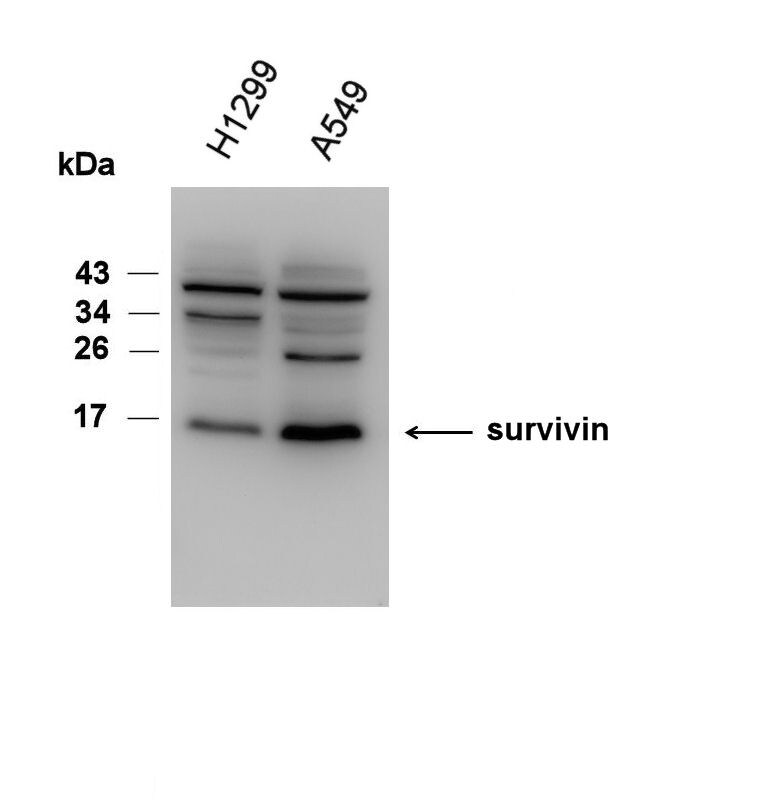
Application: Western BlotSample Tested: H1299 whole cell lysate and A549 whole cell lysateSpecies: HumanVerified Customer | Posted 01/11/2017Whole cell extracts were prepared in RIPA buffer and analyzed using immunoblot analysis. PVDF membrane. Primary antibody 1:1000 at 4°C overnight. Secondary antibody 1:20000 at 4°C for 1 hour.
-
Application: Immunohistochemistry-ParaffinSample Tested: Mouse cervixSpecies: MouseVerified Customer | Posted 05/06/2016
-
Application: Immunohistochemistry-ParaffinSample Tested: lungSpecies: RatVerified Customer | Posted 02/27/2016
-
Application: Western BlotSample Tested:Species: HumanVerified Customer | Posted 04/28/2015Survivin Antibody (NB500-201)
-
Application: Western BlotSample Tested: See PMID 23504997Species: HumanVerified Customer | Posted 12/29/2014
-
Application: Immunohistochemistry-ParaffinSample Tested: See PMID 23161837Species: HumanVerified Customer | Posted 12/29/2014
-
Application: Western BlotSample Tested: See PMID 23449974Species: HumanVerified Customer | Posted 12/23/2014
-
Application: Immunohistochemistry-ParaffinVerified Customer | Posted 11/07/2014Survivin in ovarian cancer tissue
-
Application: ImmunocytochemistrySample Tested:Species: HumanVerified Customer | Posted 06/16/2014IF Confocal analysis of HeLa cells using Survivin antibody (NB500-201, 1:10).
-
Application: ELISASample Tested:Species: HumanVerified Customer | Posted 05/20/2014
-
Application: Western BlotSample Tested:Species: HumanVerified Customer | Posted 03/27/2014Survivin in Pancreatic cancer cell P2.03.
-
Application: Western BlotSample Tested: DEndritic cell lysateSpecies: HumanVerified Customer | Posted 07/19/2013
-
Application: Western BlotSample Tested:Species: HumanVerified Customer | Posted 03/28/2013
-
Application: Western BlotVerified Customer | Posted 08/23/2012Western blot using Survivin antibody
-
Application: Western BlotVerified Customer | Posted 08/21/2012
-
Application: Western BlotSample Tested: Hela whole cell lysate, Sample Amount: 5-15 ulSpecies: HumanVerified Customer | Posted 11/19/2010
-
Application: Western BlotSample Tested: Cancer CellsSpecies: MouseVerified Customer | Posted 06/07/2010
-
Application: Western BlotSample Tested: Human smooth muscle cells, Sample Amount: 30ugSpecies: HumanVerified Customer | Posted 05/04/2010
-
Application: Western BlotSample Tested: mouse cell, Sample Amount: 50ugSpecies: MouseVerified Customer | Posted 08/31/2009
-
Application: Western BlotSample Tested: Oswald 1771 Abrams D17 Cancer Cell Lines, Sample Amount: 7-15 ugSpecies: OtherVerified Customer | Posted 07/02/2009
There are no reviews that match your criteria.
Protocols
View specific protocols for Survivin Antibody - BSA Free (NB500-201):
Survivin Antibody:
Materials
1) 1 Phosphate buffered saline (pH 7.6): NaCl 137mmol/L, KCl 2.7mmol/L, Na2HPO4 4.3mmol/L, KH2PO4 1.4 mmol/L
2) Citrate buffer, 0.01 M, pH6.0, Sodium Citrate 3g, Citric acid 0.4g
3) 3% Hydrogen peroxide
4) Primary antibody
5) Blocking serum (normal serum)
6) Biotinylated secondary antibody
7) DAB staining kit
Methods
1. Dewax and hydration of slides using xylene and EtOH:
Dry slides for 20 min in a 60 C oven
Add Xylene, 2 x 10 min
100%, 95%, 80%, and 70% EtOH, 5 min each EtOH concentration
Rinse in PBS, 5'
2 Antigen retrieval method (only for paraffin slides)
1a. High-pressure antigen retrieval procedure (recommended method)
Place slides in a glass slide holder (ensure that the slide holder is completely filled with slides, slides without sections if necessary, to ensure even heating. The entire slide holder is immersed in 1000 ml of Citrate buffer (0.01M, pH6.0) within a pressure cooker
Once steam is produced, and ONLY when steam is visible, from the pressure cooker (usually 15-20 min), the required high-pressure will have been reached, and slides will be incubated for 2 min.
Turn off heat, and allow buffer and slides to cool to room temperature
Slides are then rinsed in PBS for 5 minutes
2. Add 3% hydrogen peroxide solution, 10'at RT, then PBS, 3X5'
3. Normal blocking serum, 20'at RT
4. Incubate with Primary Ab, 4C overnight or 1.5 hours at 37C
5. Rinse with PBS, 3 X 5' each rinse
6. Add Biotin-conjugated second antibody, 10'at RT
7. Rinse with PBS, 3 X 5' each rinse
8. Add Streptavidin-Peroxidase, 10'at RT
9. Rinse with PBS, 3 X 5' each rinse
10. Staining with DAB solution, 2-5'under microscope
11. Stop the reaction by washing in tap water
12. Counterstain in Haematoxylin for 3-5 minutes
13. 75%, 80%, 95% and 100% ethanol, 5x2', xylene 2 x 10'
Survivin Antibody:
Immunoprecipitation Procedure
1) Lyse cells plated in a 60mm dish:
a) 300 ul CHAPS buffer [50mM Tris-HCl, pH 7.5/50mM NaCl/1mM EDTA/1% NP-40/0.1% CHAPS/1mM NaVO4/1mM PMSF]
b) Rock for 20 minutes at 4 degrees C
2) Harvest lysate and spin down the insoluble material at 14K rpm
3) Collect soluble fraction
4) Pre-clear lysate with 40 ul of 50:50 slurry of Protein A beads, rocking for 1 hour at 4 degrees C
5) Spin down beads at 2K rpm, at 4 degrees C
6) Collect pre-cleared lysate
7) Incubate lysate with 5-7ug of anti-Survivin (NB 500-201) overnight, rocking at 4 degrees C
8) Add 50 ul of Protein A 50:50 slurry for 2 hours, rocking at 4C
9) Wash beads with 200 ul of CHAPS buffer, three times
10) Denature immune complex by adding 2x Sample Buffer, containing 2-ME
11) Boil for 10 minutes and load onto an SDS-gel.
Western Blot Procedure
1) Cells were pelleted, washed in 1XPBS, suspended in ice water (~ 5 x 10(6) cells/ml), and placed on ice
2) Lysates were prepared with the addition of 2X lysis buffer [2% SDS/ 50mM Tris-HCl / 10% glycerol]
3) Lysates were heated to 95 degrees C for 3 minutes and then microfuged at room temperature for 10 minutes
4) 50 ug of lysate were electrophoresed (150 V) through a 4-15% PAGE
5) Proteins were transferred (60 V) onto an Immobilon-P membrane (Millipore Corp.) for 45 minutes
6) The blot was blocked overnight at 4 degrees C in blocking buffer [1XPBS, pH 7 / 5% nonfat milk / 0.1% Tween-20]
7) Washed the blot in 1XPBS / 0.1% Tween-20
8) Incubated the blot with 1 ug/ml of (NB500-201) anti-Survivin antibody, diluted in blocking buffer, for 2 hours at room temperature
9) Washed the blot in 1XPBS / 0.1% Tween-20
10) Reacted the blot with HRP-conjugated donkey anti-rabbit Ig, diluted in 1XPBS / 0.1% Tween-20, for 30 minutes at room temperature
11) Washed the blot in 1XPBS / 0.1% Tween-20
12) Visualized blot by ECL and autoradiography
NOTE: HeLa whole cell extracts (NB800-PC1) were used as a positive control for this antibody.
Find general support by application which include: protocols, troubleshooting, illustrated assays, videos and webinars.
- 7-Amino Actinomycin D (7-AAD) Cell Viability Flow Cytometry Protocol
- Antigen Retrieval Protocol (PIER)
- Antigen Retrieval for Frozen Sections Protocol
- Appropriate Fixation of IHC/ICC Samples
- Cellular Response to Hypoxia Protocols
- ChIP Protocol Video
- Chromatin Immunoprecipitation (ChIP) Protocol
- Chromatin Immunoprecipitation Protocol
- Chromogenic IHC Staining of Formalin-Fixed Paraffin-Embedded (FFPE) Tissue Protocol
- Chromogenic Immunohistochemistry Staining of Frozen Tissue
- ClariTSA™ Fluorophore Kits
- Detection & Visualization of Antibody Binding
- ELISA Sample Preparation & Collection Guide
- ELISA Troubleshooting Guide
- Extracellular Membrane Flow Cytometry Protocol
- Flow Cytometry Protocol for Cell Surface Markers
- Flow Cytometry Protocol for Staining Membrane Associated Proteins
- Flow Cytometry Staining Protocols
- Flow Cytometry Troubleshooting Guide
- Fluorescent IHC Staining of Frozen Tissue Protocol
- Graphic Protocol for Heat-induced Epitope Retrieval
- Graphic Protocol for the Preparation and Fluorescent IHC Staining of Frozen Tissue Sections
- Graphic Protocol for the Preparation and Fluorescent IHC Staining of Paraffin-embedded Tissue Sections
- Graphic Protocol for the Preparation of Gelatin-coated Slides for Histological Tissue Sections
- How to Run an R&D Systems DuoSet ELISA
- How to Run an R&D Systems Quantikine ELISA
- How to Run an R&D Systems Quantikine™ QuicKit™ ELISA
- ICC Cell Smear Protocol for Suspension Cells
- ICC Immunocytochemistry Protocol Videos
- ICC for Adherent Cells
- IHC Sample Preparation (Frozen sections vs Paraffin)
- ISH-IHC Protocol for Chromogenic Detection on Formalin Fixed Paraffin Embedded (FFPE) Tissue
- Immunocytochemistry (ICC) Protocol
- Immunocytochemistry Troubleshooting
- Immunofluorescence of Organoids Embedded in Cultrex Basement Membrane Extract
- Immunofluorescent IHC Staining of Formalin-Fixed Paraffin-Embedded (FFPE) Tissue Protocol
- Immunohistochemistry (IHC) and Immunocytochemistry (ICC) Protocols
- Immunohistochemistry Frozen Troubleshooting
- Immunohistochemistry Paraffin Troubleshooting
- Immunoprecipitation Protocol
- Intracellular Flow Cytometry Protocol Using Alcohol (Methanol)
- Intracellular Flow Cytometry Protocol Using Detergents
- Intracellular Nuclear Staining Flow Cytometry Protocol Using Detergents
- Intracellular Staining Flow Cytometry Protocol Using Alcohol Permeabilization
- Intracellular Staining Flow Cytometry Protocol Using Detergents to Permeabilize Cells
- Preparing Samples for IHC/ICC Experiments
- Preventing Non-Specific Staining (Non-Specific Binding)
- Primary Antibody Selection & Optimization
- Propidium Iodide Cell Viability Flow Cytometry Protocol
- Protocol for Heat-Induced Epitope Retrieval (HIER)
- Protocol for Liperfluo
- Protocol for Making a 4% Formaldehyde Solution in PBS
- Protocol for VisUCyte™ HRP Polymer Detection Reagent
- Protocol for the Characterization of Human Th22 Cells
- Protocol for the Characterization of Human Th9 Cells
- Protocol for the Fluorescent ICC Staining of Cell Smears - Graphic
- Protocol for the Fluorescent ICC Staining of Cultured Cells on Coverslips - Graphic
- Protocol for the Preparation & Fixation of Cells on Coverslips
- Protocol for the Preparation and Chromogenic IHC Staining of Frozen Tissue Sections
- Protocol for the Preparation and Chromogenic IHC Staining of Frozen Tissue Sections - Graphic
- Protocol for the Preparation and Chromogenic IHC Staining of Paraffin-embedded Tissue Sections
- Protocol for the Preparation and Chromogenic IHC Staining of Paraffin-embedded Tissue Sections - Graphic
- Protocol for the Preparation and Fluorescent ICC Staining of Cells on Coverslips
- Protocol for the Preparation and Fluorescent ICC Staining of Non-adherent Cells
- Protocol for the Preparation and Fluorescent ICC Staining of Stem Cells on Coverslips
- Protocol for the Preparation and Fluorescent IHC Staining of Frozen Tissue Sections
- Protocol for the Preparation and Fluorescent IHC Staining of Paraffin-embedded Tissue Sections
- Protocol for the Preparation of Gelatin-coated Slides for Histological Tissue Sections
- Protocol for the Preparation of a Cell Smear for Non-adherent Cell ICC - Graphic
- Protocol: Annexin V and PI Staining by Flow Cytometry
- Protocol: Annexin V and PI Staining for Apoptosis by Flow Cytometry
- Quantikine HS ELISA Kit Assay Principle, Alkaline Phosphatase
- Quantikine HS ELISA Kit Principle, Streptavidin-HRP Polymer
- R&D Systems Quality Control Western Blot Protocol
- Sandwich ELISA (Colorimetric) – Biotin/Streptavidin Detection Protocol
- Sandwich ELISA (Colorimetric) – Direct Detection Protocol
- TUNEL and Active Caspase-3 Detection by IHC/ICC Protocol
- The Importance of IHC/ICC Controls
- Troubleshooting Guide: ELISA
- Troubleshooting Guide: Fluorokine Flow Cytometry Kits
- Troubleshooting Guide: Immunohistochemistry
- Troubleshooting Guide: Western Blot Figures
- Western Blot Conditions
- Western Blot Protocol
- Western Blot Protocol for Cell Lysates
- Western Blot Troubleshooting
- Western Blot Troubleshooting Guide
- View all Protocols, Troubleshooting, Illustrated assays and Webinars
FAQs for Survivin Antibody - BSA Free
Showing
1
-
4 of
4 FAQs
Showing All
-
Q: Can I use this antibody with species other than those listed?
A: The species we have listed are validated and therefore have a 100% guarantee to work with this antibody. We cannot guarantee that this will work with other species.
-
Q: I hope you can help with a quick query: apologies if I have missed this information on the technical data sheet, but where on the protein has the epitope for antibody been raised?
A: Our Survivin antibody NB500-201 was raised against the full length human survivin protein (UniProt# O15392). We unfortunately do not epitope map our antibodies, so the exact epitope recognized by this antibody has not been determined.
-
Q: I would appreciate if you are able to confirm how many of the splice variants this antibody will detect (survivin wild type, survivin isoform 2 and also survivin 2-alpha). Has this been tested? If you have any data to show this, it would be beneficial.
A: NB500-201 was raised against the full-length survivin protein and is a polyclonal antibody, therefore we do not know where or how many sites the antibody detects. We have not tested for reactivity against specific splice variants, but any customer feedback regarding this would be greatly appreciated.
-
Q: We would like to ensure 3 isoforms of Survivin can all be detected before placing the order. The sample is total protein extracted from Mouse heart. May we ask if it is possible? In addition, as we know, there are 7 isoforms of Human Survivin. May we also ask the reason why there is only one band observed by HeLa (as shown on the product page)?
A: This antibody was made to the full length canonical sequence for Survivin but it has not been tested for reactivity with all isoforms of Survivin. It is possible being a polyclonal but we cannot guarantee it will react with a particular isoform.
-
Q: Can I use this antibody with species other than those listed?
A: The species we have listed are validated and therefore have a 100% guarantee to work with this antibody. We cannot guarantee that this will work with other species.
-
Q: I hope you can help with a quick query: apologies if I have missed this information on the technical data sheet, but where on the protein has the epitope for antibody been raised?
A: Our Survivin antibody NB500-201 was raised against the full length human survivin protein (UniProt# O15392). We unfortunately do not epitope map our antibodies, so the exact epitope recognized by this antibody has not been determined.
-
Q: I would appreciate if you are able to confirm how many of the splice variants this antibody will detect (survivin wild type, survivin isoform 2 and also survivin 2-alpha). Has this been tested? If you have any data to show this, it would be beneficial.
A: NB500-201 was raised against the full-length survivin protein and is a polyclonal antibody, therefore we do not know where or how many sites the antibody detects. We have not tested for reactivity against specific splice variants, but any customer feedback regarding this would be greatly appreciated.
-
Q: We would like to ensure 3 isoforms of Survivin can all be detected before placing the order. The sample is total protein extracted from Mouse heart. May we ask if it is possible? In addition, as we know, there are 7 isoforms of Human Survivin. May we also ask the reason why there is only one band observed by HeLa (as shown on the product page)?
A: This antibody was made to the full length canonical sequence for Survivin but it has not been tested for reactivity with all isoforms of Survivin. It is possible being a polyclonal but we cannot guarantee it will react with a particular isoform.
Loading...
Associated Pathways
