TGF-beta 1 (transforming growth factor beta 1) is one of three closely related mammalian members of the large TGF-beta superfamily that share a characteristic cystine knot structure. TGF-beta 1, -2 and -3 are highly pleiotropic cytokines that are proposed to act as cellular switches that regulate processes such as immune function, proliferation and epithelial-mesenchymal transition. Each TGF-beta isoform has some non-redundant functions; for TGF-beta 1, mice with targeted deletion show defects in hematopoiesis and endothelial differentiation, and die of overwhelming inflammation. Human TGF-beta 1 cDNA encodes a 390 amino acid (aa) precursor that contains a 29 aa signal peptide and a 361 aa proprotein. A furin-like convertase processes the proprotein to generate an N-terminal 249 aa latency-associated peptide (LAP) and a C-terminal 112 aa mature TGF- beta 1. Disulfide-linked homodimers of LAP and TGF-beta 1 remain non-covalently associated after secretion, forming the small latent TGF-beta 1 complex. Covalent linkage of LAP to one of three latent TGF-beta binding proteins (LTBPs) creates a large latent complex that may interact with the extracellular matrix. TGF-beta is activated from latency by pathways that include actions of the protease plasmin, matrix metalloproteases, thrombospondin 1 and a subset of integrins. Mature human TGF-beta 1 shares 100% aa identity with pig, dog and cow TGF-beta 1, and 99% aa identity with mouse, rat and horse TGF-beta 1. It demonstrates cross-species activity. TGF-beta 1 signaling begins with high-affinity binding to a type II ser/thr kinase receptor termed TGF-beta RII. This receptor then phosphorylates and activates a second ser/thr kinase receptor, TGF-beta RI (also called activin receptor-like kinase (ALK) -5), or alternatively, ALK‑1. This complex phosphorylates and activates Smad proteins that regulate transcription. Contributions of the accessory receptors betaglycan (also known as TGF-beta RIII) and endoglin, or use of Smad-independent signaling pathways, allow for disparate actions observed in response to TGF-beta in different contexts.
TGF-beta Pan Specific Antibody
R&D Systems | Catalog # AB-100-NA

Key Product Details
Species Reactivity
Validated:
Cited:
Applications
Validated:
Cited:
Label
Antibody Source
Product Specifications
Immunogen
Specificity
Clonality
Host
Isotype
Endotoxin Level
Scientific Data Images for TGF-beta Pan Specific Antibody
TGF-beta Pan Specific in Human Prostate Tissue.
TGF-beta was detected in immersion fixed paraffin-embedded sections of human prostate tissue using Rabbit Anti-TGF-beta Pan Specific Polyclonal Antibody (Catalog # AB-100-NA) at 3 µg/mL for 1 hour at room temperature followed by incubation with the Anti-Rabbit IgG VisUCyte™ HRP Polymer Antibody (Catalog # VC003). Before incubation with the primary antibody, tissue was subjected to heat-induced epitope retrieval using Antigen Retrieval Reagent-Basic (Catalog # CTS013). Tissue was stained using DAB (brown) and counterstained with hematoxylin (blue). Specific staining was localized to cytoplasm in epitjhelial cells. View our protocol for IHC Staining with VisUCyte HRP Polymer Detection Reagents.
TGF‑ beta 1 Inhibition of IL‑4-dependent Cell Proliferation and Neutralization by TGF‑ beta Antibody.
Porcine TGF-beta 1 (Catalog # 101-B1) inhibits Recombinant Mouse IL-4 (Catalog # 404-ML) induced proliferation in the HT-2 mouse T cell line in a dose-dependent manner (orange line). Inhibition of Recombinant Mouse IL-4 (7.5 ng/mL) activity elicited by Porcine TGF-beta 1 (1 ng/mL) is neutralized (green line) by increasing concentrations of TGF-beta Pan Specific Polyclonal Antibody (Catalog # AB-100-NA). The ND50range is 5-30 µg/mL.
Detection of Human TGF-beta by Flow Cytometry
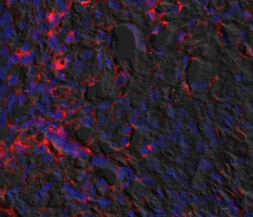
TGF-beta induces IL-9 mRNA expression in Th1 and Th2 cells.Naïve CD4+ T cells were activated with fibroblast-bound anti-CD3/CD28 under classical Th1 and Th2 conditions, with IL-12 and anti-IL-4 or IL-4, anti-IFN-gamma, and anti-IL-12 respectively for 5 days, restimulated at day 5 and split in cultures with and without TGF-beta or anti-TGF-beta for 5 more days of stimulation. (A, C and D) qRT-PCR gene expression analysis of the relative gene expression of IL-9, GATA3, and Tbet at day 10 relative to the Th0 culture. (B) CT value from qRT-PCR gene expression analysis of the gene PU.1. Each donor is represented by a specific symbol and connected with a line. Data are from four independent experiments, each with two donors. *p<0.05, **p<0.01, ***p<0.001. Image collected and cropped by CiteAb from the following open publication (https://dx.plos.org/10.1371/journal.pone.0021695), licensed under a CC-BY license. Not internally tested by R&D Systems.Applications for TGF-beta Pan Specific Antibody
Immunohistochemistry
Sample: Immersion fixed paraffin-embedded sections of human prostate tissue
Western Blot
Sample: Recombinant Human TGF-beta 1 (Catalog # 240-B)
Recombinant Human TGF-beta 2 (Catalog # 302-B2)
Recombinant Human TGF-beta 1.2 (Catalog # 304-B3)
Recombinant Human TGF-beta 3 (Catalog # 243-B3)
Recombinant Amphibian TGF-beta 5 (Catalog # 245-B5)
under non-reducing conditions only
Neutralization
Reviewed Applications
Read 3 reviews rated 4.3 using AB-100-NA in the following applications:
Formulation, Preparation, and Storage
Purification
Reconstitution
Reconstitute at 1 mg/mL in sterile PBS.
Formulation
Shipping
Stability & Storage
- 12 months from date of receipt, -20 to -70 °C as supplied.
- 1 month, 2 to 8 °C under sterile conditions after reconstitution.
- 6 months, -20 to -70 °C under sterile conditions after reconstitution.
Calculators
Background: TGF-beta
Long Name
Alternate Names
Additional TGF-beta Products
Product Documents for TGF-beta Pan Specific Antibody
Certificate of Analysis
To download a Certificate of Analysis, please enter a lot or batch number in the search box below.
Note: Certificate of Analysis not available for kit components.
Product Specific Notices for TGF-beta Pan Specific Antibody
For research use only
Citations for TGF-beta Pan Specific Antibody
 Powered by Bioz
Customer Reviews for TGF-beta Pan Specific Antibody (3)
Have you used TGF-beta Pan Specific Antibody?
Submit a review and receive an Amazon gift card!
$25/€18/£15/$25CAN/¥2500 Yen for a review with an image
$10/€7/£6/$10CAN/¥1110 Yen for a review without an image
Submit a review
Customer Images
-
Application: ImmunohistochemistrySample Tested: mouse oral tissueSpecies: MouseVerified Customer | Posted 11/23/2020

-
Application: Flow CytometrySample Tested: Blood mononuclear cells (PBMCs)Species: HumanVerified Customer | Posted 03/23/2018
-
Application: Block/NeutralizeSample Tested: Peritoneal macrophagesSpecies: MouseVerified Customer | Posted 06/06/2017
There are no reviews that match your criteria.
Protocols
Find general support by application which include: protocols, troubleshooting, illustrated assays, videos and webinars.
- Antigen Retrieval Protocol (PIER)
- Antigen Retrieval for Frozen Sections Protocol
- Appropriate Fixation of IHC/ICC Samples
- Cellular Response to Hypoxia Protocols
- Chromogenic IHC Staining of Formalin-Fixed Paraffin-Embedded (FFPE) Tissue Protocol
- Chromogenic Immunohistochemistry Staining of Frozen Tissue
- ClariTSA™ Fluorophore Kits
- Detection & Visualization of Antibody Binding
- Fluorescent IHC Staining of Frozen Tissue Protocol
- Graphic Protocol for Heat-induced Epitope Retrieval
- Graphic Protocol for the Preparation and Fluorescent IHC Staining of Frozen Tissue Sections
- Graphic Protocol for the Preparation and Fluorescent IHC Staining of Paraffin-embedded Tissue Sections
- Graphic Protocol for the Preparation of Gelatin-coated Slides for Histological Tissue Sections
- IHC Sample Preparation (Frozen sections vs Paraffin)
- Immunofluorescent IHC Staining of Formalin-Fixed Paraffin-Embedded (FFPE) Tissue Protocol
- Immunohistochemistry (IHC) and Immunocytochemistry (ICC) Protocols
- Immunohistochemistry Frozen Troubleshooting
- Immunohistochemistry Paraffin Troubleshooting
- Preparing Samples for IHC/ICC Experiments
- Preventing Non-Specific Staining (Non-Specific Binding)
- Primary Antibody Selection & Optimization
- Protocol for Heat-Induced Epitope Retrieval (HIER)
- Protocol for Making a 4% Formaldehyde Solution in PBS
- Protocol for VisUCyte™ HRP Polymer Detection Reagent
- Protocol for the Preparation & Fixation of Cells on Coverslips
- Protocol for the Preparation and Chromogenic IHC Staining of Frozen Tissue Sections
- Protocol for the Preparation and Chromogenic IHC Staining of Frozen Tissue Sections - Graphic
- Protocol for the Preparation and Chromogenic IHC Staining of Paraffin-embedded Tissue Sections
- Protocol for the Preparation and Chromogenic IHC Staining of Paraffin-embedded Tissue Sections - Graphic
- Protocol for the Preparation and Fluorescent IHC Staining of Frozen Tissue Sections
- Protocol for the Preparation and Fluorescent IHC Staining of Paraffin-embedded Tissue Sections
- Protocol for the Preparation of Gelatin-coated Slides for Histological Tissue Sections
- R&D Systems Quality Control Western Blot Protocol
- TUNEL and Active Caspase-3 Detection by IHC/ICC Protocol
- The Importance of IHC/ICC Controls
- Troubleshooting Guide: Immunohistochemistry
- Troubleshooting Guide: Western Blot Figures
- Western Blot Conditions
- Western Blot Protocol
- Western Blot Protocol for Cell Lysates
- Western Blot Troubleshooting
- Western Blot Troubleshooting Guide
- View all Protocols, Troubleshooting, Illustrated assays and Webinars
FAQs for TGF-beta Pan Specific Antibody
-
Q: Does TGF-beta Pan Specific Antibody, catalog # AB-100-NA, recognize active or latent TGF-beta (LAP)?
A: This antibody recognizes mature (active) TGF-beta. We have not tested for cross-reactivity to LAP (catalog # 246-LP) in our neutralization assay. Some cross-reactivity to latent TGF-beta may also be observed in western blot.
Associated Pathways
