TOR/mTOR [p Ser2448] Antibody - BSA Free
Novus Biologicals | Catalog # NB600-607
Loading...
Key Product Details
Validated by
Biological Validation
Species Reactivity
Validated:
Human, Rat
Cited:
Human, Rat
Applications
Validated:
Immunohistochemistry, Immunohistochemistry-Paraffin, Western Blot, ELISA
Cited:
Immunohistochemistry-Paraffin, Western Blot, IF/IHC
Label
Unconjugated
Antibody Source
Polyclonal Rabbit IgG
Format
BSA Free
Loading...
Product Specifications
Immunogen
TOR/TOR/mTOR [p Ser2448] Antibody was prepared from whole rabbit serum produced by repeated immunizations with a phosphorylated synthetic peptide corresponding a c-terminal region near Serine 2448 of human TOR/mTOR. (Uniprot: P42345)
Reactivity Notes
Reactivity against TOR/mTOR from other species has not been determined, however, reactivity with mouse is suggested based on protein sequence homologies
Modification
p Ser2448
Localization
Cell Membrane and Cytoplasmic
Specificity
Reactivity occurs with phosphorylated mTOR from human derived tissues and cells. Reactivity against mTOR from other species has not been determined, however, reactivity with mouse and rat is suggested based on protein sequence homologies.
Clonality
Polyclonal
Host
Rabbit
Isotype
IgG
Description
This is an affinity purified antibody produced by immunoaffinity chromatography using the immunizing peptide after immobilization to a solid phase
Store vial at -20C prior to opening. Aliquot contents and freeze at -20C or below for extended storage. Avoid cycles of freezing and thawing. Centrifuge product if not completely clear after standing at room temperature. This product is stable for several weeks at 4C as an undiluted liquid. Dilute only prior to immediate use.
Store vial at -20C prior to opening. Aliquot contents and freeze at -20C or below for extended storage. Avoid cycles of freezing and thawing. Centrifuge product if not completely clear after standing at room temperature. This product is stable for several weeks at 4C as an undiluted liquid. Dilute only prior to immediate use.
Scientific Data Images for TOR/mTOR [p Ser2448] Antibody - BSA Free
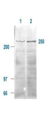
Western Blot: TOR/mTOR [p Ser2448] Antibody [NB600-607] - Affinity Purified TOR/TOR/mTOR [p Ser2448] antibody is shown to detect a 250 kDa band (indicated) corresponding to phosphorylated human TOR/mTOR present in a 293T whole cell lysates. Cells were serum-starved for 24 hours prior to harvest. ~20 ug of lysate was loaded per lane for SDS-PAGE. Untreated cells are shown in lane 1, whereas cells in lane 2 were treated with IGF-1 (100 ng/ml) for 20 min prior to harvest. Follow reaction of antibody with a 1:2000 dilution of HRP Goat-a-Rabbit IgG for visualization.
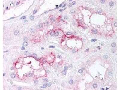
Immunohistochemistry: TOR/mTOR [p Ser2448] Antibody [NB600-607] - affinity purified TOR/TOR/mTOR [p Ser2448] antibody was used at 5 ug/ml to detect signal in a variety of tissues including multi-human, multi-brain and multi-cancer slides. This image shows moderate staining of proximal convoluted tubules of the kidney (40X). Tissue was formalin-fixed and paraffin embedded. The image shows localization of the antibody as the precipitated red signal, with a hematoxylin purple nuclear counterstain.
![Western Blot: TOR/mTOR [p Ser2448] Antibody [NB600-607]](https://resources.rndsystems.com/images/products/TOR-mTOR-[p-Ser2448]-Antibody-Western-Blot-NB600-607-img0006.jpg "Western Blot: TOR/mTOR [p Ser2448] Antibody [NB600-607]")
![Immunohistochemistry-Paraffin: TOR/mTOR [p Ser2448] Antibody [NB600-607]](https://resources.rndsystems.com/images/products/TOR-mTOR-[p-Ser2448]-Antibody-Immunohistochemistry-Paraffin-NB600-607-img0005.jpg "Immunohistochemistry-Paraffin: TOR/mTOR [p Ser2448] Antibody [NB600-607]")
Immunohistochemistry-Paraffin: TOR/mTOR [p Ser2448] Antibody [NB600-607]
Immunohistochemistry-Paraffin: TOR/mTOR [p Ser2448] Antibody [NB600-607] - Brown arrow= IBa1 positive cells, Green arrow= mTOR positive cells, Black arrow= mTOR and IBA1 positive cells. IHC-P Image submitted by a verified customer review.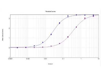
ELISA: TOR/mTOR [p Ser2448] Antibody [NB600-607] - ELISA Results of TOR/TOR/mTOR [p Ser2448] antibody tested against BSA-conjugated non-phospho [purple line] and phospho [blue line] forms of immunizing peptide. Each well was coated in duplicate with either 0.1ug of conjugate. The working dilution is 1:37,000. The starting dilution of antibody was 5ug/ml and the X-axis represents the Log10 of a 3-fold dilution. This titration is a 4-parameter curve fit where the IC50 is defined as the titer of the antibody. Assay performed using HRP conjugate stabilizer, Goat Anti-Rabbit HRP conjugated and TMB substrate.
![TOR/mTOR [p Ser2448] Antibody](https://resources.rndsystems.com/images/products/nb600-607_rabbit-polyclonal-tor-mtor-p-ser2448-antibody-235202318143142.jpg "TOR/mTOR [p Ser2448] Antibody")
TOR/mTOR [p Ser2448] Antibody
affinity purified anti-mTOR pS 2448 antibody was used at 5 ug/ml to detect signal in a variety of tissues including multi-human, multi-brain and multi-cancer slides. This image shows moderate staining of proximal convoluted tubules of the kidney (40X). Tissue was formalin-fixed and paraffin embedded. The image shows localization of the antibody as the precipitated red signal, with a hematoxylin purple nuclear counterstain. Personal Communication, Tina Roush, LifeSpanBiosciences, Seattle, WA.![TOR/mTOR [p Ser2448] Antibody](https://resources.rndsystems.com/images/products/nb600-607_rabbit-polyclonal-tor-mtor-p-ser2448-antibody-31020241535647.jpg "Western Blot: TOR/mTOR [p Ser2448] Antibody [NB600-607] -")
Western Blot: TOR/mTOR [p Ser2448] Antibody [NB600-607] -
Western Blot: TOR/mTOR [p Ser2448] Antibody [NB600-607] - Analysis of mTOR phosphorylation during microglial activation(A) Whole-cell lysates were prepared from microglial cells incubated for 2 h as indicated. Equal amounts of proteins were analyzed by western blot for phosphorylated mTOR kinase (p-mTOR), upper gel. The same blots were subsequently probed for beta -actin, lower gel. (B) Quantitation of densitometry where p-mTOR values are reflected relative to those for beta -actin. Data are expressed as means±S.E.M. of n=1 replicate for each group, each assayed in triplicates. Representative of two different experiments. Data were analyzed by one-way ANOVA followed by the Bonferroni's post hoc test. ***P<0.001 versus Control, °P<0.05 versus C-CM. Image collected & cropped by CiteAb from the following publication (https://pubmed.ncbi.nlm.nih.gov/24689533), licensed under a CC-BY license. Not internally tested by Novus Biologicals.![TOR/mTOR [p Ser2448] Antibody](https://resources.rndsystems.com/images/products/nb600-607_rabbit-polyclonal-tor-mtor-p-ser2448-antibody-310202416171492.jpg "Western Blot: TOR/mTOR [p Ser2448] Antibody [NB600-607] -")
Western Blot: TOR/mTOR [p Ser2448] Antibody [NB600-607] -
Western Blot: TOR/mTOR [p Ser2448] Antibody [NB600-607] - Analysis of mammalian target of rapamycin (mTOR) phosphorylation during microglial activation. Whole-cell lysates were prepared from microglial cells activated by control-conditioned medium (C-CM) (A) or CM from glioma cells activated with lipopolysaccharide (LPS)/IFN gamma (LI-CM) (B). Equal amounts of proteins were analyzed by western blot for phosphorylated mTOR kinase (p-mTOR), upper gel & were subsequently probed for beta -actin, lower gel. Quantitation of densitometry where p-mTOR values are reflected relative to those for beta -actin. Data are expressed as means ± standard error of the mean of n = 1 replicate for each group, each assayed in triplicate: representative of two different experiments. Data were analyzed by one-way analysis of variance followed by Bonferroni post hoc test. ***P <0.001 versus control. Image collected & cropped by CiteAb from the following publication (https://pubmed.ncbi.nlm.nih.gov/25051975), licensed under a CC-BY license. Not internally tested by Novus Biologicals.Applications for TOR/mTOR [p Ser2448] Antibody - BSA Free
Application
Recommended Usage
ELISA
1:10000-1:100000
Immunohistochemistry
5.0 ug/ml
Immunohistochemistry-Paraffin
5 ug/ml
Western Blot
1:500-1:2000
Application Notes
This affinity purified antibody has been tested for use in immunohistochemistry, ELISA and western blotting. Western blotting shows reactivity specific for phospho mTOR detecting a band at approximately 250 kDa. Reactivity in other immunoassays is unknown.
Reviewed Applications
Read 2 reviews rated 4 using NB600-607 in the following applications:
Formulation, Preparation, and Storage
Purification
Immunogen affinity purified
Formulation
0.02 M Potassium Phosphate, 0.15 M Sodium Chloride, pH 7.2
Format
BSA Free
Preservative
0.01% Sodium Azide
Concentration
Please see the vial label for concentration. If unlisted please contact technical services.
Shipping
The product is shipped with polar packs. Upon receipt, store it immediately at the temperature recommended below.
Stability & Storage
Store at -20C short term. Aliquot and store at -80C long term. Avoid freeze-thaw cycles.
Background: TOR
Long Name
Mammalian Target of Rapamycin
Alternate Names
FRAP1, FRAP2, MTOR, RAFT1
Entrez Gene IDs
2475 (Human)
Gene Symbol
MTOR
UniProt
Additional TOR Products
Product Documents for TOR/mTOR [p Ser2448] Antibody - BSA Free
Certificate of Analysis
To download a Certificate of Analysis, please enter a lot or batch number in the search box below.
Product Specific Notices for TOR/mTOR [p Ser2448] Antibody - BSA Free
This product is for research use only and is not approved for use in humans or in clinical diagnosis. Primary Antibodies are guaranteed for 1 year from date of receipt.
Related Research Areas
Citations for TOR/mTOR [p Ser2448] Antibody - BSA Free
 Powered by Bioz
Customer Reviews for TOR/mTOR [p Ser2448] Antibody - BSA Free (2)
4 out of 5
2 Customer Ratings
Have you used TOR/mTOR [p Ser2448] Antibody - BSA Free?
Submit a review and receive an Amazon gift card!
$25/€18/£15/$25CAN/¥2500 Yen for a review with an image
$10/€7/£6/$10CAN/¥1110 Yen for a review without an image
Submit a review
Customer Images
![TOR/mTOR [p Ser2448] Antibody - BSA Free NB600-607](https://resources.rndsystems.com/images/reviews/review_nb600-607_50951_0_0.jpg)
![TOR/mTOR [p Ser2448] Antibody - BSA Free NB600-607](https://resources.rndsystems.com/images/reviews/review_nb600-607_50946_0_0.jpg)
Showing
1
-
2 of
2 reviews
Showing All
Filter By:
-
Application: Western BlotSample Tested: Human microglia cell lysateSpecies: HumanVerified Customer | Posted 01/17/2020Chiavari M, Ciotti GMP, Navarra P, Lisi L. Pro-Inflammatory Activation of A New Immortalized Human Microglia Cell Line. Brain Sci. 2019;9(5):111. Published 2019 May 15. doi:10.3390/brainsci9050111
![TOR/mTOR [p Ser2448] Antibody - BSA Free NB600-607](data:image/png;base64,R0lGODlhAQABAAD/ACwAAAAAAQABAAACADs=)
-
Application: Immunohistochemistry-ParaffinSample Tested: Human Brain TissueSpecies: HumanVerified Customer | Posted 01/17/2020Brown arrow= IBa1 positive cells Green arrow= mTOR positive cells Black arrow= mTOR and IBA1 positive cells
There are no reviews that match your criteria.
Protocols
Find general support by application which include: protocols, troubleshooting, illustrated assays, videos and webinars.
- Antigen Retrieval Protocol (PIER)
- Antigen Retrieval for Frozen Sections Protocol
- Appropriate Fixation of IHC/ICC Samples
- Cellular Response to Hypoxia Protocols
- Chromogenic IHC Staining of Formalin-Fixed Paraffin-Embedded (FFPE) Tissue Protocol
- Chromogenic Immunohistochemistry Staining of Frozen Tissue
- ClariTSA™ Fluorophore Kits
- Detection & Visualization of Antibody Binding
- ELISA Sample Preparation & Collection Guide
- ELISA Troubleshooting Guide
- Fluorescent IHC Staining of Frozen Tissue Protocol
- Graphic Protocol for Heat-induced Epitope Retrieval
- Graphic Protocol for the Preparation and Fluorescent IHC Staining of Frozen Tissue Sections
- Graphic Protocol for the Preparation and Fluorescent IHC Staining of Paraffin-embedded Tissue Sections
- Graphic Protocol for the Preparation of Gelatin-coated Slides for Histological Tissue Sections
- How to Run an R&D Systems DuoSet ELISA
- How to Run an R&D Systems Quantikine ELISA
- How to Run an R&D Systems Quantikine™ QuicKit™ ELISA
- IHC Sample Preparation (Frozen sections vs Paraffin)
- Immunofluorescent IHC Staining of Formalin-Fixed Paraffin-Embedded (FFPE) Tissue Protocol
- Immunohistochemistry (IHC) and Immunocytochemistry (ICC) Protocols
- Immunohistochemistry Frozen Troubleshooting
- Immunohistochemistry Paraffin Troubleshooting
- Preparing Samples for IHC/ICC Experiments
- Preventing Non-Specific Staining (Non-Specific Binding)
- Primary Antibody Selection & Optimization
- Protocol for Heat-Induced Epitope Retrieval (HIER)
- Protocol for Making a 4% Formaldehyde Solution in PBS
- Protocol for VisUCyte™ HRP Polymer Detection Reagent
- Protocol for the Preparation & Fixation of Cells on Coverslips
- Protocol for the Preparation and Chromogenic IHC Staining of Frozen Tissue Sections
- Protocol for the Preparation and Chromogenic IHC Staining of Frozen Tissue Sections - Graphic
- Protocol for the Preparation and Chromogenic IHC Staining of Paraffin-embedded Tissue Sections
- Protocol for the Preparation and Chromogenic IHC Staining of Paraffin-embedded Tissue Sections - Graphic
- Protocol for the Preparation and Fluorescent IHC Staining of Frozen Tissue Sections
- Protocol for the Preparation and Fluorescent IHC Staining of Paraffin-embedded Tissue Sections
- Protocol for the Preparation of Gelatin-coated Slides for Histological Tissue Sections
- Quantikine HS ELISA Kit Assay Principle, Alkaline Phosphatase
- Quantikine HS ELISA Kit Principle, Streptavidin-HRP Polymer
- R&D Systems Quality Control Western Blot Protocol
- Sandwich ELISA (Colorimetric) – Biotin/Streptavidin Detection Protocol
- Sandwich ELISA (Colorimetric) – Direct Detection Protocol
- TUNEL and Active Caspase-3 Detection by IHC/ICC Protocol
- The Importance of IHC/ICC Controls
- Troubleshooting Guide: ELISA
- Troubleshooting Guide: Immunohistochemistry
- Troubleshooting Guide: Western Blot Figures
- Western Blot Conditions
- Western Blot Protocol
- Western Blot Protocol for Cell Lysates
- Western Blot Troubleshooting
- Western Blot Troubleshooting Guide
- View all Protocols, Troubleshooting, Illustrated assays and Webinars


