TRAILR2/TNFRSF10B Antibody - BSA Free
Novus Biologicals | Catalog # NB100-56618
![Western Blot: TRAILR2/TNFRSF10B Antibody [NB100-56618]](https://resources.rndsystems.com/images/products/TRAIL-R2-TNFRSF10B-Antibody-Western-Blot-NB100-56618-img0005.jpg "Western Blot: TRAILR2/TNFRSF10B Antibody [NB100-56618]")
Key Product Details
Validated by
Biological Validation
Species Reactivity
Validated:
Human, Mouse
Cited:
Human, Mouse
Applications
Validated:
Immunohistochemistry, Immunohistochemistry-Paraffin, Western Blot, Flow Cytometry, Flow (Cell Surface), Immunocytochemistry/ Immunofluorescence
Cited:
Immunohistochemistry-Paraffin, Western Blot, Flow Cytometry, Flow (Cell Surface), Immunocytochemistry/ Immunofluorescence, IF/IHC
Label
Unconjugated
Antibody Source
Polyclonal Rabbit IgG
Format
BSA Free
Loading...
Product Specifications
Immunogen
Rabbit anti-DR5 polyclonal antibody was raised against a peptide corresponding to amino acids 388 to 407 (isoform 2) of human DR5 precursor (1,2). The same sequence is found in isoform 1 at amino acids 417-436.
Reactivity Notes
Mouse reactivity reported in scientific literature (PMID: 21474767).
Clonality
Polyclonal
Host
Rabbit
Isotype
IgG
Scientific Data Images for TRAILR2/TNFRSF10B Antibody - BSA Free
Western Blot: TRAILR2/TNFRSF10B Antibody [NB100-56618]
Western Blot: TRAILR2/TNFRSF10B Antibody [NB100-56618] - TRAIL R2/TNFRSF10B Antibody [NB100-56618] - Total protein from HL-60, HepG2, HCT-116 and human placenta was separated on a 12% gel by SDS-PAGE, transferred to PVDF membrane and blocked in 5% non-fat milk in TBST. The membrane was probed with 2.0 ug/ml anti-TRAIL R2 in 1% non-fat milk in TBST and detected with an anti-rabbit HRP secondary antibody using chemiluminescence.![Immunohistochemistry-Paraffin: TRAILR2/TNFRSF10B Antibody [NB100-56618]](https://resources.rndsystems.com/images/products/TRAIL-R2-TNFRSF10B-Antibody-Immunohistochemistry-Paraffin-NB100-56618-img0007.jpg "Immunohistochemistry-Paraffin: TRAILR2/TNFRSF10B Antibody [NB100-56618]")
Immunohistochemistry-Paraffin: TRAILR2/TNFRSF10B Antibody [NB100-56618]
Immunohistochemistry-Paraffin: TRAILR2/TNFRSF10B Antibody [NB100-56618] - TRAIL R2/TNFRSF10B Antibody [NB100-56618] - Analysis of a formalin fixed paraffin-embedded (FFPE) human brain cerebellum using 1:200 conc. of TRAIL R2/TNFRSF10B antibody on a Bond Rx autostainer (Leica Biosystems). The assay involved 30 minutes of heat induced antigen retrieval (HIER) using 10mM sodium citrate buffer (pH 9.0) and endogenous peroxidase quenching with peroxide block. The sections were incubated with primary antibody for 15 minutes and Bond Polymer Refine Detection (Leica Biosystems) with DAB was used for signal development followed by counterstaining with hematoxylin. Cytoplasmic staining was observed in the Purkinje cell layer.![Immunohistochemistry: TRAILR2/TNFRSF10B Antibody [NB100-56618]](https://resources.rndsystems.com/images/products/TRAILR2-TNFRSF10B-Antibody-Immunohistochemistry-NB100-56618-img0008.jpg "Immunohistochemistry: TRAILR2/TNFRSF10B Antibody [NB100-56618]")
![Immunocytochemistry/ Immunofluorescence: TRAILR2/TNFRSF10B Antibody [NB100-56618]](https://resources.rndsystems.com/images/products/TRAIL-R2-TNFRSF10B-Antibody-Immunocytochemistry-Immunofluorescence-NB100-56618-img0004.jpg "Immunocytochemistry/ Immunofluorescence: TRAILR2/TNFRSF10B Antibody [NB100-56618]")
Immunocytochemistry/ Immunofluorescence: TRAILR2/TNFRSF10B Antibody [NB100-56618]
Immunocytochemistry/Immunofluorescence: TRAIL R2/TNFRSF10B Antibody [NB100-56618] - HepG2 cells were fixed for 10 minutes using 10% formalin and then permeabilized for 5 minutes using 1X TBS + 0.5% Triton-X100. The cells were incubated with anti-TRAIL R2 (H76) at 5ug/ml overnight at 4C and detected with an anti-rabbit Dylight 488 (Green) at a 1:500 dilution. Alpha tubulin (DM1A) NB100-690 was used as a co-stain at a 1:1000 dilution and detected with an anti-mouse Dylight 550 (Red) at a 1:500 dilution. Nuclei were counterstained with DAPI (Blue). Cells were imaged using a 40X objective.![Western Blot: TRAILR2/TNFRSF10B Antibody [NB100-56618]](https://resources.rndsystems.com/images/products/TRAIL-R2-TNFRSF10B-Antibody-Western-Blot-NB100-56618-img0003.jpg "Western Blot: TRAILR2/TNFRSF10B Antibody [NB100-56618]")
Western Blot: TRAILR2/TNFRSF10B Antibody [NB100-56618]
Western Blot: TRAILR2/TNFRSF10B Antibody [NB100-56618] - TRAIL R2/TNFRSF10B Antibody [NB100-56618] - Analysis using the Azide Free version of NB100-56618. Detection of 20 ug of whole cell lysates from HL60 cells with anti-TRAIL-R2 at 5 ug/ml.Applications for TRAILR2/TNFRSF10B Antibody - BSA Free
Application
Recommended Usage
Flow (Cell Surface)
reported in scientific literature (KOYAMA (2003); PMID 16426839)
Flow Cytometry
1:20-1:2000
Immunocytochemistry/ Immunofluorescence
1:10-1:500. Use reported in scientific literature (Georgakis et al (2005))
Immunohistochemistry
1:200. Use reported in scientific literature (PMID 27740879)
Immunohistochemistry-Paraffin
reported in scientific literature (Younes et al (2006))
Western Blot
5 ug/ml
Reviewed Applications
Read 1 review rated 5 using NB100-56618 in the following applications:
Flow Cytometry Panel Builder
Bio-Techne Knows Flow Cytometry
Save time and reduce costly mistakes by quickly finding compatible reagents using the Panel Builder Tool.
Advanced Features
- Spectra Viewer - Custom analysis of spectra from multiple fluorochromes
- Spillover Popups - Visualize the spectra of individual fluorochromes
- Antigen Density Selector - Match fluorochrome brightness with antigen density

Formulation, Preparation, and Storage
Purification
Protein G purified
Formulation
PBS
Format
BSA Free
Preservative
0.05% Sodium Azide
Concentration
1.0 mg/ml
Shipping
The product is shipped with polar packs. Upon receipt, store it immediately at the temperature recommended below.
Stability & Storage
Store at 4C short term. Aliquot and store at -20C long term. Avoid freeze-thaw cycles.
Background: TRAIL R2/TNFRSF10B
Long Name
TRAIL Receptor 2
Alternate Names
CD262, DR5, TNFRSF10B, TRAILR2
Entrez Gene IDs
8795 (Human)
Gene Symbol
TNFRSF10B
Additional TRAIL R2/TNFRSF10B Products
Product Documents for TRAILR2/TNFRSF10B Antibody - BSA Free
Certificate of Analysis
To download a Certificate of Analysis, please enter a lot or batch number in the search box below.
Product Specific Notices for TRAILR2/TNFRSF10B Antibody - BSA Free
This product is for research use only and is not approved for use in humans or in clinical diagnosis. Primary Antibodies are guaranteed for 1 year from date of receipt.
Citations for TRAILR2/TNFRSF10B Antibody - BSA Free
 Powered by Bioz
Customer Reviews for TRAILR2/TNFRSF10B Antibody - BSA Free (1)
5 out of 5
1 Customer Rating
Have you used TRAILR2/TNFRSF10B Antibody - BSA Free?
Submit a review and receive an Amazon gift card!
$25/€18/£15/$25CAN/¥2500 Yen for a review with an image
$10/€7/£6/$10CAN/¥1110 Yen for a review without an image
Submit a review
Customer Images
Showing
1
-
1 of
1 review
Showing All
Filter By:
-
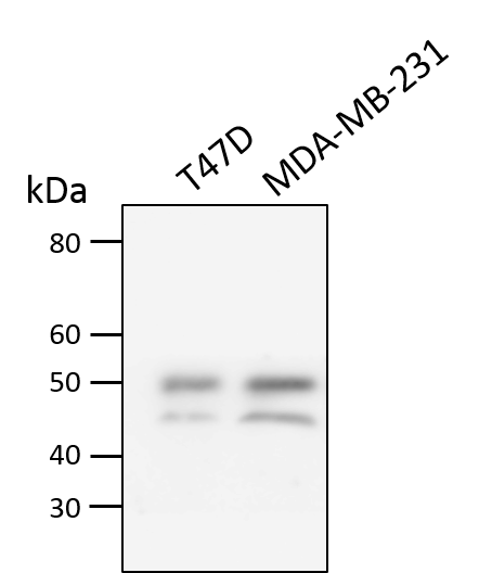
Application: Western BlotSample Tested: Breast cancer cellsSpecies: HumanVerified Customer | Posted 07/17/2018Total cell lysates from T47D and MDA-MB-231 cells were subjected to western blot. PVDF membrane were probed with 2.5 mm/ml Human TRAIL R2 (NB100-56618). A specific band was detected for TRAIL R2 at approximately 48~50 kDa. This experiment was conducted under reducing conditions

There are no reviews that match your criteria.
Protocols
Find general support by application which include: protocols, troubleshooting, illustrated assays, videos and webinars.
- 7-Amino Actinomycin D (7-AAD) Cell Viability Flow Cytometry Protocol
- Antigen Retrieval Protocol (PIER)
- Antigen Retrieval for Frozen Sections Protocol
- Appropriate Fixation of IHC/ICC Samples
- Cellular Response to Hypoxia Protocols
- Chromogenic IHC Staining of Formalin-Fixed Paraffin-Embedded (FFPE) Tissue Protocol
- Chromogenic Immunohistochemistry Staining of Frozen Tissue
- ClariTSA™ Fluorophore Kits
- Detection & Visualization of Antibody Binding
- Extracellular Membrane Flow Cytometry Protocol
- Flow Cytometry Protocol for Cell Surface Markers
- Flow Cytometry Protocol for Staining Membrane Associated Proteins
- Flow Cytometry Staining Protocols
- Flow Cytometry Troubleshooting Guide
- Fluorescent IHC Staining of Frozen Tissue Protocol
- Graphic Protocol for Heat-induced Epitope Retrieval
- Graphic Protocol for the Preparation and Fluorescent IHC Staining of Frozen Tissue Sections
- Graphic Protocol for the Preparation and Fluorescent IHC Staining of Paraffin-embedded Tissue Sections
- Graphic Protocol for the Preparation of Gelatin-coated Slides for Histological Tissue Sections
- ICC Cell Smear Protocol for Suspension Cells
- ICC Immunocytochemistry Protocol Videos
- ICC for Adherent Cells
- IHC Sample Preparation (Frozen sections vs Paraffin)
- Immunocytochemistry (ICC) Protocol
- Immunocytochemistry Troubleshooting
- Immunofluorescence of Organoids Embedded in Cultrex Basement Membrane Extract
- Immunofluorescent IHC Staining of Formalin-Fixed Paraffin-Embedded (FFPE) Tissue Protocol
- Immunohistochemistry (IHC) and Immunocytochemistry (ICC) Protocols
- Immunohistochemistry Frozen Troubleshooting
- Immunohistochemistry Paraffin Troubleshooting
- Intracellular Flow Cytometry Protocol Using Alcohol (Methanol)
- Intracellular Flow Cytometry Protocol Using Detergents
- Intracellular Nuclear Staining Flow Cytometry Protocol Using Detergents
- Intracellular Staining Flow Cytometry Protocol Using Alcohol Permeabilization
- Intracellular Staining Flow Cytometry Protocol Using Detergents to Permeabilize Cells
- Preparing Samples for IHC/ICC Experiments
- Preventing Non-Specific Staining (Non-Specific Binding)
- Primary Antibody Selection & Optimization
- Propidium Iodide Cell Viability Flow Cytometry Protocol
- Protocol for Heat-Induced Epitope Retrieval (HIER)
- Protocol for Liperfluo
- Protocol for Making a 4% Formaldehyde Solution in PBS
- Protocol for VisUCyte™ HRP Polymer Detection Reagent
- Protocol for the Characterization of Human Th22 Cells
- Protocol for the Characterization of Human Th9 Cells
- Protocol for the Fluorescent ICC Staining of Cell Smears - Graphic
- Protocol for the Fluorescent ICC Staining of Cultured Cells on Coverslips - Graphic
- Protocol for the Preparation & Fixation of Cells on Coverslips
- Protocol for the Preparation and Chromogenic IHC Staining of Frozen Tissue Sections
- Protocol for the Preparation and Chromogenic IHC Staining of Frozen Tissue Sections - Graphic
- Protocol for the Preparation and Chromogenic IHC Staining of Paraffin-embedded Tissue Sections
- Protocol for the Preparation and Chromogenic IHC Staining of Paraffin-embedded Tissue Sections - Graphic
- Protocol for the Preparation and Fluorescent ICC Staining of Cells on Coverslips
- Protocol for the Preparation and Fluorescent ICC Staining of Non-adherent Cells
- Protocol for the Preparation and Fluorescent ICC Staining of Stem Cells on Coverslips
- Protocol for the Preparation and Fluorescent IHC Staining of Frozen Tissue Sections
- Protocol for the Preparation and Fluorescent IHC Staining of Paraffin-embedded Tissue Sections
- Protocol for the Preparation of Gelatin-coated Slides for Histological Tissue Sections
- Protocol for the Preparation of a Cell Smear for Non-adherent Cell ICC - Graphic
- Protocol: Annexin V and PI Staining by Flow Cytometry
- Protocol: Annexin V and PI Staining for Apoptosis by Flow Cytometry
- R&D Systems Quality Control Western Blot Protocol
- TUNEL and Active Caspase-3 Detection by IHC/ICC Protocol
- The Importance of IHC/ICC Controls
- Troubleshooting Guide: Fluorokine Flow Cytometry Kits
- Troubleshooting Guide: Immunohistochemistry
- Troubleshooting Guide: Western Blot Figures
- Western Blot Conditions
- Western Blot Protocol
- Western Blot Protocol for Cell Lysates
- Western Blot Troubleshooting
- Western Blot Troubleshooting Guide
- View all Protocols, Troubleshooting, Illustrated assays and Webinars
Loading...
Associated Pathways

