UCH-L1/PGP9.5 Antibody (31A3)
Novus Biologicals | Catalog # NB600-1160
Key Product Details
Validated by
Biological Validation
Species Reactivity
Validated:
Human, Mouse, Rat, Porcine, Bovine, Canine, Zebrafish
Cited:
Human, Mouse, Rat, Fish - Danio rerio (Zebrafish)
Applications
Validated:
Immunohistochemistry, Immunohistochemistry-Paraffin, Immunohistochemistry-Frozen, Western Blot, Flow Cytometry, Immunocytochemistry/ Immunofluorescence, Simple Western
Cited:
Immunohistochemistry-Paraffin, Immunohistochemistry-Frozen, Western Blot, Immunocytochemistry/ Immunofluorescence, IF/IHC
Label
Unconjugated
Antibody Source
Monoclonal Mouse IgG1 kappa Clone # 31A3
Loading...
Product Specifications
Immunogen
UCH-L1/PGP9.5 protein from brain (Uniprot: P09936)
Reactivity Notes
Zebrafish reactivity reported in scientific literature (PMID: 30377377).
Localization
Cytoplasmic, Endoplasmic Reticulum membrane
Marker
pan-Neuronal Marker
Clonality
Monoclonal
Host
Mouse
Isotype
IgG1 kappa
Description
200ug/ml of antibody purified from Bioreactor Concentrate by Protein A or G. Prepared in 10 mM PBS with 0.05% BSA & 0.05% azide. Also available WITHOUT BSA & azide at 1.0 mg/ml. (NBP2-33130)
Antibody with azide - store at 2 to 8C. Antibody without azide - store at -20 to -80C.
Antibody with azide - store at 2 to 8C. Antibody without azide - store at -20 to -80C.
Scientific Data Images for UCH-L1/PGP9.5 Antibody (31A3)
![Western Blot: UCH-L1/PGP9.5 Antibody (31A3) [NB600-1160]](https://resources.rndsystems.com/images/products/UCH-L1-PGP9-5-Antibody-31A3-Western-Blot-NB600-1160-img0013.jpg "Western Blot: UCH-L1/PGP9.5 Antibody (31A3) [NB600-1160]")
Western Blot: UCH-L1/PGP9.5 Antibody (31A3) [NB600-1160]
Western Blot: UCH-L1/PGP9.5 Antibody (31A3) [NB600-1160] - Western Blot Analysis of human brain tissue lysate using UCH-L1/PGP9.5 Antibody (31A3)![Immunocytochemistry/ Immunofluorescence: UCH-L1/PGP9.5 Antibody (31A3) [NB600-1160]](https://resources.rndsystems.com/images/products/UCH-L1-PGP9-5-Antibody-31A3-Immunocytochemistry-Immunofluorescence-NB600-1160-img0012.jpg "Immunocytochemistry/ Immunofluorescence: UCH-L1/PGP9.5 Antibody (31A3) [NB600-1160]")
Immunocytochemistry/ Immunofluorescence: UCH-L1/PGP9.5 Antibody (31A3) [NB600-1160]
Immunocytochemistry/Immunofluorescence: UCH-L1/PGP9.5 Antibody (31A3) [NB600-1160] - Immunofluorescence Analysis of T98G cells labeling Pgp9.5 with UCH-L1/PGP9.5 Antibody (31A3) followed by Goat anti-Mouse IgG-CF488 (Green). The nuclear counterstain is Nucspot (Red)![Immunohistochemistry-Paraffin: UCH-L1/PGP9.5 Antibody (31A3) [NB600-1160]](https://resources.rndsystems.com/images/products/UCH-L1-PGP9-5-Antibody-31A3-Immunohistochemistry-Paraffin-NB600-1160-img0004.jpg "Immunohistochemistry-Paraffin: UCH-L1/PGP9.5 Antibody (31A3) [NB600-1160]")
Immunohistochemistry-Paraffin: UCH-L1/PGP9.5 Antibody (31A3) [NB600-1160]
Immunohistochemistry-Paraffin: UCH-L1/PGP9.5 Antibody (31A3) [NB600-1160] - Formalin-fixed, paraffin-embedded human brain stained with UchL1 antibody (PGP9.5) (1:500), peroxidase-conjugate and DAB chromogen. Staining seen in cytoplasm, ER and membrane. Fixation in 95% ethanol/5% acetic acid for 2-3 hours prior to paraffin embedding is recommended. Specimens which have not been fixed in acetic acid/alcohol require pre-treatment with citrate buffer: for example, unmask of the target epitope by boiling the tissue sections in 10mM citrate buffer, pH 6.0, for 10 min followed by cooling at room temp for 20 min.![Flow Cytometry: UCH-L1/PGP9.5 Antibody (31A3) [NB600-1160]](https://resources.rndsystems.com/images/products/UCH-L1-PGP9-5-Antibody-31A3-Flow-Cytometry-NB600-1160-img0011.jpg "Flow Cytometry: UCH-L1/PGP9.5 Antibody (31A3) [NB600-1160]")
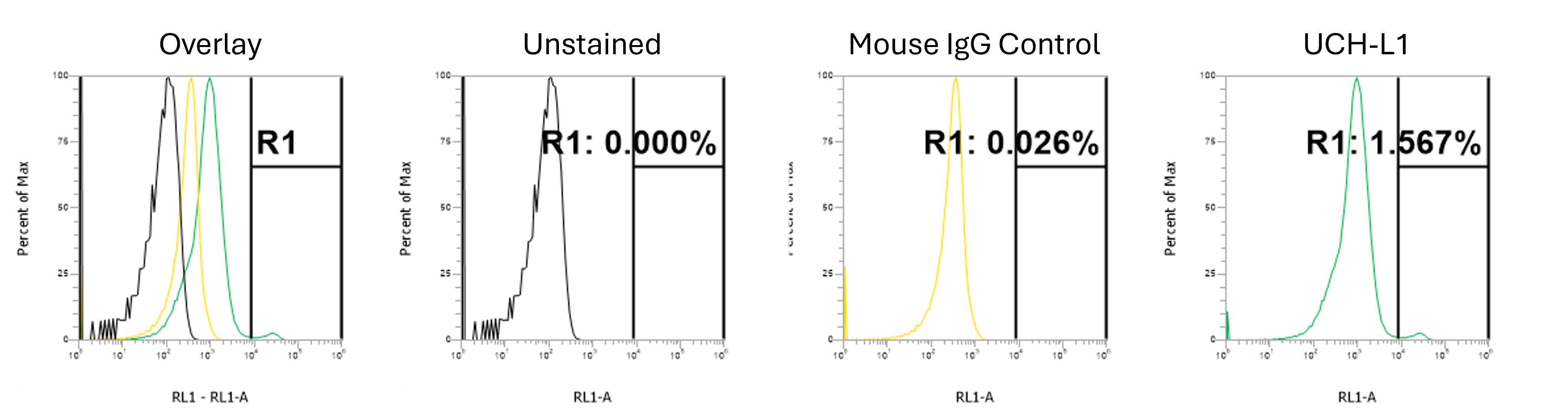
Flow Cytometry: UCH-L1/PGP9.5 Antibody (31A3) [NB600-1160]
Flow Cytometry: UCH-L1/PGP9.5 Antibody (31A3) [NB600-1160] - Flow Cytometric Analysis of T98G cells using UCH-L1/PGP9.5 Antibody (31A3)followed by Goat anti-Mouse IgG-CF488 (Blue); Isotype Control (Red).![Western Blot: UCH-L1/PGP9.5 Antibody (31A3) [NB600-1160]](https://resources.rndsystems.com/images/products/UCH-L1-PGP9-5-Antibody-31A3-Western-Blot-NB600-1160-img0003.jpg "Western Blot: UCH-L1/PGP9.5 Antibody (31A3) [NB600-1160]")
Western Blot: UCH-L1/PGP9.5 Antibody (31A3) [NB600-1160]
Western Blot: UCH-L1/PGP9.5 Antibody (31A3) [NB600-1160] - analysis of UchL1 in 1) human, 2) mouse and 3) rat brain lysate using UchL1 antibody at 1 ug/ml. goat anti-mouse Ig HRP secondary antibody and ECL substrate solution were used for this test.![Immunocytochemistry/ Immunofluorescence: UCH-L1/PGP9.5 Antibody (31A3) [NB600-1160]](https://resources.rndsystems.com/images/products/UCH-L1-PGP9-5-Antibody-31A3-Immunocytochemistry-Immunofluorescence-NB600-1160-img0005.jpg "Immunocytochemistry/ Immunofluorescence: UCH-L1/PGP9.5 Antibody (31A3) [NB600-1160]")
Immunocytochemistry/ Immunofluorescence: UCH-L1/PGP9.5 Antibody (31A3) [NB600-1160]
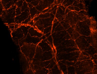
Immunocytochemistry/Immunofluorescence: UCH-L1/PGP9.5 Antibody (31A3) [NB600-1160] - analysis of PGP9.5 in rat mesenteric artery using anti-PGP9.5 antibody. Image from verified customer review.![Simple Western: UCH-L1/PGP9.5 Antibody (31A3) [NB600-1160]](https://resources.rndsystems.com/images/products/UCH-L1-PGP9-5-Antibody-31A3-Simple-Western-NB600-1160-img0006.jpg "Simple Western: UCH-L1/PGP9.5 Antibody (31A3) [NB600-1160]")
Simple Western: UCH-L1/PGP9.5 Antibody (31A3) [NB600-1160]
Simple Western: UCH-L1/PGP9.5 Antibody (31A3) [NB600-1160] - Simple Western lane view shows a specific band for PGP9.5 / UCHL-1 in 0.2 mg/ml of h. Cerebellum (left) and IMR-32 (right) lysate(s). This experiment was performed under reducing conditions using the 12-230 kDa separation system.![Simple Western: UCH-L1/PGP9.5 Antibody (31A3) [NB600-1160]](https://resources.rndsystems.com/images/products/UCH-L1-PGP9-5-Antibody-31A3-Simple-Western-NB600-1160-img0010.jpg "Simple Western: UCH-L1/PGP9.5 Antibody (31A3) [NB600-1160]")
Simple Western: UCH-L1/PGP9.5 Antibody (31A3) [NB600-1160]
Simple Western: UCH-L1/PGP9.5 Antibody (31A3) [NB600-1160] - Electropherogram images of the corresponding Simple Western lane. PGP9.5 / UCHL-1 antibody was used at 10 ug/ml dilution of h. Cerebellum and IMR-32 lysates(s) respectively. [NB600-1160]")
Immunohistochemistry-Paraffin: Mouse Monoclonal UCH-L1/PGP9.5 Antibody (31A3) [NB600-1160]
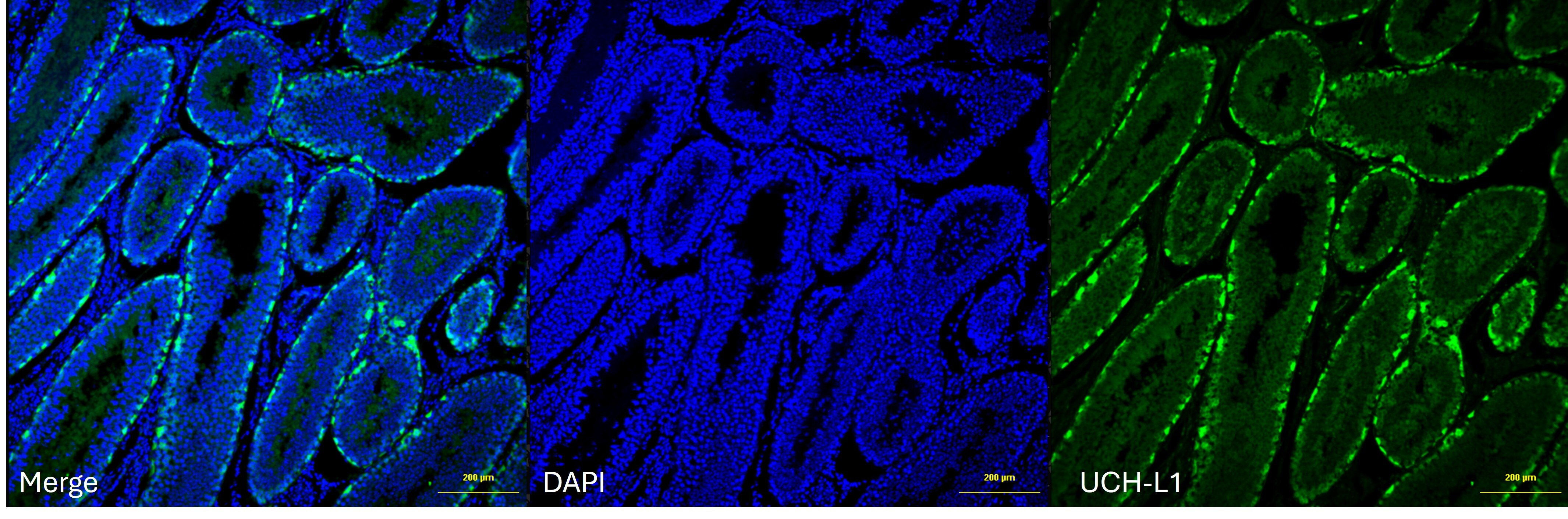
UCH-L1/PGP9.5 Antibody on bovine adult testis at 1:100. Image from a verified customer review. [NB600-1160]")
Flow Cytometry: Mouse Monoclonal UCH-L1/PGP9.5 Antibody (31A3) [NB600-1160]
Flow on single cell suspension of adult bovine testes, UCH-L1/PGP9.5 was used at 1:50 for 30min on ice. Secondary was Alexa Fluor 647 at 1:500 for 20min on ice. Image from a verified customer review.Applications for UCH-L1/PGP9.5 Antibody (31A3)
Application
Recommended Usage
Flow Cytometry
1-2 ug/million cells
Immunocytochemistry/ Immunofluorescence
1-2 ug/ml
Immunohistochemistry
0.5-1.0ug/ml
Immunohistochemistry-Frozen
0.5-1.0ug/ml
Immunohistochemistry-Paraffin
1-2 ug/ml
Simple Western
10 ug/mL
Western Blot
1-2 ug/ml
Application Notes
Immunohistochemistry (Formalin-fixed): 1-2ug/ml for 30 minutes at RT. Staining of formalin-fixed tissues requires heating tissue sections in 10mM Tris with 1mM EDTA, pH 9.0, for 45 min at 95C followed by cooling at RT for 20 minutes.
Optimal dilution for a specific application should be determined.
See Simple Western Antibody Database for Simple Western validation: Tested in Human Cerebellum and IMR-32 lysates, separated by Size, antibody dilution of 10 ug/mL, apparent MW was 31 kDa
Optimal dilution for a specific application should be determined.
See Simple Western Antibody Database for Simple Western validation: Tested in Human Cerebellum and IMR-32 lysates, separated by Size, antibody dilution of 10 ug/mL, apparent MW was 31 kDa
Reviewed Applications
Read 3 reviews rated 5 using NB600-1160 in the following applications:
Flow Cytometry Panel Builder
Bio-Techne Knows Flow Cytometry
Save time and reduce costly mistakes by quickly finding compatible reagents using the Panel Builder Tool.
Advanced Features
- Spectra Viewer - Custom analysis of spectra from multiple fluorochromes
- Spillover Popups - Visualize the spectra of individual fluorochromes
- Antigen Density Selector - Match fluorochrome brightness with antigen density

Formulation, Preparation, and Storage
Purification
Protein A or G purified
Formulation
10 mM PBS with 0.05% BSA
Preservative
0.05% Sodium Azide
Concentration
0.2 mg/ml
Shipping
The product is shipped with polar packs. Upon receipt, store it immediately at the temperature recommended below.
Stability & Storage
Store at 4C.
Background: UCH-L1/PGP9.5
Functionally, UchL1 antibody is a thiol protease enzyme that recognizes and hydrolyzes a peptide bond at the C-terminal glycine of ubiquitin. UchL1 antibody also binds to free monoubiquitin and inhibits monoubiquitin degradation in lysosomes; it may function to stabilize monoubiquitin within neurons. A mutation in the UchL1 gene has been found to cause a form of Parkinson's disease, and this discovery has spurred considerable research interest in UchL1 and to its alternative name of PARK5.
Antibody to UchL1 is widely used as an immunohistochemical marker of nerves and neuroendocrine cells. The UchL1 clone 31A3 antibody stains neuronal cell bodies and axons in the central and peripheral nervous systems as well as small nerve fibers in peripheral tissues, including epidermal tissues (Day & Thompson, 2010). The antibody also stains neuroendrocrine cells in the kidney, pituitary, thyroid, pancreas, gastrointestinal tract, and tumors of the diffuse neuroendocrine system. This antibody has also identified UchL1 expression in renal tubule spermatogonia, testis, ovary, and both pregnant and non-pregnant corpus luteum. In western blots, the antibody identifies UchL1 as a band of approximately 20-30 kDa. The antibody is specific to UchL1 and does not cross-react with the closely related UchL3 protein (Yi et al, 2007).
Long Name
Ubiquitin C-terminal Hydrolase L1
Alternate Names
PARK5, PGP9.5, UCHL1
Gene Symbol
UCHL1
UniProt
Additional UCH-L1/PGP9.5 Products
Product Documents for UCH-L1/PGP9.5 Antibody (31A3)
Certificate of Analysis
To download a Certificate of Analysis, please enter a lot or batch number in the search box below.
Product Specific Notices for UCH-L1/PGP9.5 Antibody (31A3)
This product is for research use only and is not approved for use in humans or in clinical diagnosis. Primary Antibodies are guaranteed for 1 year from date of receipt.
Related Research Areas
Citations for UCH-L1/PGP9.5 Antibody (31A3)
 Powered by Bioz
Customer Reviews for UCH-L1/PGP9.5 Antibody (31A3) (3)
5 out of 5
3 Customer Ratings
Have you used UCH-L1/PGP9.5 Antibody (31A3)?
Submit a review and receive an Amazon gift card!
$25/€18/£15/$25CAN/¥2500 Yen for a review with an image
$10/€7/£6/$10CAN/¥1110 Yen for a review without an image
Submit a review
Customer Images
Showing
1
-
3 of
3 reviews
Showing All
Filter By:
-
Application: Flow CytometrySample Tested: Adult testisSpecies: BovineVerified Customer | Posted 01/15/2026Flow on single cell suspension of adult bovine testes, UCH-L1 was used at 1:50 for 30min on ice. Secondary was Alexa Fluor 647 at 1:500 for 20min on ice.

-
Application: Immunohistochemistry-ParaffinSample Tested: Adult testisSpecies: BovineVerified Customer | Posted 12/23/2025Worked well with IHC-Paraffin on bovine adult testis at 1:100.
-
Application: ImmunocytochemistrySample Tested: Mesenteric artery-wholeSpecies: RatVerified Customer | Posted 03/26/2015Mesenteric Artery, PGP 9.5 1:250/
There are no reviews that match your criteria.
Protocols
Find general support by application which include: protocols, troubleshooting, illustrated assays, videos and webinars.
- 7-Amino Actinomycin D (7-AAD) Cell Viability Flow Cytometry Protocol
- Antigen Retrieval Protocol (PIER)
- Antigen Retrieval for Frozen Sections Protocol
- Appropriate Fixation of IHC/ICC Samples
- Cellular Response to Hypoxia Protocols
- Chromogenic IHC Staining of Formalin-Fixed Paraffin-Embedded (FFPE) Tissue Protocol
- Chromogenic Immunohistochemistry Staining of Frozen Tissue
- ClariTSA™ Fluorophore Kits
- Detection & Visualization of Antibody Binding
- Extracellular Membrane Flow Cytometry Protocol
- Flow Cytometry Protocol for Cell Surface Markers
- Flow Cytometry Protocol for Staining Membrane Associated Proteins
- Flow Cytometry Staining Protocols
- Flow Cytometry Troubleshooting Guide
- Fluorescent IHC Staining of Frozen Tissue Protocol
- Graphic Protocol for Heat-induced Epitope Retrieval
- Graphic Protocol for the Preparation and Fluorescent IHC Staining of Frozen Tissue Sections
- Graphic Protocol for the Preparation and Fluorescent IHC Staining of Paraffin-embedded Tissue Sections
- Graphic Protocol for the Preparation of Gelatin-coated Slides for Histological Tissue Sections
- ICC Cell Smear Protocol for Suspension Cells
- ICC Immunocytochemistry Protocol Videos
- ICC for Adherent Cells
- IHC Sample Preparation (Frozen sections vs Paraffin)
- Immunocytochemistry (ICC) Protocol
- Immunocytochemistry Troubleshooting
- Immunofluorescence of Organoids Embedded in Cultrex Basement Membrane Extract
- Immunofluorescent IHC Staining of Formalin-Fixed Paraffin-Embedded (FFPE) Tissue Protocol
- Immunohistochemistry (IHC) and Immunocytochemistry (ICC) Protocols
- Immunohistochemistry Frozen Troubleshooting
- Immunohistochemistry Paraffin Troubleshooting
- Intracellular Flow Cytometry Protocol Using Alcohol (Methanol)
- Intracellular Flow Cytometry Protocol Using Detergents
- Intracellular Nuclear Staining Flow Cytometry Protocol Using Detergents
- Intracellular Staining Flow Cytometry Protocol Using Alcohol Permeabilization
- Intracellular Staining Flow Cytometry Protocol Using Detergents to Permeabilize Cells
- Preparing Samples for IHC/ICC Experiments
- Preventing Non-Specific Staining (Non-Specific Binding)
- Primary Antibody Selection & Optimization
- Propidium Iodide Cell Viability Flow Cytometry Protocol
- Protocol for Heat-Induced Epitope Retrieval (HIER)
- Protocol for Liperfluo
- Protocol for Making a 4% Formaldehyde Solution in PBS
- Protocol for VisUCyte™ HRP Polymer Detection Reagent
- Protocol for the Characterization of Human Th22 Cells
- Protocol for the Characterization of Human Th9 Cells
- Protocol for the Fluorescent ICC Staining of Cell Smears - Graphic
- Protocol for the Fluorescent ICC Staining of Cultured Cells on Coverslips - Graphic
- Protocol for the Preparation & Fixation of Cells on Coverslips
- Protocol for the Preparation and Chromogenic IHC Staining of Frozen Tissue Sections
- Protocol for the Preparation and Chromogenic IHC Staining of Frozen Tissue Sections - Graphic
- Protocol for the Preparation and Chromogenic IHC Staining of Paraffin-embedded Tissue Sections
- Protocol for the Preparation and Chromogenic IHC Staining of Paraffin-embedded Tissue Sections - Graphic
- Protocol for the Preparation and Fluorescent ICC Staining of Cells on Coverslips
- Protocol for the Preparation and Fluorescent ICC Staining of Non-adherent Cells
- Protocol for the Preparation and Fluorescent ICC Staining of Stem Cells on Coverslips
- Protocol for the Preparation and Fluorescent IHC Staining of Frozen Tissue Sections
- Protocol for the Preparation and Fluorescent IHC Staining of Paraffin-embedded Tissue Sections
- Protocol for the Preparation of Gelatin-coated Slides for Histological Tissue Sections
- Protocol for the Preparation of a Cell Smear for Non-adherent Cell ICC - Graphic
- Protocol: Annexin V and PI Staining by Flow Cytometry
- Protocol: Annexin V and PI Staining for Apoptosis by Flow Cytometry
- R&D Systems Quality Control Western Blot Protocol
- TUNEL and Active Caspase-3 Detection by IHC/ICC Protocol
- The Importance of IHC/ICC Controls
- Troubleshooting Guide: Fluorokine Flow Cytometry Kits
- Troubleshooting Guide: Immunohistochemistry
- Troubleshooting Guide: Western Blot Figures
- Western Blot Conditions
- Western Blot Protocol
- Western Blot Protocol for Cell Lysates
- Western Blot Troubleshooting
- Western Blot Troubleshooting Guide
- View all Protocols, Troubleshooting, Illustrated assays and Webinars
Loading...