VCAM-1/CD106 Antibody (1.G11B1) - Azide and BSA Free
Novus Biologicals | Catalog # NBP1-28404
Clone 1.G11B1 was used by HLDA to establish CD designation.
![Western Blot: VCAM-1/CD106 Antibody (1.G11B1) [NBP1-28404]](https://resources.rndsystems.com/images/products/VCAM-1-CD106-Antibody-1-G11B1-Western-Blot-NBP1-28404-img0007.jpg "Western Blot: VCAM-1/CD106 Antibody (1.G11B1) [NBP1-28404]")
Key Product Details
Validated by
Biological Validation
Species Reactivity
Validated:
Human, Porcine
Cited:
Human
Applications
Validated:
Immunohistochemistry, Immunohistochemistry-Paraffin, Western Blot, Flow Cytometry
Cited:
Western Blot
Label
Unconjugated
Antibody Source
Monoclonal Mouse IgG1 kappa Clone # 1.G11B1
Format
Azide and BSA Free
Loading...
Product Specifications
Immunogen
The immunogen is a peptide made to the amino acid region VCAM1
Reactivity Notes
Porcine reactivity reported from a verified customer review.
Specificity
Human CD106/VCAM-1
Clonality
Monoclonal
Host
Mouse
Isotype
IgG1 kappa
Scientific Data Images for VCAM-1/CD106 Antibody (1.G11B1) - Azide and BSA Free
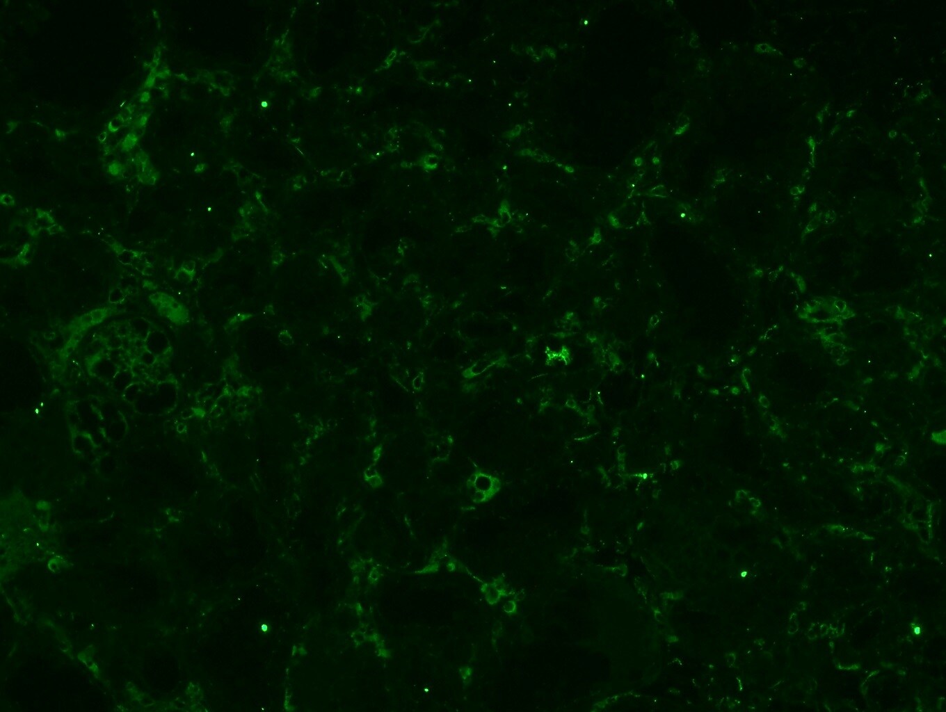
![Immunohistochemistry-Paraffin: VCAM-1/CD106 Antibody (1.G11B1) [NBP1-28404]](https://resources.rndsystems.com/images/products/VCAM-1-CD106-Antibody-1-G11B1-Immunohistochemistry-Paraffin-NBP1-28404-img0005.jpg "Immunohistochemistry-Paraffin: VCAM-1/CD106 Antibody (1.G11B1) [NBP1-28404]")
Immunohistochemistry-Paraffin: VCAM-1/CD106 Antibody (1.G11B1) [NBP1-28404]
Immunohistochemistry-Paraffin: VCAM-1/CD106 Antibody (1.G11B1) [NBP1-28404] - Veins and venules in the interalveolar stroma of porcine mammary gland are positive for the human VCAM-1. Antigen retrieval by Proteinase K. AlexaFluor 488 anti-mouse antibody was used as a secondary detection antibody. Image from verified customer review.![Flow Cytometry: VCAM-1/CD106 Antibody (1.G11B1) [NBP1-28404]](https://resources.rndsystems.com/images/products/VCAM-1-CD106-Antibody-1-G11B1-Flow-Cytometry-NBP1-28404-img0006.jpg "Flow Cytometry: VCAM-1/CD106 Antibody (1.G11B1) [NBP1-28404]")
Flow Cytometry: VCAM-1/CD106 Antibody (1.G11B1) [NBP1-28404]
Flow Cytometry: VCAM-1/CD106 Antibody (1.G11B1) [NBP1-28404] - TNF alpha stimulated human endothelial cell line HUV-EC-C was stained with Mouse Anti-Human CD106-UNLB followed by Goat Anti-Mouse IgG1, Human ads-FITC.![Flow Cytometry: VCAM-1/CD106 Antibody (1.G11B1) [NBP1-28404]](https://resources.rndsystems.com/images/products/VCAM-1-CD106-Antibody-1-G11B1-Flow-Cytometry-NBP1-28404-img0003.jpg "Flow Cytometry: VCAM-1/CD106 Antibody (1.G11B1) [NBP1-28404]")
Flow Cytometry: VCAM-1/CD106 Antibody (1.G11B1) [NBP1-28404]
Flow Cytometry: VCAM-1/CD106 Antibody (1.G11B1) [NBP1-28404] - Analysis using the FITC conjugate of NBP1-28404. Staining of TNF alpha stimulated endothelial cell line HUV-EC-C. [NBP1-28404] -")
Western Blot: VCAM-1/CD106 Antibody (1.G11B1) [NBP1-28404] -
Western Blot: VCAM-1/CD106 Antibody (1.G11B1) [NBP1-28404] - Knockdown of RPN2 decreased luminal B BC cell dissemination in vivo. Luminal B ER+ T47D cells, transfected with negative control siRNA (siRNA-C) or siRNA targeting RPN2 (siRNA-RPN2) were injected + estradiol (E2) ± neutrophils (Neu) into zebrafish transgenic embryos with green fluorescent blood vessels & analyzed as described in materials & methods. (A) Migration in vitro (n = 6). (B)In vivo dissemination of transfected T47D in presence of E2 ± Neu (n = 23–26). Scale bar = 100 µm. (n = 23–26). (C) Western blot analysis for confirmation of siRNA-RPN2 transfection & VCAM-1, ICAM-1, & MUC-1 expression. (D) Focal adhesions area (n = 5–6). Scale bar = 10 µm. (E) Proliferation (n = 6). Representative images of zebrafish embryos with disseminated luminal B T47D BC cells & immunocytochemistry analysis of vinculin expression are shown. Arrows show disseminated T47D & arrowheads show focal adhesions. BV = blood vessels. Data are presented as mean ± SEM. Two-tailed Student’s t-test *P < 0.05, ***P < 0.001, ns, not significant. Data are represented of at least two independent experiments. Image collected & cropped by CiteAb from the following publication (https://pubmed.ncbi.nlm.nih.gov/33330095), licensed under a CC-BY license. Not internally tested by Novus Biologicals. [NBP1-28404] -")
Western Blot: VCAM-1/CD106 Antibody (1.G11B1) [NBP1-28404] -
Western Blot: VCAM-1/CD106 Antibody (1.G11B1) [NBP1-28404] - Knockdown of U2AF1 decreased luminal A BC cell dissemination in vivo. Luminal A ER+ MCF-7 cells, transfected with negative control siRNA (siRNA-C) or siRNA targeting U2AF1 (siRNA-U2AF1) were injected in presence of estradiol (E2) ± neutrophils (Neu) into zebrafish transgenic embryos, with green fluorescent blood vessels, & analyzed as described in materials & methods. (A) Migration in vitro (n = 6–12). (B)In vivo dissemination in presence of E2 ± Neu (n = 38–41). Scale bar = 100 µm. (C) Western blot analysis for confirmation of siRNA-U2AF1 transfection & ICAM-1, VCAM-1, & MUC-1 expression. (D) Focal adhesion area (n = 7). Scale bar = 10 µm. (E) Proliferation in vitro (n = 12). Representative images of zebrafish embryos with disseminated MCF-7 cells & immunocytochemistry analysis of vinculin expression are shown. Arrows show disseminated MCF-7 cells & arrowheads show focal adhesions. BV = blood vessels. Data are presented as mean ± SEM. Two-tailed Student’s t-test *P < 0.05, **P < 0.01, ns, not significant. Data are represented of at least two independent experiments. Image collected & cropped by CiteAb from the following publication (https://pubmed.ncbi.nlm.nih.gov/33330095), licensed under a CC-BY license. Not internally tested by Novus Biologicals.Applications for VCAM-1/CD106 Antibody (1.G11B1) - Azide and BSA Free
Application
Recommended Usage
Flow Cytometry
1 ug/10^6 cells
Immunohistochemistry-Paraffin
Validated from a verified customer review
Western Blot
1:100 - 1:2000
Reviewed Applications
Read 1 review rated 3 using NBP1-28404 in the following applications:
Flow Cytometry Panel Builder
Bio-Techne Knows Flow Cytometry
Save time and reduce costly mistakes by quickly finding compatible reagents using the Panel Builder Tool.
Advanced Features
- Spectra Viewer - Custom analysis of spectra from multiple fluorochromes
- Spillover Popups - Visualize the spectra of individual fluorochromes
- Antigen Density Selector - Match fluorochrome brightness with antigen density

Formulation, Preparation, and Storage
Purification
Protein A or G purified
Formulation
Borate buffered saline, pH 8.2
Format
Azide and BSA Free
Preservative
No Preservative
Concentration
0.1 mg/ml
Shipping
The product is shipped with polar packs. Upon receipt, store it immediately at the temperature recommended below.
Stability & Storage
Store at 4C. Do not freeze.
Background: VCAM-1/CD106
Long Name
Vascular Cell Adhesion Molecule 1
Alternate Names
CD106, VCAM1
Gene Symbol
VCAM1
Additional VCAM-1/CD106 Products
Product Documents for VCAM-1/CD106 Antibody (1.G11B1) - Azide and BSA Free
Certificate of Analysis
To download a Certificate of Analysis, please enter a lot or batch number in the search box below.
Product Specific Notices for VCAM-1/CD106 Antibody (1.G11B1) - Azide and BSA Free
This product is for research use only and is not approved for use in humans or in clinical diagnosis. Primary Antibodies are guaranteed for 1 year from date of receipt.
Related Research Areas
Citations for VCAM-1/CD106 Antibody (1.G11B1) - Azide and BSA Free
 Powered by Bioz
Customer Reviews for VCAM-1/CD106 Antibody (1.G11B1) - Azide and BSA Free (1)
3 out of 5
1 Customer Rating
Have you used VCAM-1/CD106 Antibody (1.G11B1) - Azide and BSA Free?
Submit a review and receive an Amazon gift card!
$25/€18/£15/$25CAN/¥2500 Yen for a review with an image
$10/€7/£6/$10CAN/¥1110 Yen for a review without an image
Submit a review
Customer Images
Showing
1
-
1 of
1 review
Showing All
Filter By:
-
Application: Immunohistochemistry-ParaffinSample Tested: Mammary gland tissueSpecies: PorcineVerified Customer | Posted 06/28/2019Veins and venules in the interalveolar stroma of porcine mammary gland are positive for the human VCAM-1.Antigen retrieval by proteinase K. Alexa Fluor 488 anti-mouse antibody was used as a secondary detection antibody.

There are no reviews that match your criteria.
Protocols
Find general support by application which include: protocols, troubleshooting, illustrated assays, videos and webinars.
- 7-Amino Actinomycin D (7-AAD) Cell Viability Flow Cytometry Protocol
- Antigen Retrieval Protocol (PIER)
- Antigen Retrieval for Frozen Sections Protocol
- Appropriate Fixation of IHC/ICC Samples
- Cellular Response to Hypoxia Protocols
- Chromogenic IHC Staining of Formalin-Fixed Paraffin-Embedded (FFPE) Tissue Protocol
- Chromogenic Immunohistochemistry Staining of Frozen Tissue
- ClariTSA™ Fluorophore Kits
- Detection & Visualization of Antibody Binding
- Extracellular Membrane Flow Cytometry Protocol
- Flow Cytometry Protocol for Cell Surface Markers
- Flow Cytometry Protocol for Staining Membrane Associated Proteins
- Flow Cytometry Staining Protocols
- Flow Cytometry Troubleshooting Guide
- Fluorescent IHC Staining of Frozen Tissue Protocol
- Graphic Protocol for Heat-induced Epitope Retrieval
- Graphic Protocol for the Preparation and Fluorescent IHC Staining of Frozen Tissue Sections
- Graphic Protocol for the Preparation and Fluorescent IHC Staining of Paraffin-embedded Tissue Sections
- Graphic Protocol for the Preparation of Gelatin-coated Slides for Histological Tissue Sections
- IHC Sample Preparation (Frozen sections vs Paraffin)
- Immunofluorescent IHC Staining of Formalin-Fixed Paraffin-Embedded (FFPE) Tissue Protocol
- Immunohistochemistry (IHC) and Immunocytochemistry (ICC) Protocols
- Immunohistochemistry Frozen Troubleshooting
- Immunohistochemistry Paraffin Troubleshooting
- Intracellular Flow Cytometry Protocol Using Alcohol (Methanol)
- Intracellular Flow Cytometry Protocol Using Detergents
- Intracellular Nuclear Staining Flow Cytometry Protocol Using Detergents
- Intracellular Staining Flow Cytometry Protocol Using Alcohol Permeabilization
- Intracellular Staining Flow Cytometry Protocol Using Detergents to Permeabilize Cells
- Preparing Samples for IHC/ICC Experiments
- Preventing Non-Specific Staining (Non-Specific Binding)
- Primary Antibody Selection & Optimization
- Propidium Iodide Cell Viability Flow Cytometry Protocol
- Protocol for Heat-Induced Epitope Retrieval (HIER)
- Protocol for Liperfluo
- Protocol for Making a 4% Formaldehyde Solution in PBS
- Protocol for VisUCyte™ HRP Polymer Detection Reagent
- Protocol for the Characterization of Human Th22 Cells
- Protocol for the Characterization of Human Th9 Cells
- Protocol for the Preparation & Fixation of Cells on Coverslips
- Protocol for the Preparation and Chromogenic IHC Staining of Frozen Tissue Sections
- Protocol for the Preparation and Chromogenic IHC Staining of Frozen Tissue Sections - Graphic
- Protocol for the Preparation and Chromogenic IHC Staining of Paraffin-embedded Tissue Sections
- Protocol for the Preparation and Chromogenic IHC Staining of Paraffin-embedded Tissue Sections - Graphic
- Protocol for the Preparation and Fluorescent IHC Staining of Frozen Tissue Sections
- Protocol for the Preparation and Fluorescent IHC Staining of Paraffin-embedded Tissue Sections
- Protocol for the Preparation of Gelatin-coated Slides for Histological Tissue Sections
- Protocol: Annexin V and PI Staining by Flow Cytometry
- Protocol: Annexin V and PI Staining for Apoptosis by Flow Cytometry
- R&D Systems Quality Control Western Blot Protocol
- TUNEL and Active Caspase-3 Detection by IHC/ICC Protocol
- The Importance of IHC/ICC Controls
- Troubleshooting Guide: Fluorokine Flow Cytometry Kits
- Troubleshooting Guide: Immunohistochemistry
- Troubleshooting Guide: Western Blot Figures
- Western Blot Conditions
- Western Blot Protocol
- Western Blot Protocol for Cell Lysates
- Western Blot Troubleshooting
- Western Blot Troubleshooting Guide
- View all Protocols, Troubleshooting, Illustrated assays and Webinars
Loading...
Associated Pathways


