VCAM-1/CD106 Antibody (6G9) - BSA Free
Novus Biologicals | Catalog # NBP1-47491
Key Product Details
Species Reactivity
Validated:
Human, Mouse
Cited:
Human, Mouse
Applications
Validated:
Immunohistochemistry, Immunohistochemistry-Paraffin, Western Blot, Immunoblotting, ELISA, Simple Western, Immunoprecipitation
Cited:
Immunohistochemistry-Paraffin, Western Blot, Immunoblotting, Immunocytochemistry/ Immunofluorescence, Immunoprecipitation
Label
Unconjugated
Antibody Source
Monoclonal Mouse IgG1 Clone # 6G9
Format
BSA Free
Loading...
Product Specifications
Immunogen
This VCAM-1/CD106 Antibody (6G9) was developed against a purified recombinant fragment of human VCAM-1 expressed in E. coli. [Uniprot: P19320]
Reactivity Notes
Use in Mouse reported in scientific literature (PMID: 32243809).
Localization
Membrane. Single-pass type I membrane protein.
Clonality
Monoclonal
Host
Mouse
Isotype
IgG1
Theoretical MW
81 kDa.
Disclaimer note: The observed molecular weight of the protein may vary from the listed predicted molecular weight due to post translational modifications, post translation cleavages, relative charges, and other experimental factors.
Disclaimer note: The observed molecular weight of the protein may vary from the listed predicted molecular weight due to post translational modifications, post translation cleavages, relative charges, and other experimental factors.
Scientific Data Images for VCAM-1/CD106 Antibody (6G9) - BSA Free
![Western Blot: VCAM-1/CD106 Antibody (6G9)BSA Free [NBP1-47491]](https://resources.rndsystems.com/images/products/VCAM-1-CD106-Antibody-6G9-Western-Blot-NBP1-47491-img0006.jpg "Western Blot: VCAM-1/CD106 Antibody (6G9)BSA Free [NBP1-47491]")
Western Blot: VCAM-1/CD106 Antibody (6G9)BSA Free [NBP1-47491]
Western Blot: VCAM-1/CD106 Antibody (6G9) [NBP1-47491] - Using VCAM1 mouse mAb against (1) HUVEC and (2) EC cell lysate.![Immunohistochemistry: VCAM-1/CD106 Antibody (6G9) - BSA Free [NBP1-47491]](https://resources.rndsystems.com/images/products/VCAM-1-CD106-Antibody-6G9-Immunohistochemistry-NBP1-47491-img0010.jpg "Immunohistochemistry: VCAM-1/CD106 Antibody (6G9) - BSA Free [NBP1-47491]")
Immunohistochemistry: VCAM-1/CD106 Antibody (6G9) - BSA Free [NBP1-47491]
VCAM-1-CD106-Antibody-6G9-Immunohistochemistry-NBP1-47491-img0010.jpg![Immunohistochemistry-Paraffin: VCAM-1/CD106 Antibody (6G9) - BSA Free [NBP1-47491]](https://resources.rndsystems.com/images/products/VCAM-1-CD106-Antibody-6G9-Immunohistochemistry-Paraffin-NBP1-47491-img0004.jpg "Immunohistochemistry-Paraffin: VCAM-1/CD106 Antibody (6G9) - BSA Free [NBP1-47491]")
Immunohistochemistry-Paraffin: VCAM-1/CD106 Antibody (6G9) - BSA Free [NBP1-47491]
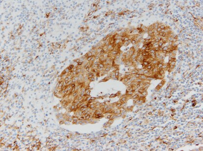
Immunohistochemistry-Paraffin: VCAM-1/CD106 Antibody (6G9) [NBP1-47491] - (A) human lung cancer and (B) colon cancer using VCAM1 mouse mAb with DAB staining.![Immunohistochemistry-Paraffin: VCAM-1/CD106 Antibody (6G9) - BSA Free [NBP1-47491]](https://resources.rndsystems.com/images/products/VCAM-1-CD106-Antibody-6G9-Immunohistochemistry-Paraffin-NBP1-47491-img0005.jpg "Immunohistochemistry-Paraffin: VCAM-1/CD106 Antibody (6G9) - BSA Free [NBP1-47491]")
Immunohistochemistry-Paraffin: VCAM-1/CD106 Antibody (6G9) - BSA Free [NBP1-47491]
Immunohistochemistry-Paraffin: VCAM-1/CD106 Antibody (6G9) [NBP1-47491] - Human placenta tissues using VCAM1 mouse mAb.![Immunohistochemistry: VCAM-1/CD106 Antibody (6G9) - BSA Free [NBP1-47491]](https://resources.rndsystems.com/images/products/VCAM-1-CD106-Antibody-6G9-Immunohistochemistry-NBP1-47491-img0007.jpg "Immunohistochemistry: VCAM-1/CD106 Antibody (6G9) - BSA Free [NBP1-47491]")
Immunohistochemistry: VCAM-1/CD106 Antibody (6G9) - BSA Free [NBP1-47491]
Immunohistochemistry: VCAM-1/CD106 Antibody (6G9) [NBP1-47491] - Breast carcinoma, cytoplasmic staining. IHC image submitted by a verified customer review.![Immunohistochemistry-Paraffin: VCAM-1/CD106 Antibody (6G9) - BSA Free [NBP1-47491]](https://resources.rndsystems.com/images/products/VCAM-1-CD106-Antibody-6G9-Immunohistochemistry-Paraffin-NBP1-47491-img0009.jpg "Immunohistochemistry-Paraffin: VCAM-1/CD106 Antibody (6G9) - BSA Free [NBP1-47491]")
Immunohistochemistry-Paraffin: VCAM-1/CD106 Antibody (6G9) - BSA Free [NBP1-47491]
Immunohistochemistry-Paraffin: VCAM-1/CD106 Antibody (6G9) [NBP1-47491] - FFPE section of human lung cancer. Image at 20X magnification. IHC-P image submitted by a verified customer review.![Simple Western: VCAM-1/CD106 Antibody (6G9)BSA Free [NBP1-47491]](https://resources.rndsystems.com/images/products/VCAM-1-CD106-Antibody-6G9-Simple-Western-NBP1-47491-img0008.jpg "Simple Western: VCAM-1/CD106 Antibody (6G9)BSA Free [NBP1-47491]")
Simple Western: VCAM-1/CD106 Antibody (6G9)BSA Free [NBP1-47491]
Simple Western: VCAM-1/CD106 Antibody (6G9) [NBP1-47491] - Lane view shows a specific band for CD106/VCAM1 in 0.5 mg/mL of HUVEC lysate. This experiment was performed under reducing conditions using the 12-230 kDa separation system. - BSA Free [NBP1-47491] -")
Immunohistochemistry: VCAM-1/CD106 Antibody (6G9) - BSA Free [NBP1-47491] -
Photomicrographs of two separate gut sections from a patient with EHEC colitis.Panels (A) and (B) are stained with CD31 to enumerate endothelium lining the vessels (40× magnification). (C) and (D) are stained to show VCAM-1 expression in endothelium, indicating inflammatory activation (40× magnification). - BSA Free [NBP1-47491] -")
Immunocytochemistry/ Immunofluorescence: VCAM-1/CD106 Antibody (6G9) - BSA Free [NBP1-47491] -
Immunocytochemistry/ Immunofluorescence: VCAM-1/CD106 Antibody (6G9) - BSA Free [NBP1-47491] - Enhanced expression of endothelial SENP1 & GATA2 correlates w/ graft arteriosclerosis (GA) progression.Similarly sized human coronary arteries w/ GA from chronically rejecting heart allografts or w/out disease from non-transplanted hearts collected & evaluated by histological analysis. (a,b) Dramatically increased expression of endothelial SENP1 detected in the diseased vessel wall. Endothelial SENP1 expression is demonstrated by immunofluorescence analysis of coronary artery cross-sections that stained for SENP1 & the endothelial marker PECAM-1 w/ DAPI labelling of the nuclei. Representative images are shown in (a) w/ quantification data in (b). Bar represents 50 μm. (c) Induction of endothelial adhesion molecules resulted in a similar augmented pattern as endothelial SENP1. Representative images of immunofluorescence staining for VCAM-1 or P-selectin & PECAM-1 in coronary arteries w/ DAPI counterstaining are shown. Bar represents 50 μm. (d,e) Expression of endothelial GATA2 elevated w/ the progression of aggravated rejection. Representative images of the immunofluorescence staining of coronary arteries for GATA2 & PECAM-1 together w/ DAPI nuclear staining are shown in (d) w/ quantification data in (e). Negative SENP1 & GATA2 staining in the isotype or blocking peptide controls are also shown in (a) & (d). Bar represents 50 μm. Data presented in (b,e) are the mean±s.e.m. from five separate clinical specimens per group as indicated. *P<0.05, **P<0.01 & ***P<0.0001; one-way ANOVA followed by Bonferroni test. GA, graft arteriosclerosis; PECAM, platelet/endothelial cell adhesion molecule; SENP, sentrin-specific protease; VCAM, vascular cell adhesion molecule. Image collected & cropped by CiteAb from the following publication (https://www.nature.com/articles/ncomms15426), licensed under a CC-BY license. Not internally tested by Novus Biologicals. - BSA Free [NBP1-47491] -")
Immunocytochemistry/ Immunofluorescence: VCAM-1/CD106 Antibody (6G9) - BSA Free [NBP1-47491] -
Immunocytochemistry/ Immunofluorescence: VCAM-1/CD106 Antibody (6G9) - BSA Free [NBP1-47491] - Loss of endothelial SENP1 inhibits EC activation.(a) Grafts from WT or SENP1-ecKO mice were harvested 3 days post-transplantation. The induction of endothelial adhesion molecules was demonstrated by immunofluorescence staining of ICAM-1, VCAM-1, or P-selectin & PECAM-1 with DAPI labelling of the nuclei. Bar represents 50 μm. (b–e) Attenuated induction of adhesion molecules in SENP1-ecKO MAECs. Flow cytometry analysis of ICAM-1, VCAM-1 & P-selectin in MAECs isolated from WT or SENP1-ecKO mice after TNF or IL-1 beta treatment. Representative histograms are shown in (b) with the quantification of mean intensity in (c–e). (f–h) Overexpression of the catalytically inactive form of SENP1 (SENP1-Mut) inhibits the induction of adhesion molecules in HUVECs. HUVECs were infected by Ad-SENP1-Mut or vector control (Ad-LacZ) for 24 h, treated with pro-inflammatory cytokines & analysed by flow cytometry in the same way as MAECs. Representative histograms of ICAM-1 & VCAM-1 are shown in (f) with the quantification of mean intensity in (g,h). Data are presented as the mean±s.e.m. from at least three independent experiments. *P<0.05 & **P<0.01; two-way ANOVA followed by Bonferroni post-test. MAEC, mouse aortic endothelial cell. Image collected & cropped by CiteAb from the following publication (https://www.nature.com/articles/ncomms15426), licensed under a CC-BY license. Not internally tested by Novus Biologicals.Applications for VCAM-1/CD106 Antibody (6G9) - BSA Free
Application
Recommended Usage
ELISA
1:10000
Immunoblotting
reported in scientific literature (PMID 28569748)
Immunohistochemistry
1:200-1:1000
Immunohistochemistry-Paraffin
1:200-1:1000
Immunoprecipitation
reported in scientific literature (PMID 28569748)
Simple Western
1:1000
Western Blot
1:500-1:2000
Application Notes
In Simple Western only 10 - 15 uL of the recommended dilution is used per data point.
See Simple Western Antibody Database for Simple Western validation: Tested in HUVEC lysate 0.5 mg/mL, separated by Size, antibody dilution of 1:1000, apparent MW was 56 kDa. Separated by Size-Wes, Sally Sue/Peggy Sue.
See Simple Western Antibody Database for Simple Western validation: Tested in HUVEC lysate 0.5 mg/mL, separated by Size, antibody dilution of 1:1000, apparent MW was 56 kDa. Separated by Size-Wes, Sally Sue/Peggy Sue.
Reviewed Applications
Read 2 reviews rated 4.5 using NBP1-47491 in the following applications:
Formulation, Preparation, and Storage
Purification
Ammonium sulfate precipitation
Formulation
PBS
Format
BSA Free
Preservative
0.03% Sodium Azide
Concentration
1.0 mg/ml
Shipping
The product is shipped with polar packs. Upon receipt, store it immediately at the temperature recommended below.
Stability & Storage
Store at 4C short term. Aliquot and store at -20C long term. Avoid freeze-thaw cycles.
Background: VCAM-1/CD106
Long Name
Vascular Cell Adhesion Molecule 1
Alternate Names
CD106, VCAM1
Entrez Gene IDs
7412 (Human)
Gene Symbol
VCAM1
UniProt
Additional VCAM-1/CD106 Products
Product Documents for VCAM-1/CD106 Antibody (6G9) - BSA Free
Certificate of Analysis
To download a Certificate of Analysis, please enter a lot or batch number in the search box below.
Product Specific Notices for VCAM-1/CD106 Antibody (6G9) - BSA Free
This product is for research use only and is not approved for use in humans or in clinical diagnosis. Primary Antibodies are guaranteed for 1 year from date of receipt.
Related Research Areas
Citations for VCAM-1/CD106 Antibody (6G9) - BSA Free
 Powered by Bioz
Customer Reviews for VCAM-1/CD106 Antibody (6G9) - BSA Free (2)
4.5 out of 5
2 Customer Ratings
Have you used VCAM-1/CD106 Antibody (6G9) - BSA Free?
Submit a review and receive an Amazon gift card!
$25/€18/£15/$25CAN/¥2500 Yen for a review with an image
$10/€7/£6/$10CAN/¥1110 Yen for a review without an image
Submit a review
Customer Images
-(01-ml)_NBP1-47491_6901.jpg)
Showing
1
-
2 of
2 reviews
Showing All
Filter By:
-
Application: Immunohistochemistry-ParaffinSample Tested: FFPE and human lung cancerSpecies: HumanVerified Customer | Posted 10/13/202020X

-
Application: ImmunohistochemistrySample Tested:Species: HumanVerified Customer | Posted 04/21/2014Breast carcinoma: Cytoplasmic staining
There are no reviews that match your criteria.
Protocols
Find general support by application which include: protocols, troubleshooting, illustrated assays, videos and webinars.
- Antigen Retrieval Protocol (PIER)
- Antigen Retrieval for Frozen Sections Protocol
- Appropriate Fixation of IHC/ICC Samples
- Cellular Response to Hypoxia Protocols
- Chromogenic IHC Staining of Formalin-Fixed Paraffin-Embedded (FFPE) Tissue Protocol
- Chromogenic Immunohistochemistry Staining of Frozen Tissue
- ClariTSA™ Fluorophore Kits
- Detection & Visualization of Antibody Binding
- ELISA Sample Preparation & Collection Guide
- ELISA Troubleshooting Guide
- Fluorescent IHC Staining of Frozen Tissue Protocol
- Graphic Protocol for Heat-induced Epitope Retrieval
- Graphic Protocol for the Preparation and Fluorescent IHC Staining of Frozen Tissue Sections
- Graphic Protocol for the Preparation and Fluorescent IHC Staining of Paraffin-embedded Tissue Sections
- Graphic Protocol for the Preparation of Gelatin-coated Slides for Histological Tissue Sections
- How to Run an R&D Systems DuoSet ELISA
- How to Run an R&D Systems Quantikine ELISA
- How to Run an R&D Systems Quantikine™ QuicKit™ ELISA
- IHC Sample Preparation (Frozen sections vs Paraffin)
- Immunofluorescent IHC Staining of Formalin-Fixed Paraffin-Embedded (FFPE) Tissue Protocol
- Immunohistochemistry (IHC) and Immunocytochemistry (ICC) Protocols
- Immunohistochemistry Frozen Troubleshooting
- Immunohistochemistry Paraffin Troubleshooting
- Immunoprecipitation Protocol
- Preparing Samples for IHC/ICC Experiments
- Preventing Non-Specific Staining (Non-Specific Binding)
- Primary Antibody Selection & Optimization
- Protocol for Heat-Induced Epitope Retrieval (HIER)
- Protocol for Making a 4% Formaldehyde Solution in PBS
- Protocol for VisUCyte™ HRP Polymer Detection Reagent
- Protocol for the Preparation & Fixation of Cells on Coverslips
- Protocol for the Preparation and Chromogenic IHC Staining of Frozen Tissue Sections
- Protocol for the Preparation and Chromogenic IHC Staining of Frozen Tissue Sections - Graphic
- Protocol for the Preparation and Chromogenic IHC Staining of Paraffin-embedded Tissue Sections
- Protocol for the Preparation and Chromogenic IHC Staining of Paraffin-embedded Tissue Sections - Graphic
- Protocol for the Preparation and Fluorescent IHC Staining of Frozen Tissue Sections
- Protocol for the Preparation and Fluorescent IHC Staining of Paraffin-embedded Tissue Sections
- Protocol for the Preparation of Gelatin-coated Slides for Histological Tissue Sections
- Quantikine HS ELISA Kit Assay Principle, Alkaline Phosphatase
- Quantikine HS ELISA Kit Principle, Streptavidin-HRP Polymer
- R&D Systems Quality Control Western Blot Protocol
- Sandwich ELISA (Colorimetric) – Biotin/Streptavidin Detection Protocol
- Sandwich ELISA (Colorimetric) – Direct Detection Protocol
- TUNEL and Active Caspase-3 Detection by IHC/ICC Protocol
- The Importance of IHC/ICC Controls
- Troubleshooting Guide: ELISA
- Troubleshooting Guide: Immunohistochemistry
- Troubleshooting Guide: Western Blot Figures
- Western Blot Conditions
- Western Blot Protocol
- Western Blot Protocol for Cell Lysates
- Western Blot Troubleshooting
- Western Blot Troubleshooting Guide
- View all Protocols, Troubleshooting, Illustrated assays and Webinars
Loading...
Associated Pathways


