VEGF Antibody (VG76e) - BSA Free
Novus Biologicals | Catalog # NB100-648
Key Product Details
Species Reactivity
Validated:
Human, Rat, Porcine, Bovine, Goat, Sheep
Cited:
Human, Rat, Porcine, Bovine
Applications
Validated:
Immunohistochemistry, Immunohistochemistry-Paraffin, Western Blot, ELISA, Immunocytochemistry/ Immunofluorescence, Radioimmunoassay
Cited:
Immunohistochemistry-Paraffin, Western Blot, Radioimmunoassay, IF/IHC
Label
Unconjugated
Antibody Source
Monoclonal Mouse IgG1 Clone # VG76e
Format
BSA Free
Loading...
Product Specifications
Immunogen
Recombinant human VEGF189 expressed in E.coli [UniProt# P15692]
Reactivity Notes
Mixed results in Cow and Pig (IHC only). Customers have reported success staining sheep tissue (see review). Goat reactivity reported from a verified customer review.
Specificity
Using recombinant proteins, it was demonstrated that VG76e recognizes the 121, 165, and 189 isoforms of VEGF
Clonality
Monoclonal
Host
Mouse
Isotype
IgG1
Theoretical MW
40 kDa.
Disclaimer note: The observed molecular weight of the protein may vary from the listed predicted molecular weight due to post translational modifications, post translation cleavages, relative charges, and other experimental factors.
Disclaimer note: The observed molecular weight of the protein may vary from the listed predicted molecular weight due to post translational modifications, post translation cleavages, relative charges, and other experimental factors.
Scientific Data Images for VEGF Antibody (VG76e) - BSA Free
![Immunohistochemistry-Paraffin: VEGF Antibody (VG76e) - BSA Free [NB100-648]](https://resources.rndsystems.com/images/products/VEGF-Antibody-VG76e-Immunohistochemistry-Paraffin-NB100-648-img0004.jpg "Immunohistochemistry-Paraffin: VEGF Antibody (VG76e) - BSA Free [NB100-648]")
Immunohistochemistry-Paraffin: VEGF Antibody (VG76e) - BSA Free [NB100-648]
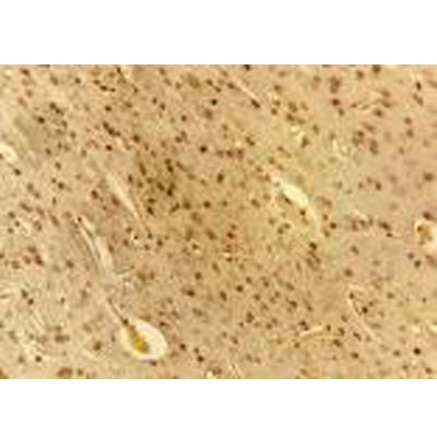
Immunohistochemistry-Paraffin: VEGF Antibody (VG76e) [NB100-648] - IHC analysis of VEGF in fetal sheep brain tissue. Image courtesy of anonymous customer review.![Western Blot: VEGF Antibody (VG76e)BSA Free [NB100-648]](https://resources.rndsystems.com/images/products/VEGF-Antibody-VG76e-Western-Blot-NB100-648-img0005.jpg "Western Blot: VEGF Antibody (VG76e)BSA Free [NB100-648]")
Western Blot: VEGF Antibody (VG76e)BSA Free [NB100-648]
Western Blot: VEGF Antibody (VG76e) [NB100-648] - Analysis of VEGF in human kidney protein (30ug).![Immunocytochemistry/ Immunofluorescence: VEGF Antibody (VG76e) - BSA Free [NB100-648]](https://resources.rndsystems.com/images/products/VEGF-Antibody-VG76e-Immunocytochemistry-Immunofluorescence-NB100-648-img0007.jpg "Immunocytochemistry/ Immunofluorescence: VEGF Antibody (VG76e) - BSA Free [NB100-648]")
Immunocytochemistry/ Immunofluorescence: VEGF Antibody (VG76e) - BSA Free [NB100-648]
Immunocytochemistry/Immunofluorescence: VEGF Antibody (VG76e) [NB100-648] - Immunofluorescent analysis of VEGF using NB100-648. This image submitted through a verified customer review.![Immunohistochemistry-Paraffin: VEGF Antibody (VG76e) - BSA Free [NB100-648]](https://resources.rndsystems.com/images/products/VEGF-Antibody-VG76e-Immunohistochemistry-Paraffin-NB100-648-img0006.jpg "Immunohistochemistry-Paraffin: VEGF Antibody (VG76e) - BSA Free [NB100-648]")
Immunohistochemistry-Paraffin: VEGF Antibody (VG76e) - BSA Free [NB100-648]
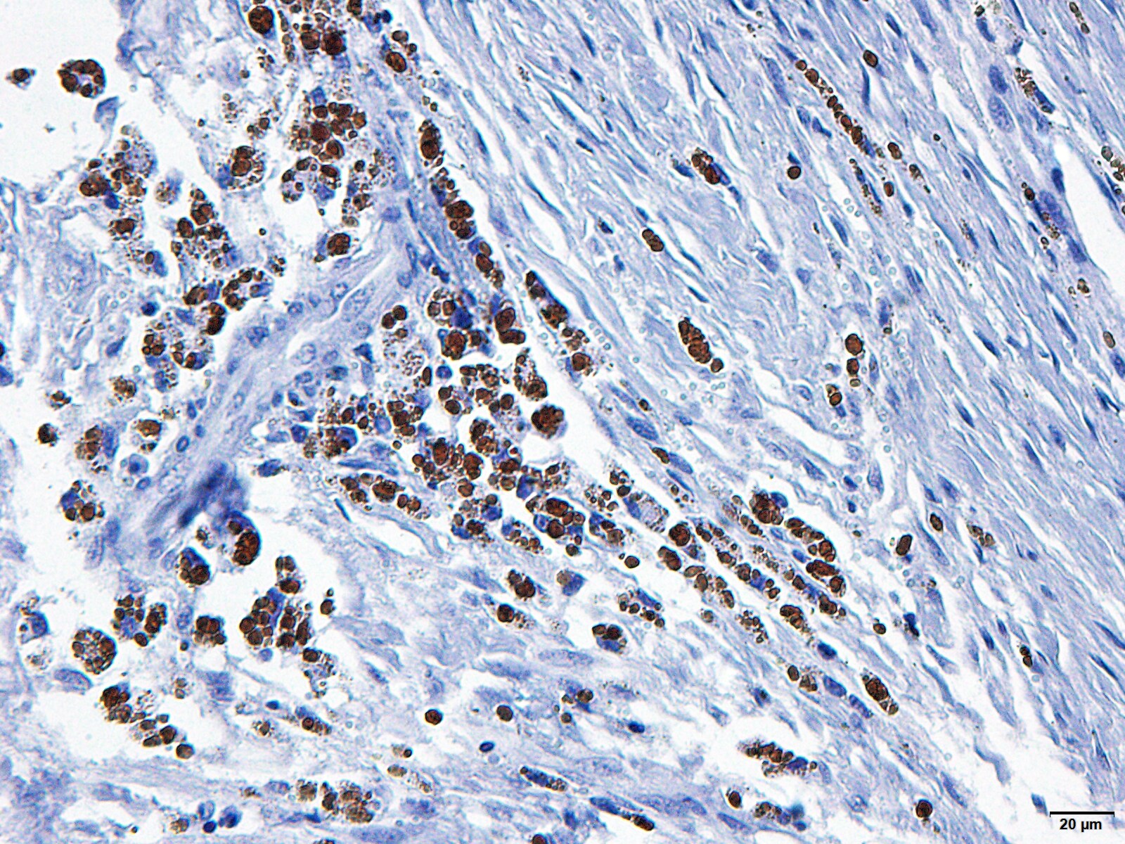
Immunohistochemistry-Paraffin: VEGF Antibody (VG76e) [NB100-648] - IHC-P analysis of VEGF in sheep tonsil using NB100-648. This image was submitted through a verified customer review. [NB100-648]")
Western Blot: Mouse Monoclonal VEGF Antibody (VG76e) [NB100-648]
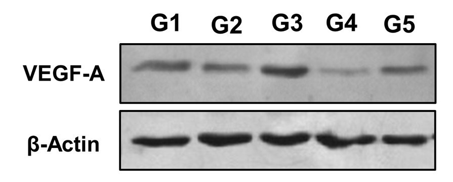
Western Blot: Mouse Monoclonal VEGF Antibody (VG76e) [NB100-648] - Bone lysate homogenate from goats. SDS-PAGE with 0.3 mg of total protein from the homogenate per well. VEGF, NB100-648, was used at a dilution of 1:1000 in TBS Tween and 5% milk powder. The protein was labeled at 40 kDa. Quantification of VEGFA in bone graft in the tibia of intrailiac cellularized goats at different times compared with tibial autograft and untreated bone defect. Evaluation of pre-cellularized graft in iliac crest in the treatment of bone defects in the tibia of goats. G1: cellularized graft for 4 weeks applied to tibial bone defect, G2: cellularized graft for 6 weeks applied to tibial bone defect, G3: cellularized graft for 8 weeks applied to tibial bone defect, G4: tibial autograft and G5: untreated bone defect. All groups were evaluated 120 days after graft implantation. Image from a verified customer review. [NB100-648]")
Immunohistochemistry-Paraffin: Mouse Monoclonal VEGF Antibody (VG76e) [NB100-648]
The sections of histological sections of goat fragments were deparaffinized, peroxidase blocked in methanol and 30% hydrogen peroxide (9:1), and antigen retrieval was performed with porcine gastric pepsin and HCl at pH 3.0 in an oven at 37°C for 30 minutes. Then, blocking with BSA was performed for 1 hour followed by incubation with primary antibody VEGF antibody NB100-648 (1:100) overnight. Subsequently, excess primary antibody was removed with PBS pH 7.6 and incubated with secondary antibody (goat anti-mouse IgG - NB7539 (1:200) for 1 hour and application of polymer for 30 minutes. Finally, 150 μL of DAB was applied for five minutes and counterstained with hematoxylin. Image from a verified customer review.Applications for VEGF Antibody (VG76e) - BSA Free
Application
Recommended Usage
ELISA
1:100-1:2000
Immunocytochemistry/ Immunofluorescence
1:50-1:200
Immunohistochemistry
1:200
Immunohistochemistry-Paraffin
1:200
Radioimmunoassay
reported in scientific literature
Western Blot
0.5-2ug/ml
Application Notes
In Western blot a band is observed between 38-44 kDa. With IHC-paraffin sections, microwave treatment with Tris HCl pH 10 buffer (NOT citrate buffer) is necessary. VEGF-A is a hetero dimer, consisting of 2 chains of approximately 24kDa each.
Reviewed Applications
Read 7 reviews rated 4.1 using NB100-648 in the following applications:
Formulation, Preparation, and Storage
Purification
Protein G purified
Formulation
PBS
Format
BSA Free
Preservative
0.02% Sodium Azide
Concentration
1 mg/ml
Shipping
The product is shipped with polar packs. Upon receipt, store it immediately at the temperature recommended below.
Stability & Storage
Store at 4C short term. Aliquot and store at -20C long term. Avoid freeze-thaw cycles.
Background: VEGF
References
1. Melincovici CS, Bosca AB, susman S, et al. Vascular endothelial growth factor (VEGF) - key factor in normal and pathological angiogenesis. Rom J Morphol Embryol. 2018;59(2):455-467.
2. Shaik F, Cuthbert GA, Homer-Vanniasinkam S, Muench SP, Ponnambalam S, Harrison MA. Structural Basis for Vascular Endothelial Growth Factor Receptor Activation and Implications for Disease Therapy. Biomolecules. 2020;10(12):1673. https://doi.org/10.3390/biom10121673
3. Apte RS, Chen DS, Ferrara N. VEGF in Signaling and Disease: Beyond Discovery and Development. Cell. 2019;176(6):1248-1264. https://doi.org/10.1016/j.cell.2019.01.021
4. Matsumoto K, Ema M. Roles of VEGF-A signalling in development, regeneration, and tumours. J Biochem. 2014;156(1):1-10. https://doi.org/10.1093/jb/mvu031
5. Itatani Y, Kawada K, Yamamoto T, Sakai Y. Resistance to Anti-Angiogenic Therapy in Cancer-Alterations to Anti-VEGF Pathway. Int J Mol Sci. 2018;19(4):1232. Published 2018 Apr 18. doi:10.3390/ijms19041232
6. Uniprot (P15692)
7. Hamilton JL, Nagao M, Levine BR, Chen D, Olsen BR, Im HJ. Targeting VEGF and Its Receptors for the Treatment of Osteoarthritis and Associated Pain. J Bone Miner Res. 2016;31(5):911-924. https://doi.org/10.1002/jbmr.2828
Long Name
Vascular Endothelial Growth Factor
Alternate Names
MVCD1, VAS, Vasculotropin, VEGF-A, VEGFA, VPF
Entrez Gene IDs
7422 (Human)
Gene Symbol
VEGFA
UniProt
Additional VEGF Products
Product Documents for VEGF Antibody (VG76e) - BSA Free
Certificate of Analysis
To download a Certificate of Analysis, please enter a lot or batch number in the search box below.
Product Specific Notices for VEGF Antibody (VG76e) - BSA Free
This product is for research use only and is not approved for use in humans or in clinical diagnosis. Primary Antibodies are guaranteed for 1 year from date of receipt.
Citations for VEGF Antibody (VG76e) - BSA Free
 Powered by Bioz
Customer Reviews for VEGF Antibody (VG76e) - BSA Free (7)
4.1 out of 5
7 Customer Ratings
Have you used VEGF Antibody (VG76e) - BSA Free?
Submit a review and receive an Amazon gift card!
$25/€18/£15/$25CAN/¥2500 Yen for a review with an image
$10/€7/£6/$10CAN/¥1110 Yen for a review without an image
Submit a review
Customer Images
_NB100-648_9086.jpg)
-(01-ml)_NB100-648_7551.jpg)
Showing
1
-
5 of
7 reviews
Showing All
Filter By:
-
Application: Immunohistochemistry-ParaffinSample Tested: Bone and Bone ExtractsSpecies: GoatVerified Customer | Posted 07/29/2025The f histological goat bone fragments were incubated with the primary antibody VEGF NB100-648 overnight. Subsequently, the secondary antibody goat anti-mouse IgG - NB7539 was applied and incubated for 1 hour.Validation of an immunohistochemistry technique for goats

-
Application: Western BlotSample Tested: Bone and Bone ExtractsSpecies: GoatVerified Customer | Posted 05/13/2025Bone lysate homogenate from goats. SDS-PAGE with 0.3 mg of total protein from the homogenate per well. VEGF, NB100-648, was used at a dilution of 1:1000 in TBS Tween and 5% milk powder. The protein was labeled at 40 kDa.Quantification of VEGFA in bone graft in the tibia of intrailiac cellularized goats at different times compared with tibial autograft and untreated bone defect. Evaluation of pre-cellularized graft in iliac crest in the treatment of bone defects in the tibia of goats. G1: cellularized graft for 4 weeks applied to tibial bone defect, G2: cellularized graft for 6 weeks applied to tibial bone defect, G3: cellularized graft for 8 weeks applied to tibial bone defect, G4: tibial autograft and G5: untreated bone defect. All groups were evaluated 120 days after graft implantation.
Bio-Techne ResponseThis review reflects a new species or application tested on a primary antibody.
-
Application: Western BlotSample Tested: Bovine LysateSpecies: OtherVerified Customer | Posted 09/14/2014
-
Application: Immunohistochemistry-ParaffinSample Tested: sheep tonsilSpecies: OtherVerified Customer | Posted 07/29/2014Vegf in sheep tonsil
-
Application: ImmunofluorescenceSample Tested:Species: OtherVerified Customer | Posted 05/13/2014
-
Application: ImmunocytochemistrySample Tested: pig endothelial cellsSpecies: OtherVerified Customer | Posted 05/13/2014
-
Application: Immunohistochemistry-ParaffinSample Tested: Fetal Sheep Brain TissueSpecies: OtherVerified Customer | Posted 05/04/2011
There are no reviews that match your criteria.
Protocols
Find general support by application which include: protocols, troubleshooting, illustrated assays, videos and webinars.
- Antigen Retrieval Protocol (PIER)
- Antigen Retrieval for Frozen Sections Protocol
- Appropriate Fixation of IHC/ICC Samples
- Cellular Response to Hypoxia Protocols
- Chromogenic IHC Staining of Formalin-Fixed Paraffin-Embedded (FFPE) Tissue Protocol
- Chromogenic Immunohistochemistry Staining of Frozen Tissue
- ClariTSA™ Fluorophore Kits
- Detection & Visualization of Antibody Binding
- ELISA Sample Preparation & Collection Guide
- ELISA Troubleshooting Guide
- Fluorescent IHC Staining of Frozen Tissue Protocol
- Graphic Protocol for Heat-induced Epitope Retrieval
- Graphic Protocol for the Preparation and Fluorescent IHC Staining of Frozen Tissue Sections
- Graphic Protocol for the Preparation and Fluorescent IHC Staining of Paraffin-embedded Tissue Sections
- Graphic Protocol for the Preparation of Gelatin-coated Slides for Histological Tissue Sections
- How to Run an R&D Systems DuoSet ELISA
- How to Run an R&D Systems Quantikine ELISA
- How to Run an R&D Systems Quantikine™ QuicKit™ ELISA
- ICC Cell Smear Protocol for Suspension Cells
- ICC Immunocytochemistry Protocol Videos
- ICC for Adherent Cells
- IHC Sample Preparation (Frozen sections vs Paraffin)
- Immunocytochemistry (ICC) Protocol
- Immunocytochemistry Troubleshooting
- Immunofluorescence of Organoids Embedded in Cultrex Basement Membrane Extract
- Immunofluorescent IHC Staining of Formalin-Fixed Paraffin-Embedded (FFPE) Tissue Protocol
- Immunohistochemistry (IHC) and Immunocytochemistry (ICC) Protocols
- Immunohistochemistry Frozen Troubleshooting
- Immunohistochemistry Paraffin Troubleshooting
- Preparing Samples for IHC/ICC Experiments
- Preventing Non-Specific Staining (Non-Specific Binding)
- Primary Antibody Selection & Optimization
- Protocol for Heat-Induced Epitope Retrieval (HIER)
- Protocol for Making a 4% Formaldehyde Solution in PBS
- Protocol for VisUCyte™ HRP Polymer Detection Reagent
- Protocol for the Fluorescent ICC Staining of Cell Smears - Graphic
- Protocol for the Fluorescent ICC Staining of Cultured Cells on Coverslips - Graphic
- Protocol for the Preparation & Fixation of Cells on Coverslips
- Protocol for the Preparation and Chromogenic IHC Staining of Frozen Tissue Sections
- Protocol for the Preparation and Chromogenic IHC Staining of Frozen Tissue Sections - Graphic
- Protocol for the Preparation and Chromogenic IHC Staining of Paraffin-embedded Tissue Sections
- Protocol for the Preparation and Chromogenic IHC Staining of Paraffin-embedded Tissue Sections - Graphic
- Protocol for the Preparation and Fluorescent ICC Staining of Cells on Coverslips
- Protocol for the Preparation and Fluorescent ICC Staining of Non-adherent Cells
- Protocol for the Preparation and Fluorescent ICC Staining of Stem Cells on Coverslips
- Protocol for the Preparation and Fluorescent IHC Staining of Frozen Tissue Sections
- Protocol for the Preparation and Fluorescent IHC Staining of Paraffin-embedded Tissue Sections
- Protocol for the Preparation of Gelatin-coated Slides for Histological Tissue Sections
- Protocol for the Preparation of a Cell Smear for Non-adherent Cell ICC - Graphic
- Quantikine HS ELISA Kit Assay Principle, Alkaline Phosphatase
- Quantikine HS ELISA Kit Principle, Streptavidin-HRP Polymer
- R&D Systems Quality Control Western Blot Protocol
- Sandwich ELISA (Colorimetric) – Biotin/Streptavidin Detection Protocol
- Sandwich ELISA (Colorimetric) – Direct Detection Protocol
- TUNEL and Active Caspase-3 Detection by IHC/ICC Protocol
- The Importance of IHC/ICC Controls
- Troubleshooting Guide: ELISA
- Troubleshooting Guide: Immunohistochemistry
- Troubleshooting Guide: Western Blot Figures
- Western Blot Conditions
- Western Blot Protocol
- Western Blot Protocol for Cell Lysates
- Western Blot Troubleshooting
- Western Blot Troubleshooting Guide
- View all Protocols, Troubleshooting, Illustrated assays and Webinars
FAQs for VEGF Antibody (VG76e) - BSA Free
Showing
1
-
4 of
4 FAQs
Showing All
-
Q: Can you tell me if NB100-648 was raised against a peptide sequence of the mature VEGF protein, or against the whole protein? Do you know what aspect of the protein structure of VEGF that the antibody recognizes? Can you tell me if the antibody will detect VEGF-A isoforms such as 110,121,145,148, etc?
A: The lab have informed me that the immunogen used was full length VEGF (subtype 189) protein. Unfortunately we have not tested if this detects all isoforms of VEGF-A.
-
Q: I have a question about your VEGF (AF214570) and its antibody (NB100-648). How long are their expiry date?
A: We guarantee our antibodies for 6 months from the date of purchase, but they should function much longer than that.
-
Q: What is the heparin binding activity of FGF and VEGF?
A: These proteins are not assayed for their ability to bind Heparin. More information about the FGF family of growth factors is available in this review article: Basilico, C. (1992) Adv. Can. Res. 59:115.
-
Q: Why is the molecular weight of VEGF different from the similar antibody, for some companies the the molecular weight is 40KD)?
A: I can't comment on another company's antibody because I don't have any information about their products. I can tell you that VEGF is expressed in a variety of isoforms and is subject to various post-translational modifications that influence its apparent molecular weight in an SDS-PAGE gel compared to the theoretical molecular weight.
-
Q: Can you tell me if NB100-648 was raised against a peptide sequence of the mature VEGF protein, or against the whole protein? Do you know what aspect of the protein structure of VEGF that the antibody recognizes? Can you tell me if the antibody will detect VEGF-A isoforms such as 110,121,145,148, etc?
A: The lab have informed me that the immunogen used was full length VEGF (subtype 189) protein. Unfortunately we have not tested if this detects all isoforms of VEGF-A.
-
Q: I have a question about your VEGF (AF214570) and its antibody (NB100-648). How long are their expiry date?
A: We guarantee our antibodies for 6 months from the date of purchase, but they should function much longer than that.
-
Q: What is the heparin binding activity of FGF and VEGF?
A: These proteins are not assayed for their ability to bind Heparin. More information about the FGF family of growth factors is available in this review article: Basilico, C. (1992) Adv. Can. Res. 59:115.
-
Q: Why is the molecular weight of VEGF different from the similar antibody, for some companies the the molecular weight is 40KD)?
A: I can't comment on another company's antibody because I don't have any information about their products. I can tell you that VEGF is expressed in a variety of isoforms and is subject to various post-translational modifications that influence its apparent molecular weight in an SDS-PAGE gel compared to the theoretical molecular weight.
Loading...
Associated Pathways


