Vimentin Antibody (V9) - BSA Free
Novus Biologicals | Catalog # NBP1-97670
![Immunocytochemistry/ Immunofluorescence: Vimentin Antibody (V9) - BSA Free [NBP1-97670]](https://resources.rndsystems.com/images/products/Vimentin-Antibody-V9-Immunocytochemistry-Immunofluorescence-NBP1-97670-img0006.jpg "Immunocytochemistry/ Immunofluorescence: Vimentin Antibody (V9) - BSA Free [NBP1-97670]")
Key Product Details
Validated by
Knockout/Knockdown
Species Reactivity
Validated:
Human, Rat, Bovine, Canine, Chicken, Equine, Feline, Sheep
Cited:
Human, Canine
Applications
Validated:
Immunohistochemistry, Immunohistochemistry-Paraffin, Immunohistochemistry-Frozen, Western Blot, Flow Cytometry, Immunocytochemistry/ Immunofluorescence, CyTOF-ready
Cited:
Flow Cytometry, Immunocytochemistry/ Immunofluorescence
Label
Unconjugated
Antibody Source
Monoclonal Mouse IgG1 Clone # V9
Format
BSA Free
Loading...
Product Specifications
Immunogen
This antibody is a mouse monoclonal IgG1 antibody derived by fusion of PAI Mouse myeloma cells with spleen cells from a BALB/c Mouse immunized with vimentin isolated from porcine lens.
Reactivity Notes
Reacts with Potoroo. Canine reactivity reported in scientific literature (PMID: 24664167). Feline, Bovine, and Equine reactivity reported from a verified customer review.
Marker
Mesenchymal Cells Marker
Specificity
This antibody reacts exclusively with vimentin, which is expressed in mesenchymal cells and mesenchymal derived tumors e.g. lymphoma, sarcoma and melanoma.
Clonality
Monoclonal
Host
Mouse
Isotype
IgG1
Theoretical MW
53.6 kDa.
Disclaimer note: The observed molecular weight of the protein may vary from the listed predicted molecular weight due to post translational modifications, post translation cleavages, relative charges, and other experimental factors.
Disclaimer note: The observed molecular weight of the protein may vary from the listed predicted molecular weight due to post translational modifications, post translation cleavages, relative charges, and other experimental factors.
Description
The antibody is shipped at ambient temperature and may be stored at 4C. For prolonged storage prepare appropriate aliquots and store at or below -20C. Prior to use, an aliquot is thawed slowly in the dark at ambient temperature, spun down again and used to prepare working dilutions by adding sterile phosphate buffered saline (PBS, pH 7.2). Repeated thawing and freezing should be avoided. Working dilutions should be stored at 4C, not refrozen, and preferably used the same day. If a slight precipitation occurs upon storage, this should be removed by centrifugation. It will not affect the performance or the concentration of the product.
Scientific Data Images for Vimentin Antibody (V9) - BSA Free
![Immunohistochemistry-Frozen: Vimentin Antibody (V9) - BSA Free [NBP1-97670]](https://resources.rndsystems.com/images/products/Vimentin-Antibody-V9-Immunohistochemistry-Frozen-NBP1-97670-img0005.jpg "Immunohistochemistry-Frozen: Vimentin Antibody (V9) - BSA Free [NBP1-97670]")
Immunohistochemistry-Frozen: Vimentin Antibody (V9) - BSA Free [NBP1-97670]
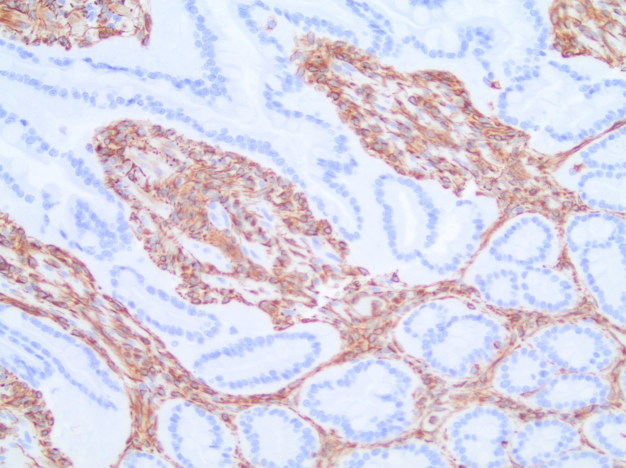
Immunohistochemistry-Frozen: Vimentin Antibody (V9) [NBP1-97670] - Strong specific staining of connective tissue cells in a frozen tissue section of human colon. No reactivity in epithelial cells.![Flow Cytometry: Vimentin Antibody (V9) - BSA Free [NBP1-97670]](https://resources.rndsystems.com/images/products/Vimentin-Antibody-V9-Flow-Cytometry-NBP1-97670-img0002.jpg "Flow Cytometry: Vimentin Antibody (V9) - BSA Free [NBP1-97670]")
Flow Cytometry: Vimentin Antibody (V9) - BSA Free [NBP1-97670]
Flow Cytometry: Vimentin Antibody (V9) [NBP1-97670] - An intracellular stain was performed on Jurkat cells with Vimentin antibody (V9) NBP1-97670PE (blue) and a matched isotype control NBP2-27287PE (orange). Cells were fixed with 4% PFA and then permeablized with 0.1% saponin. Cells were incubated in an antibody dilution of 1 ug/mL for 30 minutes at room temperature. Both antibodies were conjugated to phycoerythrin.![Immunocytochemistry/ Immunofluorescence: Vimentin Antibody (V9) - BSA Free [NBP1-97670]](https://resources.rndsystems.com/images/products/Vimentin-Antibody-V9-Immunocytochemistry-Immunofluorescence-NBP1-97670-img0003.jpg "Immunocytochemistry/ Immunofluorescence: Vimentin Antibody (V9) - BSA Free [NBP1-97670]")
Immunocytochemistry/ Immunofluorescence: Vimentin Antibody (V9) - BSA Free [NBP1-97670]
Immunocytochemistry/Immunofluorescence: Vimentin Antibody (V9) [NBP1-97670] - Strong specific staining of glomeruli and connetive tissue in human kidney with in a 1:200 dilution. No reactivity in epithelial cells.![Immunocytochemistry/ Immunofluorescence: Vimentin Antibody (V9) - BSA Free [NBP1-97670]](https://resources.rndsystems.com/images/products/Vimentin-Antibody-V9-Immunocytochemistry-Immunofluorescence-NBP1-97670-img0004.jpg "Immunocytochemistry/ Immunofluorescence: Vimentin Antibody (V9) - BSA Free [NBP1-97670]")
Immunocytochemistry/ Immunofluorescence: Vimentin Antibody (V9) - BSA Free [NBP1-97670]
Immunocytochemistry/Immunofluorescence: Vimentin Antibody (V9) [NBP1-97670] - Strong specific staining of connective tissue cells in swine colon with in a 1:200 dilution. No reactivity in epithelial cells.![Flow Cytometry: Vimentin Antibody (V9) - BSA Free [NBP1-97670]](https://resources.rndsystems.com/images/products/Vimentin-Antibody-V9-Flow-Cytometry-NBP1-97670-img0001.jpg "Flow Cytometry: Vimentin Antibody (V9) - BSA Free [NBP1-97670]")
Flow Cytometry: Vimentin Antibody (V9) - BSA Free [NBP1-97670]
Flow Cytometry: Vimentin Antibody (V9) [NBP1-97670] - Analysis of PE conjugate of NBP1-97670. An intracellular stain was performed on THP-1 cells with Vimentin antibody (V9) NBP1-97670PE (blue) and a matched isotype control NBP2-27287PE (orange). Cells were fixed with 4% PFA and then permeablized with 0.1% s [NBP1-97670] -")
Immunohistochemistry-Paraffin: Vimentin Antibody (V9) [NBP1-97670] -
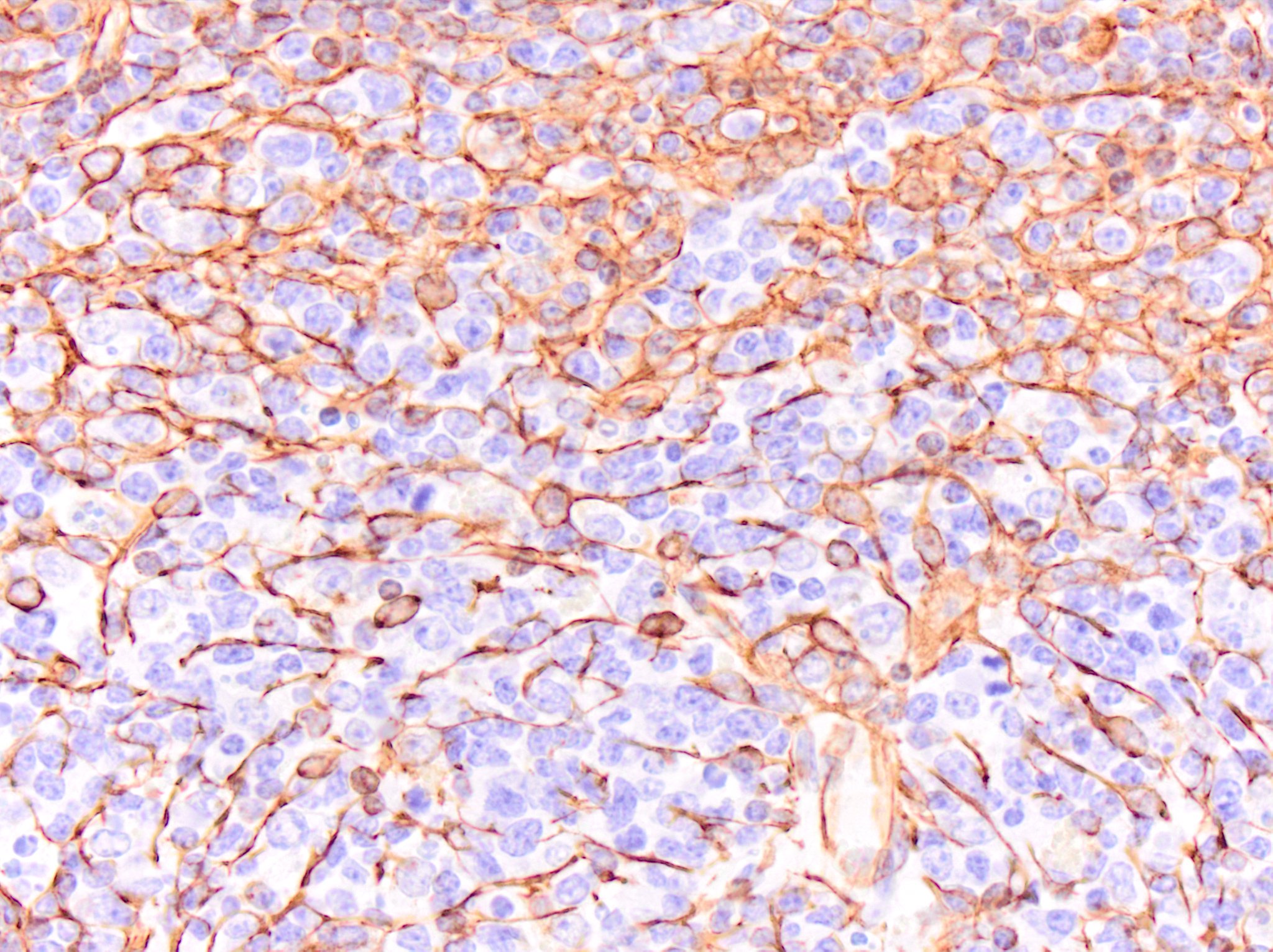
Immunohistochemistry-Paraffin: Vimentin Antibody (V9) [NBP1-97670] - Vimentin immunoreactivity in human tonsil. NBP1-97670 was diluted to 0.5ug/mL and was left on sections for 30m at room temperature. Secondary antibody was Horse-Anti Mouse HRP Veterinary Reagent. Image from verified customer review. [NBP1-97670] -")
Immunohistochemistry-Paraffin: Vimentin Antibody (V9) [NBP1-97670] -
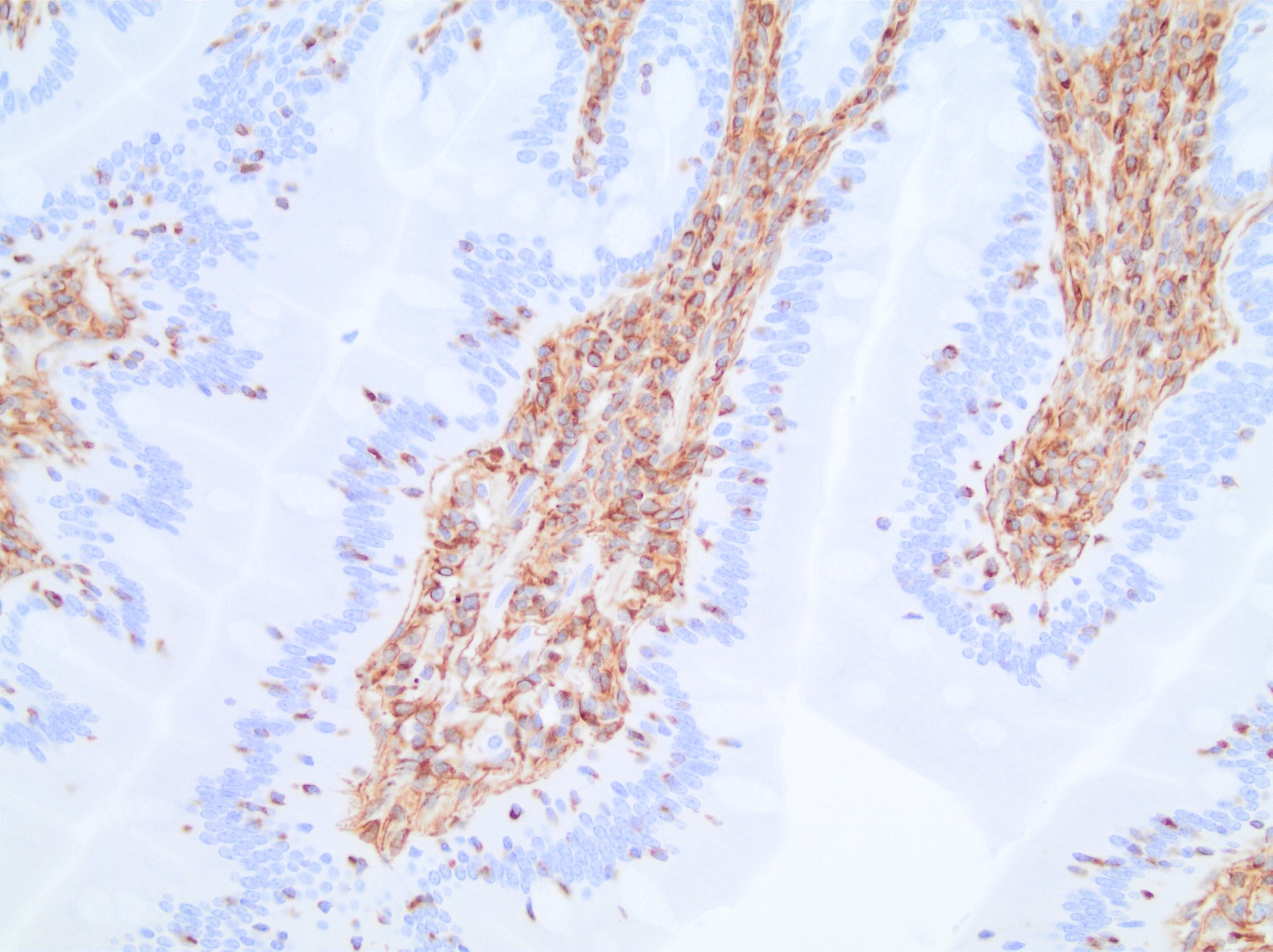
Immunohistochemistry-Paraffin: Vimentin Antibody (V9) [NBP1-97670] - Vimentin immunoreactivity in horse intestine. NBP1-97670 was used at a concentration of 0.5ug/mL and was left on slides for 30m at room temperature. Secondary antibody was Horse Anti-Mouse HRP Veterinary Reagent. Image from verified customer review. [NBP1-97670] -")
Immunohistochemistry-Paraffin: Vimentin Antibody (V9) [NBP1-97670] -
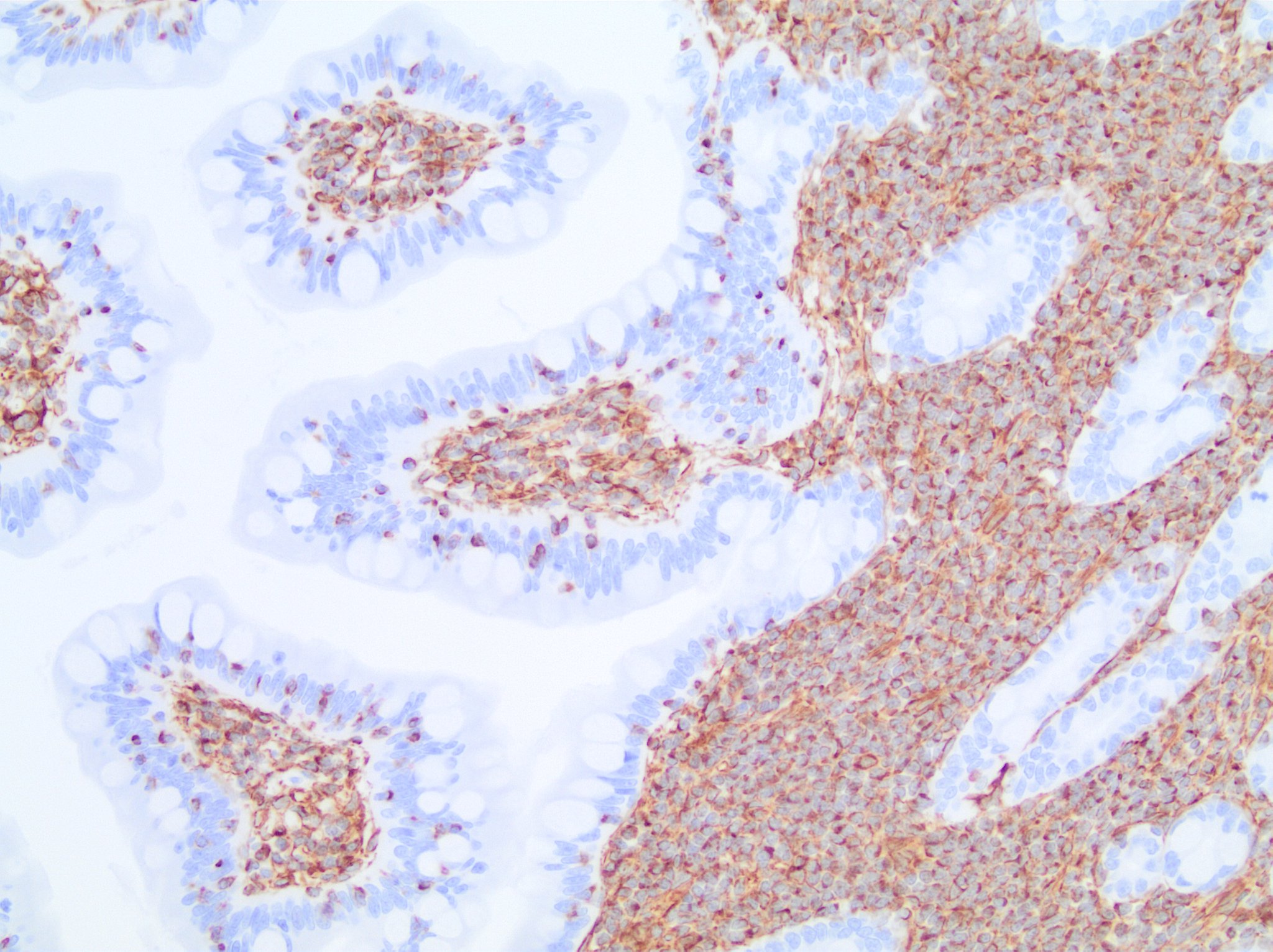
Immunohistochemistry-Paraffin: Vimentin Antibody (V9) [NBP1-97670] - Vimentin immunoreactivity in feline intestine. NBP1-97670 was used at a concentration of 0.5ug/mL. Secondary antibody was Horse Anti-Mouse HRP Veterinary Reagent. Image from verified customer review. [NBP1-97670] -")
Immunohistochemistry-Paraffin: Vimentin Antibody (V9) [NBP1-97670] -
Immunohistochemistry-Paraffin: Vimentin Antibody (V9) [NBP1-97670] - Vimentin immunoreactivity in canine intestine. NBP1-97670 was used at a concentration of 0.5ug/mL and was left on tissue sections for 30m at room temperature. Secondary was Horse Anti-Mouse Veterinary Reagent. Image from verified customer review. [NBP1-97670] -")
Immunohistochemistry-Paraffin: Mouse Monoclonal Vimentin Antibody (V9) [NBP1-97670] -
Immunohistochemistry-Paraffin: Mouse Monoclonal Vimentin Antibody (V9) [NBP1-97670] - Vimentin was stained in human FFPE tonsil tissue. HIER antigen retrieval at pH 9 for 20min was performed. Concentration used: 1:100. AF750 conjugated version of the antibody was used (Catalog # NBP1-97670AF750). Image from a verified customer review. [NBP1-97670]")
Immunohistochemistry-Paraffin: Mouse Monoclonal Vimentin Antibody (V9) [NBP1-97670]
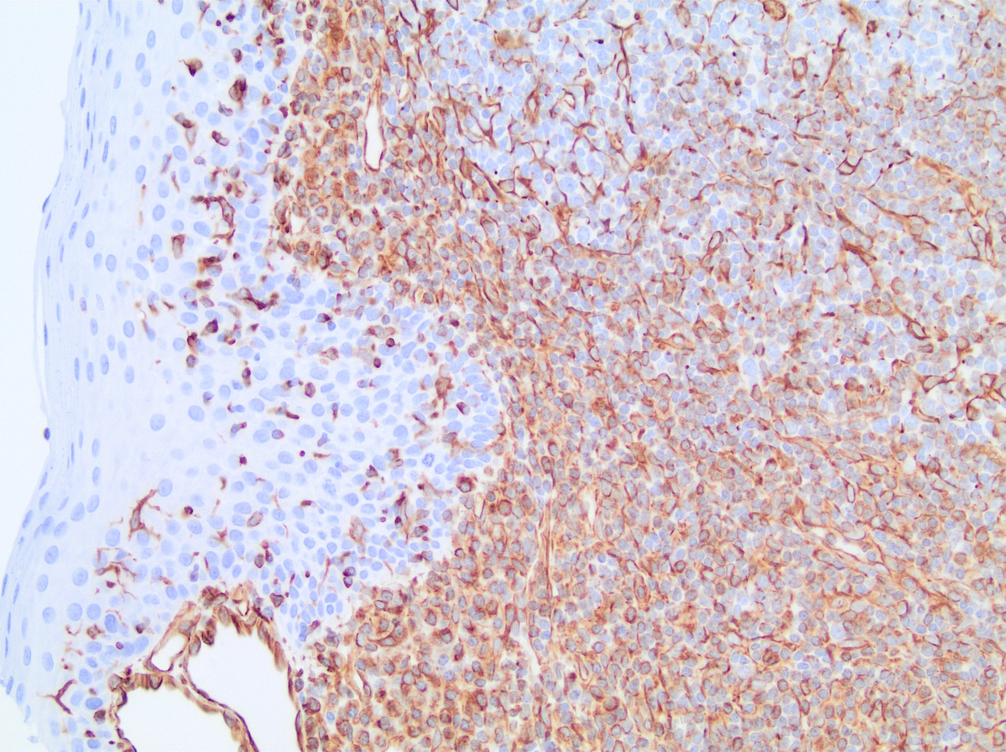
Vimentin immunoreactivity in an FFPE section of cow lymph node. NBP1-97670 was diluted to 0.5ug/mL and was left on tissue sections for 30min at room temperature. Image from a verified customer review. - BSA Free [NBP1-97670] -")
Immunocytochemistry/ Immunofluorescence: Vimentin Antibody (V9) - BSA Free [NBP1-97670] -
The effect of N-BLR knockdown on invasion by specific siRNAs. a N-BLR abundance is decreased in stably silenced clones. b Invasion assays at 36 h show significant reduction of stably silenced N-BLR invading cells. c Migration assay at 24 h identified also significant reduction in migration of stably silenced N-BLR clones. d The 12 most significantly differentially expressed genes for both upregulated and downregulated genes. The data originated from 44 K Agilent microarray where HCT116 stable shRNA N-BLR clones #3-1 and #4-7 were compared with HCT116 empty vector control clone. The probes recognizing E-cadherin and vimentin are in red and blue, respectively. e Confirmation of microarray data by real time PCR shows that E-cadherin is increased and vimentin is markedly decreased in stably silenced clones (#3-1 and #4-7). f E-cadherin, vimentin, and ZEB1 were identified in vitro by immunofluorescence with specific antibodies. Immunofluorescence signal of E-cadherin (green color) was markedly increased in both clones. The ZEB1 signal was present in cells with empty vector (green color) but not in clones #3-1 and #4-7. Blue color indicate nuclei. Single green, blue, and merged channel images of ZEB1 are reported in Additional file 3: Figure S9B. g ZEB1 mRNA downregulation in HCT116 stable shRNA N-BLR clones #3-1 and #4-7 compared with control HCT116 empty vector clone. h Western blotting for E-cadherin and ZEB1 measured in the same clones; vinculin was used as loading control. (Student’s t-test; *p < 0.05; **p < 0.01; ***p < 0.001; ****p < 0.0001) Image collected and cropped by CiteAb from the following open publication (https://pubmed.ncbi.nlm.nih.gov/28535802), licensed under a CC-BY license. Not internally tested by Novus Biologicals.Applications for Vimentin Antibody (V9) - BSA Free
Application
Recommended Usage
Flow Cytometry
1:25-1:200
Immunocytochemistry/ Immunofluorescence
1:10-1:500
Immunohistochemistry
1:10-1:500
Immunohistochemistry-Frozen
1:25-1:200
Immunohistochemistry-Paraffin
1:25-1:200
Western Blot
1:100-1:1000
Application Notes
For staining paraffin-embedded tissues pretreatment by boiling tissue sections for 10 minutes in 10 mM citrate buffer (pH 6.0) is required. Optimal antibody dilution should be determined by titration; recommended range is 1:25 - 1:200 for flow cytometry, and for immunohistochemistry with avidin-biotinylated Horseradish peroxidase complex (ABC) as detection reagent, and 1:100 - 1:1000 for immunoblotting applications.
This antibody is CyTOF ready..
This antibody is CyTOF ready..
Reviewed Applications
Read 5 reviews rated 5 using NBP1-97670 in the following applications:
Flow Cytometry Panel Builder
Bio-Techne Knows Flow Cytometry
Save time and reduce costly mistakes by quickly finding compatible reagents using the Panel Builder Tool.
Advanced Features
- Spectra Viewer - Custom analysis of spectra from multiple fluorochromes
- Spillover Popups - Visualize the spectra of individual fluorochromes
- Antigen Density Selector - Match fluorochrome brightness with antigen density

Formulation, Preparation, and Storage
Purification
Protein A or G purified
Formulation
PBS, pH 7.2
Format
BSA Free
Preservative
0.09% Sodium Azide
Concentration
1 mg/ml
Shipping
The product is shipped with polar packs. Upon receipt, store it immediately at the temperature recommended below.
Stability & Storage
Store at 4C short term. Aliquot and store at -20C long term. Avoid freeze-thaw cycles.
Background: Vimentin
Activated macrophages have been shown to secrete phosphorylated vimentin which can be stimulated by a variety of pathophysiological factors including oxidized low-density lipoproteins and TNF-alpha or inhibited by IL-10 (1). The vimentin protein is often expressed at the cell surface playing a role in cell-cell interactions, tissue damage and repair, immune response, and pathogen recognition (1). Vimentin functions in many cytoskeletal processes including cell migration, which is highlighted by its upregulation during epithelial-to-mesenchymal transition (EMT) (4,5). Vimentin is a commonly used marker for EMT and is expressed by many tumor types (5). For example, high metastasis of oral squamous cell carcinomas also showed high vimentin positive expression in immunohistochemical staining analysis (5). A number of vimentin targeting compounds are in cancer-related clinical trials, however, given the multifunctional role of vimentin, the effect of inhibition on non-malignant cells needs to be thoroughly examined (5).
References
1. Ramos, I., Stamatakis, K., Oeste, C. L., & Perez-Sala, D. (2020). Vimentin as a Multifaceted Player and Potential Therapeutic Target in Viral Infections. International Journal of Molecular Sciences. https://doi.org/10.3390/ijms21134675
2. Uniprot (P08670)
3. Morrow, C. S., & Moore, D. L. (2020). Vimentin's side gig: Regulating cellular proteostasis in mammalian systems. Cytoskeleton (Hoboken, N.J.). https://doi.org/10.1002/cm.21645
4. van Bodegraven, E. J., & Etienne-Manneville, S. (2020). Intermediate filaments against actomyosin: the david and goliath of cell migration. Current Opinion in Cell Biology. https://doi.org/10.1016/j.ceb.2020.05.006
5. Strouhalova, K., Prechova, M., Gandalovicova, A., Brabek, J., Gregor, M., & Rosel, D. (2020). Vimentin Intermediate Filaments as Potential Target for Cancer Treatment. Cancers. https://doi.org/10.3390/cancers12010184
Alternate Names
VIM
Gene Symbol
VIM
Additional Vimentin Products
Product Documents for Vimentin Antibody (V9) - BSA Free
Certificate of Analysis
To download a Certificate of Analysis, please enter a lot or batch number in the search box below.
Product Specific Notices for Vimentin Antibody (V9) - BSA Free
This product is for research use only and is not approved for use in humans or in clinical diagnosis. Primary Antibodies are guaranteed for 1 year from date of receipt.
Citations for Vimentin Antibody (V9) - BSA Free
 Powered by Bioz
Customer Reviews for Vimentin Antibody (V9) - BSA Free (5)
5 out of 5
5 Customer Ratings
Have you used Vimentin Antibody (V9) - BSA Free?
Submit a review and receive an Amazon gift card!
$25/€18/£15/$25CAN/¥2500 Yen for a review with an image
$10/€7/£6/$10CAN/¥1110 Yen for a review without an image
Submit a review
Customer Images
Showing
1
-
5 of
5 reviews
Showing All
Filter By:
-
Application: Immunohistochemistry-ParaffinSample Tested: Lymph NodeSpecies: CowVerified Customer | Posted 06/30/2025Vimentin immunoreactivity in an FFPE section of cow lymph node. NBP1-97670 was diluted to 0.5ug/mL and was left on tissue sections for 30min at room temperature.
 Bio-Techne ResponseThis review reflects a new species or application tested on a primary antibody.
 -
Application: Immunohistochemistry-ParaffinSample Tested: TonsilSpecies: HumanVerified Customer | Posted 06/05/2023Vimentin immunoreactivity in human tonsil. NBP1-97670 was diluted to 0.5ug/mL and was left on sections for 30m at room temperature. Secondary antibody was Horse-Anti Mouse HRP Veterinary Reagent.
-
Application: Immunohistochemistry-ParaffinSample Tested: IntestineSpecies: EquineVerified Customer | Posted 06/05/2023Vimentin immunoreactivity in horse intestine. NBP1-97670 was used at a concentration of 0.5ug/mL and was left on slides for 30m at room temperature. Secondary antibody was Horse Anti-Mouse HRP Veterinary Reagent.
Bio-Techne ResponseThis review was submitted through the legacy Novus Innovators Program, reflecting a new species or application tested on a primary antibody.
-
Application: Immunohistochemistry-ParaffinSample Tested: IntestineSpecies: FelineVerified Customer | Posted 06/05/2023Vimentin immunoreactivity in feline intestine. NBP1-97670 was used at a concentration of 0.5ug/mL. Secondary antibody was Horse Anti-Mouse HRP Veterinary Reagent.Requires heat-induced epitope retrieval. I used Target Retrieval Solution heated to 95 degrees Celsius using a vegetable steamer. Slides were left in pre-heated solution for 20m.
Bio-Techne ResponseThis review was submitted through the legacy Novus Innovators Program, reflecting a new species or application tested on a primary antibody.
-
Application: Immunohistochemistry-ParaffinSample Tested: IntestineSpecies: CanineVerified Customer | Posted 06/05/2023Vimentin immunoreactivity in canine intestine. NBP1-97670 was used at a concentration of 0.5ug/mL and was left on tissue sections for 30m at room temperature. Secondary was Horse Anti-Mouse Veterinary Reagent.
There are no reviews that match your criteria.
Protocols
Find general support by application which include: protocols, troubleshooting, illustrated assays, videos and webinars.
- 7-Amino Actinomycin D (7-AAD) Cell Viability Flow Cytometry Protocol
- Antigen Retrieval Protocol (PIER)
- Antigen Retrieval for Frozen Sections Protocol
- Appropriate Fixation of IHC/ICC Samples
- Cellular Response to Hypoxia Protocols
- Chromogenic IHC Staining of Formalin-Fixed Paraffin-Embedded (FFPE) Tissue Protocol
- Chromogenic Immunohistochemistry Staining of Frozen Tissue
- ClariTSA™ Fluorophore Kits
- Detection & Visualization of Antibody Binding
- Extracellular Membrane Flow Cytometry Protocol
- Flow Cytometry Protocol for Cell Surface Markers
- Flow Cytometry Protocol for Staining Membrane Associated Proteins
- Flow Cytometry Staining Protocols
- Flow Cytometry Troubleshooting Guide
- Fluorescent IHC Staining of Frozen Tissue Protocol
- Graphic Protocol for Heat-induced Epitope Retrieval
- Graphic Protocol for the Preparation and Fluorescent IHC Staining of Frozen Tissue Sections
- Graphic Protocol for the Preparation and Fluorescent IHC Staining of Paraffin-embedded Tissue Sections
- Graphic Protocol for the Preparation of Gelatin-coated Slides for Histological Tissue Sections
- ICC Cell Smear Protocol for Suspension Cells
- ICC Immunocytochemistry Protocol Videos
- ICC for Adherent Cells
- IHC Sample Preparation (Frozen sections vs Paraffin)
- Immunocytochemistry (ICC) Protocol
- Immunocytochemistry Troubleshooting
- Immunofluorescence of Organoids Embedded in Cultrex Basement Membrane Extract
- Immunofluorescent IHC Staining of Formalin-Fixed Paraffin-Embedded (FFPE) Tissue Protocol
- Immunohistochemistry (IHC) and Immunocytochemistry (ICC) Protocols
- Immunohistochemistry Frozen Troubleshooting
- Immunohistochemistry Paraffin Troubleshooting
- Intracellular Flow Cytometry Protocol Using Alcohol (Methanol)
- Intracellular Flow Cytometry Protocol Using Detergents
- Intracellular Nuclear Staining Flow Cytometry Protocol Using Detergents
- Intracellular Staining Flow Cytometry Protocol Using Alcohol Permeabilization
- Intracellular Staining Flow Cytometry Protocol Using Detergents to Permeabilize Cells
- Preparing Samples for IHC/ICC Experiments
- Preventing Non-Specific Staining (Non-Specific Binding)
- Primary Antibody Selection & Optimization
- Propidium Iodide Cell Viability Flow Cytometry Protocol
- Protocol for Heat-Induced Epitope Retrieval (HIER)
- Protocol for Liperfluo
- Protocol for Making a 4% Formaldehyde Solution in PBS
- Protocol for VisUCyte™ HRP Polymer Detection Reagent
- Protocol for the Characterization of Human Th22 Cells
- Protocol for the Characterization of Human Th9 Cells
- Protocol for the Fluorescent ICC Staining of Cell Smears - Graphic
- Protocol for the Fluorescent ICC Staining of Cultured Cells on Coverslips - Graphic
- Protocol for the Preparation & Fixation of Cells on Coverslips
- Protocol for the Preparation and Chromogenic IHC Staining of Frozen Tissue Sections
- Protocol for the Preparation and Chromogenic IHC Staining of Frozen Tissue Sections - Graphic
- Protocol for the Preparation and Chromogenic IHC Staining of Paraffin-embedded Tissue Sections
- Protocol for the Preparation and Chromogenic IHC Staining of Paraffin-embedded Tissue Sections - Graphic
- Protocol for the Preparation and Fluorescent ICC Staining of Cells on Coverslips
- Protocol for the Preparation and Fluorescent ICC Staining of Non-adherent Cells
- Protocol for the Preparation and Fluorescent ICC Staining of Stem Cells on Coverslips
- Protocol for the Preparation and Fluorescent IHC Staining of Frozen Tissue Sections
- Protocol for the Preparation and Fluorescent IHC Staining of Paraffin-embedded Tissue Sections
- Protocol for the Preparation of Gelatin-coated Slides for Histological Tissue Sections
- Protocol for the Preparation of a Cell Smear for Non-adherent Cell ICC - Graphic
- Protocol: Annexin V and PI Staining by Flow Cytometry
- Protocol: Annexin V and PI Staining for Apoptosis by Flow Cytometry
- R&D Systems Quality Control Western Blot Protocol
- TUNEL and Active Caspase-3 Detection by IHC/ICC Protocol
- The Importance of IHC/ICC Controls
- Troubleshooting Guide: Fluorokine Flow Cytometry Kits
- Troubleshooting Guide: Immunohistochemistry
- Troubleshooting Guide: Western Blot Figures
- Western Blot Conditions
- Western Blot Protocol
- Western Blot Protocol for Cell Lysates
- Western Blot Troubleshooting
- Western Blot Troubleshooting Guide
- View all Protocols, Troubleshooting, Illustrated assays and Webinars

