The human ANPEP gene encodes aminopeptidase N (APN), which is also known as microsomal aminopeptidase, alanyl aminopeptidase, aminopeptidase M, CD13, or membrane protein p161 (1‑3). The deduced amino acid sequence of human APN consists of a short cytoplasmic tail (residues 2 to 8), a transmembrane region (residue 9 to 32), a Ser/Thr rich region and a zinc metalloprotease domain (residues 69 to 966). The amino acid sequence of human APN is 78% and 77% identical to that of rat and mouse, respectively. Widely expressed in many cells, tissues and species, APN cleaves the N-terminal amino acids from bioactive peptides, leading to their inactivation or degradation. The roles of APN in many fields, such as neuroscience, hematopoeitic cells, immune system, angiogenesis, cancer and viral infection, have been reviewed (3).
Human Aminopeptidase N/CD13 Antibody
R&D Systems | Catalog # MAB3815

Key Product Details
Validated by
Knockout/Knockdown
Species Reactivity
Validated:
Human
Cited:
Human
Applications
Validated:
Knockout Validated, Immunohistochemistry, Western Blot, Immunocytochemistry, Simple Western
Cited:
Immunohistochemistry, Immunohistochemistry-Paraffin, Western Blot, Immunocytochemistry, Co-Immunoprecipitation
Label
Unconjugated
Antibody Source
Monoclonal Mouse IgG2A Clone # 498001
Loading...
Product Specifications
Immunogen
Mouse myeloma cell line NS0-derived recombinant human Aminopeptidase N/CD13
Lys69-Lys967
Accession # AAA51719
Lys69-Lys967
Accession # AAA51719
Specificity
Detects human Aminopeptidase N/CD13 in direct ELISAs and Western blots. In direct ELISAs and Western blots, no cross-reactivity with recombinant mouse Aminopeptidase N is observed.
Clonality
Monoclonal
Host
Mouse
Isotype
IgG2A
Scientific Data Images for Human Aminopeptidase N/CD13 Antibody
Detection of Human Aminopeptidase N/CD13 by Western Blot.
Western blot shows lysates of human kidney tissue and human prostate tissue. PVDF membrane was probed with 2 µg/mL of Mouse Anti-Human Aminopeptidase N/CD13 Monoclonal Antibody (Catalog # MAB3815) followed by HRP-conjugated Anti-Mouse IgG Secondary Antibody (Catalog # HAF018). A specific band was detected for Aminopeptidase N/CD13 at approximately 150 kDa (as indicated). This experiment was conducted under reducing conditions and using Immunoblot Buffer Group 1.
Aminopeptidase N/CD13 in SH-SY5Y Human Cell Line.
Aminopeptidase N/CD13 was detected in immersion fixed SH-SY5Y human neuroblastoma cell line using Mouse Anti-Human Aminopeptidase N/CD13 Monoclonal Antibody (Catalog # MAB3815) at 8 µg/mL for 3 hours at room temperature. Cells were stained using the NorthernLights™ 557-conjugated Anti-Mouse IgG Secondary Antibody (red; Catalog # NL007) and counterstained with DAPI (blue). Specific staining was localized to cytoplasm and plasma membranes. View our protocol for Fluorescent ICC Staining of Cells on Coverslips.
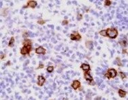
Aminopeptidase N/CD13 in Human Liver Cancer Tissue.
Aminopeptidase N/CD13 was detected in immersion fixed paraffin-embedded sections of human liver cancer tissue using 25 µg/mL Mouse Anti-Human Aminopeptidase N/CD13 Monoclonal Antibody (Catalog # MAB3815) overnight at 4 °C. Tissue was stained with the Anti-Mouse HRP-DAB Cell & Tissue Staining Kit (brown; Catalog # CTS002) and counterstained with hematoxylin (blue). View our protocol for Chromogenic IHC Staining of Paraffin-embedded Tissue Sections.
Aminopeptidase N/CD13 in Human Brain.
Aminopeptidase N/CD13 was detected in immersion fixed paraffin-embedded sections of human brain (cortex) using Mouse Anti-Human Aminopeptidase N/CD13 Monoclonal Antibody (Catalog # MAB3815) at 1.7 µg/mL overnight at 4 °C. Tissue was stained using the Anti-Mouse HRP-DAB Cell & Tissue Staining Kit (brown; Catalog # CTS002) and counterstained with hematoxylin (blue). Specific staining was localized to blood vessels. View our protocol for Chromogenic IHC Staining of Paraffin-embedded Tissue Sections.
Detection of Human Aminopeptidase N/CD13 by Simple WesternTM.
Simple Western lane view shows lysates of human small intestine tissue and human prostate tissue, loaded at 0.5 mg/mL. A specific band was detected for Aminopeptidase N/CD13 at approximately 204 kDa (as indicated) using 40 µg/mL of Mouse Anti-Human Aminopeptidase N/CD13 Monoclonal Antibody (Catalog # MAB3815). This experiment was conducted under reducing conditions and using the 12-230 kDa separation system.
Western Blot Shows Human Aminopeptidase N/CD13 Specificity by Using Knockout Cell Line.
Western blot shows lysates of U937 human histiocytic lymphoma cell line and human APN knockout U937 human histiocytic lymphoma cell line (KO). PVDF membrane was probed with 2 µg/mL of Mouse Anti-Human Aminopeptidase N/CD13 Monoclonal Antibody (Catalog # MAB3815) followed by HRP-conjugated Anti-Mouse IgG Secondary Antibody (HAF018). A specific band was detected for Aminopeptidase N/CD13 at approximately 150 kDa (as indicated) in the parental U937 human histiocytic lymphoma cell line, but is not detectable in knockout U937 human histiocytic lymphoma cell line. GAPDH (MAB5718) is shown as a loading control. This experiment was conducted under reducing conditions and using Western Blot Buffer Group 1.Applications for Human Aminopeptidase N/CD13 Antibody
Application
Recommended Usage
Immunocytochemistry
5-25 µg/mL
Sample: Immersion fixed SH‑SY5Y human neuroblastoma cell line
Sample: Immersion fixed SH‑SY5Y human neuroblastoma cell line
Immunohistochemistry
1-25 µg/mL
Sample: Immersion fixed paraffin-embedded sections of human liver cancer tissue and immersion fixed paraffin-embedded sections of human brain (cortex)
Sample: Immersion fixed paraffin-embedded sections of human liver cancer tissue and immersion fixed paraffin-embedded sections of human brain (cortex)
Knockout Validated
Aminopeptidase N/CD13
is specifically detected in U937 human histiocytic lymphoma parental cell
line but is not detectable in Aminopeptidase N/CD13 knockout U937
human histiocytic lymphoma cell line.
Simple Western
40 µg/mL
Sample: Human small intestine tissue and Human prostate tissue
Sample: Human small intestine tissue and Human prostate tissue
Western Blot
2 µg/mL
Sample: Human kidney tissue and human prostate tissue
Sample: Human kidney tissue and human prostate tissue
Reviewed Applications
Read 1 review rated 5 using MAB3815 in the following applications:
Formulation, Preparation, and Storage
Purification
Protein A or G purified from hybridoma culture supernatant
Reconstitution
Reconstitute at 0.5 mg/mL in sterile PBS. For liquid material, refer to CoA for concentration.
Loading...
Formulation
Lyophilized from a 0.2 μm filtered solution in PBS with Trehalose. *Small pack size (SP) is supplied either lyophilized or as a 0.2 µm filtered solution in PBS.
Shipping
Lyophilized product is shipped at ambient temperature. Liquid small pack size (-SP) is shipped with polar packs. Upon receipt, store immediately at the temperature recommended below.
Stability & Storage
Use a manual defrost freezer and avoid repeated freeze-thaw cycles.
- 12 months from date of receipt, -20 to -70 °C as supplied.
- 1 month, 2 to 8 °C under sterile conditions after reconstitution.
- 6 months, -20 to -70 °C under sterile conditions after reconstitution.
Calculators
Background: Aminopeptidase N/CD13
References
- Olsen, J. et al. (1988) FEBS Lett. 238:307.
- Look, A.T. et al. (1989) J. Clin. Invest. 83:1299.
- Turner, A.J. (2004) in Handbook of Proteolytic Enzymes (ed. Barrett, et al.) pp. 289, Academic Press, San Diego.
Alternate Names
Aminopeptidase M, ANPEP, APN, CD13, gp150, PEPN
Gene Symbol
ANPEP
UniProt
Additional Aminopeptidase N/CD13 Products
Product Documents for Human Aminopeptidase N/CD13 Antibody
Certificate of Analysis
To download a Certificate of Analysis, please enter a lot or batch number in the search box below.
Note: Certificate of Analysis not available for kit components.
Product Specific Notices for Human Aminopeptidase N/CD13 Antibody
For research use only
Citations for Human Aminopeptidase N/CD13 Antibody
 Powered by Bioz
Customer Reviews for Human Aminopeptidase N/CD13 Antibody (1)
5 out of 5
1 Customer Rating
Have you used Human Aminopeptidase N/CD13 Antibody?
Submit a review and receive an Amazon gift card!
$25/€18/£15/$25CAN/¥2500 Yen for a review with an image
$10/€7/£6/$10CAN/¥1110 Yen for a review without an image
Submit a review
Customer Images
Showing
1
-
1 的
1 review
Showing All
Filter By:
-
Application: ImmunohistochemistrySample Tested: Liver cancer tissueSpecies: HumanVerified Customer | Posted 01/26/2022

There are no reviews that match your criteria.
Protocols
Find general support by application which include: protocols, troubleshooting, illustrated assays, videos and webinars.
- Antigen Retrieval Protocol (PIER)
- Antigen Retrieval for Frozen Sections Protocol
- Appropriate Fixation of IHC/ICC Samples
- Cellular Response to Hypoxia Protocols
- Chromogenic IHC Staining of Formalin-Fixed Paraffin-Embedded (FFPE) Tissue Protocol
- Chromogenic Immunohistochemistry Staining of Frozen Tissue
- ClariTSA™ Fluorophore Kits
- Detection & Visualization of Antibody Binding
- Fluorescent IHC Staining of Frozen Tissue Protocol
- Graphic Protocol for Heat-induced Epitope Retrieval
- Graphic Protocol for the Preparation and Fluorescent IHC Staining of Frozen Tissue Sections
- Graphic Protocol for the Preparation and Fluorescent IHC Staining of Paraffin-embedded Tissue Sections
- Graphic Protocol for the Preparation of Gelatin-coated Slides for Histological Tissue Sections
- ICC Cell Smear Protocol for Suspension Cells
- ICC Immunocytochemistry Protocol Videos
- ICC for Adherent Cells
- IHC Sample Preparation (Frozen sections vs Paraffin)
- Immunocytochemistry (ICC) Protocol
- Immunocytochemistry Troubleshooting
- Immunofluorescence of Organoids Embedded in Cultrex Basement Membrane Extract
- Immunofluorescent IHC Staining of Formalin-Fixed Paraffin-Embedded (FFPE) Tissue Protocol
- Immunohistochemistry (IHC) and Immunocytochemistry (ICC) Protocols
- Immunohistochemistry Frozen Troubleshooting
- Immunohistochemistry Paraffin Troubleshooting
- Preparing Samples for IHC/ICC Experiments
- Preventing Non-Specific Staining (Non-Specific Binding)
- Primary Antibody Selection & Optimization
- Protocol for Heat-Induced Epitope Retrieval (HIER)
- Protocol for Making a 4% Formaldehyde Solution in PBS
- Protocol for VisUCyte™ HRP Polymer Detection Reagent
- Protocol for the Fluorescent ICC Staining of Cell Smears - Graphic
- Protocol for the Fluorescent ICC Staining of Cultured Cells on Coverslips - Graphic
- Protocol for the Preparation & Fixation of Cells on Coverslips
- Protocol for the Preparation and Chromogenic IHC Staining of Frozen Tissue Sections
- Protocol for the Preparation and Chromogenic IHC Staining of Frozen Tissue Sections - Graphic
- Protocol for the Preparation and Chromogenic IHC Staining of Paraffin-embedded Tissue Sections
- Protocol for the Preparation and Chromogenic IHC Staining of Paraffin-embedded Tissue Sections - Graphic
- Protocol for the Preparation and Fluorescent ICC Staining of Cells on Coverslips
- Protocol for the Preparation and Fluorescent ICC Staining of Non-adherent Cells
- Protocol for the Preparation and Fluorescent ICC Staining of Stem Cells on Coverslips
- Protocol for the Preparation and Fluorescent IHC Staining of Frozen Tissue Sections
- Protocol for the Preparation and Fluorescent IHC Staining of Paraffin-embedded Tissue Sections
- Protocol for the Preparation of Gelatin-coated Slides for Histological Tissue Sections
- Protocol for the Preparation of a Cell Smear for Non-adherent Cell ICC - Graphic
- R&D Systems Quality Control Western Blot Protocol
- TUNEL and Active Caspase-3 Detection by IHC/ICC Protocol
- The Importance of IHC/ICC Controls
- Troubleshooting Guide: Immunohistochemistry
- Troubleshooting Guide: Western Blot Figures
- Western Blot Conditions
- Western Blot Protocol
- Western Blot Protocol for Cell Lysates
- Western Blot Troubleshooting
- Western Blot Troubleshooting Guide
- View all Protocols, Troubleshooting, Illustrated assays and Webinars
Loading...
Associated Pathways
