
CEACAM-5, also known as CEA and CD66e, belongs to the large family of CEACAM and pregnancy specific glycoproteins. CEACAM molecules are either transmembrane or GPI-linked, and are differentially expressed between species (1, 2). Orthologs of human CEACAM‑5 have not been described in other species. CEACAM-5, which is expressed primarily by epithelial cells, consists of an N-terminal Ig-like V-set domain followed by six Ig-like C2-set domains and a GPI anchor (2‑4). CEACAM-5 is synthesized as a 180 kDa, variably glycosylated molecule of which approximately 60% is carbohydrate (5). CEACAM-5 functions as a calcium‑independent adhesion molecule through homophilic and heterophilic interactions with CEACAM-1 (6‑8). CEACAM-5 is restricted to the apical face of intestinal epithelial cells in the adult but is more diffuse during embryonic development and in tumors (7). This is consistent with a role in the development and maintenance of epithelial architecture. CEACAM-5 is upregulated in a wide variety of human tumors and is a commonly used cancer marker (9). It promotes tumor cell migration, invasion, adhesion, and metastasis (10). It also contributes to tumor formation by maintaining cellular proliferation in the presence of differentiation stimuli, and by blocking apoptosis following loss of ECM anchorage (anoikis) (11, 12). The GPI anchoring of CEACAM-5 can be released by GPI-PLD, resulting in a soluble molecule that also promotes tumor metastasis (13). Cell surface expression of CEACAM-5 on tumor cells prevents the adhesion of CEACAM-1 expressing NK cells and provides protection from NK‑mediated lysis (6). CEACAM-5 also binds a subset of Neisseria opacity proteins (Opa) and E. coli adhesion proteins (14‑16). These interactions trigger clustering of the lipid raft-localized CEACAM-5 to sites of pathogen contact (15, 16).
Human CEACAM-5/CD66e Antibody (487609)
R&D Systems | Catalog # MAB41281

Scientific images may be resized, cropped, or adjusted for brightness or contrast for presentation purposes. These updates are limited to presentation and do not affect the underlying scientific interpretation of the data. Where available, additional source data may be provided upon request.
Key Product Details
Species Reactivity
Validated:
Cited:
Applications
Validated:
Cited:
Label
Antibody Source
Product Specifications
Immunogen
Lys35-Ala685
Accession # ABM87752
Specificity
Clonality
Host
Isotype
Scientific Data Images for Human CEACAM-5/CD66e Antibody (487609)
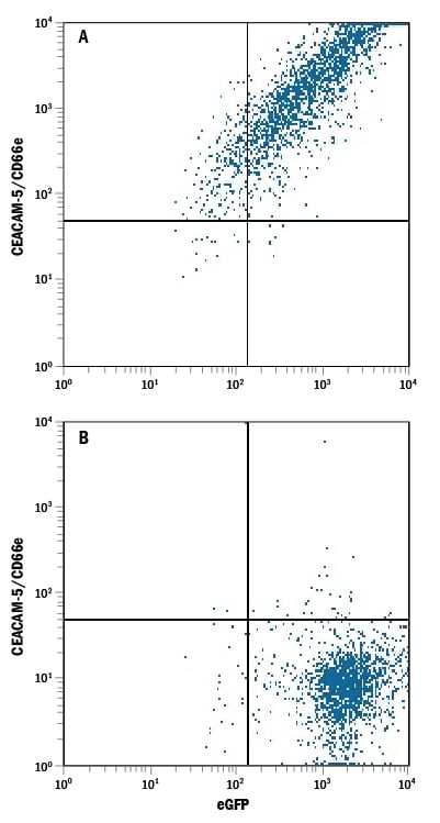
Detection of CEACAM‑5/CD66e in HEK293 Human Cell Line Transfected with Human CEACAM-5/CD66e and eGFP by Flow Cytometry.
HEK293 human embryonic kidney cell line transfected with either (A) human CEACAM-5/CD66e or (B) human CEACAM-8/CD66b and eGFP was stained with Mouse Anti-Human CEACAM-5/CD66e Monoclonal Antibody (Catalog # MAB41281) followed by Allophycocyanin-conjugated Anti-Mouse IgG Secondary Antibody (Catalog # F0101B). Quadrant markers were set based on control antibody staining (Catalog # MAB003).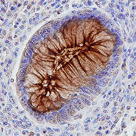
CEACAM‑5/CD66e in Human Colon.
CEACAM-5/CD66e was detected in immersion fixed paraffin-embedded sections of human colon using Mouse Anti-Human CEACAM-5/CD66e Monoclonal Antibody (Catalog # MAB41281) at 10 µg/mL for 1 hour at room temperature followed by incubation with the Anti-Mouse IgG VisUCyte™ HRP Polymer Antibody (Catalog # VC001). Tissue was stained using DAB (brown) and counterstained with hematoxylin (blue). Specific staining was localized to cell membranes. View our protocol for IHC Staining with VisUCyte HRP Polymer Detection Reagents.Applications for Human CEACAM-5/CD66e Antibody (487609)
CyTOF-ready
Flow Cytometry
Sample: HEK293 human embryonic kidney cell line transfected with human CEACAM-5/CD66e and eGFP
Immunohistochemistry
Sample: Immersion fixed paraffin-embedded sections of human colon
Western Blot
Sample: Recombinant Human CEACAM‑5/CD66e (Catalog # 4128-CM)
Reviewed Applications
Read 5 reviews rated 4.8 using MAB41281 in the following applications:
Flow Cytometry Panel Builder
Bio-Techne Knows Flow Cytometry
Save time and reduce costly mistakes by quickly finding compatible reagents using the Panel Builder Tool.
Advanced Features
- Spectra Viewer - Custom analysis of spectra from multiple fluorochromes
- Spillover Popups - Visualize the spectra of individual fluorochromes
- Antigen Density Selector - Match fluorochrome brightness with antigen density

Formulation, Preparation, and Storage
Purification
Reconstitution
Reconstitute at 0.5 mg/mL in sterile PBS. For liquid material, refer to CoA for concentration.
Formulation
Shipping
Stability & Storage
- 12 months from date of receipt, -20 to -70 °C as supplied.
- 1 month, 2 to 8 °C under sterile conditions after reconstitution.
- 6 months, -20 to -70 °C under sterile conditions after reconstitution.
Calculators
Background: CEACAM-5/CD66e
References
- Zebhauser, R. et al. (2005) Genomics 86:566.
- Hammarstrom, S. (1999) Semin. Cancer Biol. 9:67.
- Schrewe H. et al. (1990) Mol. Cell. Biol. 10:2738.
- Hefta, S.A. et al. (1988) Proc. Natl. Acad. Sci. 85:4648.
- Garcia, M. et al. (1991) Cancer Res. 51:5679.
- Stern, N. et al. (2005) J. Immunol. 174:6692.
- Benchimol, S. et al. (1989) Cell 57:327.
- Zhou, H. et al. (1993) J. Cell Biol. 122:951.
- Goldenberg, D.M. et al. (1976) J. Natl. Cancer Inst. 57:11.
- Blumenthal, R.D. et al. (2005) Cancer Res. 65:8809.
- Screaton, R.A. et al. (1997) J. Cell Biol. 137:939.
- Ordonez, C. et al. (2000) Cancer Res. 60:3419.
- Yamamoto, Y. et al. (2005) Biochem. Biophys. Res. Commun. 333:223.
- Chen, T. et al. (1997) J. Exp. Med. 185:1557.
- Bos, M.P. et al. (1997) Infect. Immun. 65:2353.
- Berger, C.N. et al. (2004) Mol. Microbiol. 52:963.
Long Name
Alternate Names
Gene Symbol
UniProt
Additional CEACAM-5/CD66e Products
Product Documents for Human CEACAM-5/CD66e Antibody (487609)
Certificate of Analysis
To download a Certificate of Analysis, please enter a lot or batch number in the search box below.
Note: Certificate of Analysis not available for kit components.
Product Specific Notices for Human CEACAM-5/CD66e Antibody (487609)
For research use only
Citations for Human CEACAM-5/CD66e Antibody (487609)
 Powered by Bioz
Customer Reviews for Human CEACAM-5/CD66e Antibody (487609) (5)
Have you used Human CEACAM-5/CD66e Antibody (487609)?
Submit a review and receive an Amazon gift card!
$25/€18/£15/$25CAN/¥2500 for a review with an image
$10/€7/£6/$10CAN/¥1110 for a review without an image
Submit a review
Customer Images
-
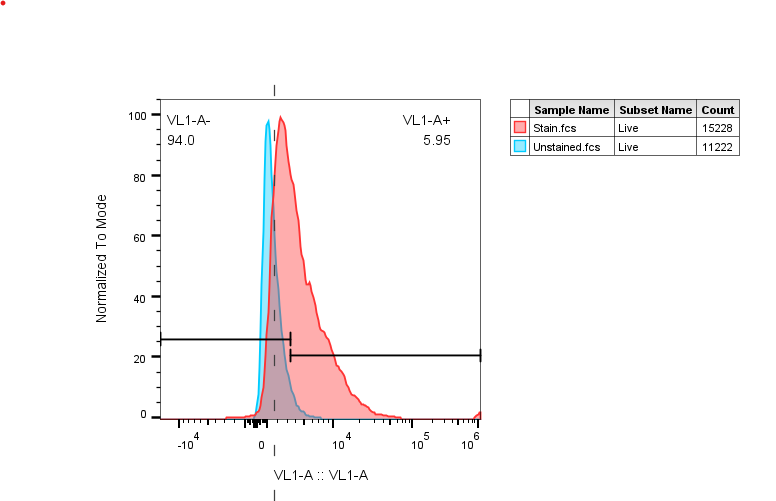
Application: Flow CytometrySample Tested: Colon cancer cell lineSpecies: HumanVerified Customer | Posted 07/29/2025Unstained vs stained in mid-CEA-expressing cell line

-
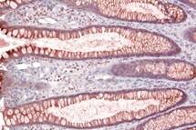
Application: ImmunohistochemistrySample Tested: Colon tissueSpecies: HumanVerified Customer | Posted 10/02/2021
-
Application: Western BlotSample Tested: Tumor cell lineSpecies: HumanVerified Customer | Posted 08/16/2021
-
Application: ELISASample Tested: tumor cell linesSpecies: HumanVerified Customer | Posted 03/23/2018
-
Application: ELISASample Tested: Human recombinant antibodySpecies: HumanVerified Customer | Posted 10/31/2017
There are no reviews that match your criteria.
Protocols
Find general support by application which include: protocols, troubleshooting, illustrated assays, videos and webinars.
- 7-Amino Actinomycin D (7-AAD) Cell Viability Flow Cytometry Protocol
- Antigen Retrieval Protocol (PIER)
- Antigen Retrieval for Frozen Sections Protocol
- Appropriate Fixation of IHC/ICC Samples
- Cellular Response to Hypoxia Protocols
- Chromogenic IHC Staining of Formalin-Fixed Paraffin-Embedded (FFPE) Tissue Protocol
- Chromogenic Immunohistochemistry Staining of Frozen Tissue
- ClariTSA™ Fluorophore Kits
- Detection & Visualization of Antibody Binding
- Extracellular Membrane Flow Cytometry Protocol
- Flow Cytometry Protocol for Cell Surface Markers
- Flow Cytometry Protocol for Staining Membrane Associated Proteins
- Flow Cytometry Staining Protocols
- Flow Cytometry Troubleshooting Guide
- Fluorescent IHC Staining of Frozen Tissue Protocol
- Graphic Protocol for Heat-induced Epitope Retrieval
- Graphic Protocol for the Preparation and Fluorescent IHC Staining of Frozen Tissue Sections
- Graphic Protocol for the Preparation and Fluorescent IHC Staining of Paraffin-embedded Tissue Sections
- Graphic Protocol for the Preparation of Gelatin-coated Slides for Histological Tissue Sections
- IHC Sample Preparation (Frozen sections vs Paraffin)
- Immunofluorescent IHC Staining of Formalin-Fixed Paraffin-Embedded (FFPE) Tissue Protocol
- Immunohistochemistry (IHC) and Immunocytochemistry (ICC) Protocols
- Immunohistochemistry Frozen Troubleshooting
- Immunohistochemistry Paraffin Troubleshooting
- Intracellular Flow Cytometry Protocol Using Alcohol (Methanol)
- Intracellular Flow Cytometry Protocol Using Detergents
- Intracellular Nuclear Staining Flow Cytometry Protocol Using Detergents
- Intracellular Staining Flow Cytometry Protocol Using Alcohol Permeabilization
- Intracellular Staining Flow Cytometry Protocol Using Detergents to Permeabilize Cells
- Preparing Samples for IHC/ICC Experiments
- Preventing Non-Specific Staining (Non-Specific Binding)
- Primary Antibody Selection & Optimization
- Propidium Iodide Cell Viability Flow Cytometry Protocol
- Protocol for Heat-Induced Epitope Retrieval (HIER)
- Protocol for Liperfluo
- Protocol for Making a 4% Formaldehyde Solution in PBS
- Protocol for VisUCyte™ HRP Polymer Detection Reagent
- Protocol for the Characterization of Human Th22 Cells
- Protocol for the Characterization of Human Th9 Cells
- Protocol for the Preparation & Fixation of Cells on Coverslips
- Protocol for the Preparation and Chromogenic IHC Staining of Frozen Tissue Sections
- Protocol for the Preparation and Chromogenic IHC Staining of Frozen Tissue Sections - Graphic
- Protocol for the Preparation and Chromogenic IHC Staining of Paraffin-embedded Tissue Sections
- Protocol for the Preparation and Chromogenic IHC Staining of Paraffin-embedded Tissue Sections - Graphic
- Protocol for the Preparation and Fluorescent IHC Staining of Frozen Tissue Sections
- Protocol for the Preparation and Fluorescent IHC Staining of Paraffin-embedded Tissue Sections
- Protocol for the Preparation of Gelatin-coated Slides for Histological Tissue Sections
- Protocol: Annexin V and PI Staining by Flow Cytometry
- Protocol: Annexin V and PI Staining for Apoptosis by Flow Cytometry
- R&D Systems Quality Control Western Blot Protocol
- TUNEL and Active Caspase-3 Detection by IHC/ICC Protocol
- The Importance of IHC/ICC Controls
- Troubleshooting Guide: Fluorokine Flow Cytometry Kits
- Troubleshooting Guide: Immunohistochemistry
- Troubleshooting Guide: Western Blot Figures
- Western Blot Conditions
- Western Blot Protocol
- Western Blot Protocol for Cell Lysates
- Western Blot Troubleshooting
- Western Blot Troubleshooting Guide
- View all Protocols, Troubleshooting, Illustrated assays and Webinars
Associated Pathways
