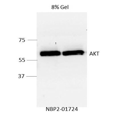
![Western Blot: AKT1 Antibody (OTI4D6) [NBP2-01724]](https://resources.rndsystems.com/images/products/AKT1-Antibody-4D6-Western-Blot-NBP2-01724-img0009.jpg "Western Blot: AKT1 Antibody (OTI4D6) [NBP2-01724]")
Key Product Details
Species Reactivity
Validated:
Human, Mouse, Rat, Canine, Monkey
Cited:
Mouse, Rat
Applications
Validated:
Immunohistochemistry, Immunohistochemistry-Paraffin, Western Blot, Flow Cytometry, Immunocytochemistry/ Immunofluorescence, Simple Western
Cited:
Western Blot
Label
Unconjugated
Antibody Source
Monoclonal Mouse IgG1 Clone # OTI4D6
Loading...
Product Specifications
Immunogen
This AKT1 antibody is made to the full length human recombinant protein of human AKT1(NP_005154) produced in HEK293T cell.
Reactivity Notes
Please note that this antibody is reactive to Mouse and derived from the same host, Mouse. Mouse-On-Mouse blocking reagent may be needed for IHC and ICC experiments to reduce high background signal. You can find these reagents under catalog numbers PK-2200-NB and MP-2400-NB. Please contact Technical Support if you have any questions.
Specificity
This antibody also cross-reacts with AKT3.
Clonality
Monoclonal
Host
Mouse
Isotype
IgG1
Theoretical MW
55.7 kDa.
Disclaimer note: The observed molecular weight of the protein may vary from the listed predicted molecular weight due to post translational modifications, post translation cleavages, relative charges, and other experimental factors.
Disclaimer note: The observed molecular weight of the protein may vary from the listed predicted molecular weight due to post translational modifications, post translation cleavages, relative charges, and other experimental factors.
Scientific Data Images for AKT1 Antibody (OTI4D6)
Western Blot: AKT1 Antibody (OTI4D6) [NBP2-01724]
Western Blot: AKT1 Antibody (OTI4D6) [NBP2-01724] - WB analysis of extracts (35 ug) from 9 different cell lines using AKT1 antibody.![Western Blot: AKT1 Antibody (OTI4D6) [NBP2-01724]](https://resources.rndsystems.com/images/products/AKT1-Antibody-4D6-Western-Blot-NBP2-01724-img0010.jpg "Western Blot: AKT1 Antibody (OTI4D6) [NBP2-01724]")
Western Blot: AKT1 Antibody (OTI4D6) [NBP2-01724]
Western Blot: AKT1 Antibody (OTI4D6) [NBP2-01724] - WB detection of AKT1 in human MD-MB-231 cells using AKT1 antibody NBP2-01724 at a dilution of 1:2000. WB image submitted by a verified customer review.![Immunocytochemistry/ Immunofluorescence: AKT1 Antibody (OTI4D6) [NBP2-01724]](https://resources.rndsystems.com/images/products/AKT1-Antibody-4D6-Immunocytochemistry-Immunofluorescence-NBP2-01724-img0008.jpg "Immunocytochemistry/ Immunofluorescence: AKT1 Antibody (OTI4D6) [NBP2-01724]")
Immunocytochemistry/ Immunofluorescence: AKT1 Antibody (OTI4D6) [NBP2-01724]
Immunocytochemistry/Immunofluorescence: AKT1 Antibody (OTI4D6) [NBP2-01724] - ICC/IF of COS7 cells transiently transfected by pCMV6-ENTRY AKT1.![Western Blot: AKT1 Antibody (OTI4D6) [NBP2-01724]](https://resources.rndsystems.com/images/products/Akt1-Antibody-4D6-Western-Blot-NBP2-01724-img0012.jpg "Western Blot: AKT1 Antibody (OTI4D6) [NBP2-01724]")
Western Blot: AKT1 Antibody (OTI4D6) [NBP2-01724]
Western Blot: AKT1 Antibody (OTI4D6) [NBP2-01724] - WB analysis of extracts (10 ug) from a mouse cell line and 3 different mouse tissues using AKT1 antibody at a 1:200 dilution.![Immunohistochemistry-Paraffin: AKT1 Antibody (OTI4D6) [NBP2-01724]](https://resources.rndsystems.com/images/products/AKT1-Antibody-4D6-Immunohistochemistry-Paraffin-NBP2-01724-img0007.jpg "Immunohistochemistry-Paraffin: AKT1 Antibody (OTI4D6) [NBP2-01724]")
Immunohistochemistry-Paraffin: AKT1 Antibody (OTI4D6) [NBP2-01724]
Immunohistochemistry-Paraffin: AKT1 Antibody (OTI4D6) [NBP2-01724] - IHC-P staining of paraffin-embedded human prostate tissue using anti-AKT1 antibody. Heat-induced epitope retrieval in 10 mM citric buffer, pH 6.0, 100C for 10 min.![Flow Cytometry: AKT1 Antibody (OTI4D6) [NBP2-01724]](https://resources.rndsystems.com/images/products/AKT1-Antibody-4D6-Flow-Cytometry-NBP2-01724-img0001.jpg "Flow Cytometry: AKT1 Antibody (OTI4D6) [NBP2-01724]")
Flow Cytometry: AKT1 Antibody (OTI4D6) [NBP2-01724]
Flow Cytometry: AKT1 Antibody (OTI4D6) [NBP2-01724] - Flow cytometric analysis of Jurkat cells using anti-AKT1 antibody (red), compared to a nonspecific negative control antibody (blue).![Western Blot: AKT1 Antibody (OTI4D6) [NBP2-01724]](https://resources.rndsystems.com/images/products/AKT1-Antibody-4D6-Western-Blot-NBP2-01724-img0002.jpg "Western Blot: AKT1 Antibody (OTI4D6) [NBP2-01724]")
Western Blot: AKT1 Antibody (OTI4D6) [NBP2-01724]
Western Blot: AKT1 Antibody (OTI4D6) [NBP2-01724] - HEK293T cells were transfected with the pCMV6-ENTRY control (left lane) or pCMV6-ENTRY AKT1 (right lane) cDNA for 48 hrs and lysed. Equivalent amounts of cell lysates (5 ug per lane) were separated by SDS-PAGE and immunoblotted with anti-AKT1.![Immunohistochemistry-Paraffin: AKT1 Antibody (OTI4D6) [NBP2-01724]](https://resources.rndsystems.com/images/products/AKT1-Antibody-4D6-Immunohistochemistry-Paraffin-NBP2-01724-img0003.jpg "Immunohistochemistry-Paraffin: AKT1 Antibody (OTI4D6) [NBP2-01724]")
Immunohistochemistry-Paraffin: AKT1 Antibody (OTI4D6) [NBP2-01724]
Immunohistochemistry-Paraffin: AKT1 Antibody (OTI4D6) [NBP2-01724] - IHC-P staining of paraffin-embedded adenocarcinoma of human endometrium tissue using anti-AKT1 antibody. Heat-induced epitope retrieval in10 mM citric buffer, pH 6.0, 100C for 10 min.![Immunohistochemistry-Paraffin: AKT1 Antibody (OTI4D6) [NBP2-01724]](https://resources.rndsystems.com/images/products/AKT1-Antibody-4D6-Immunohistochemistry-Paraffin-NBP2-01724-img0004.jpg "Immunohistochemistry-Paraffin: AKT1 Antibody (OTI4D6) [NBP2-01724]")
Immunohistochemistry-Paraffin: AKT1 Antibody (OTI4D6) [NBP2-01724]
Immunohistochemistry-Paraffin: AKT1 Antibody (OTI4D6) [NBP2-01724] - IHC-P staining of paraffin-embedded carcinoma of human bladder tissue using anti-AKT1 antibody. Heat-induced epitope retrieval in 10 mM citric buffer, pH 6.0, 100C for 10 min.![Immunohistochemistry-Paraffin: AKT1 Antibody (OTI4D6) [NBP2-01724]](https://resources.rndsystems.com/images/products/AKT1-Antibody-4D6-Immunohistochemistry-Paraffin-NBP2-01724-img0005.jpg "Immunohistochemistry-Paraffin: AKT1 Antibody (OTI4D6) [NBP2-01724]")
Immunohistochemistry-Paraffin: AKT1 Antibody (OTI4D6) [NBP2-01724]
Immunohistochemistry-Paraffin: AKT1 Antibody (OTI4D6) [NBP2-01724] - IHC-P staining of paraffin-embedded carcinoma of human kidney tissue using AKT1 antibody. Heat-induced epitope retrieval in 10 mM citric buffer, pH 6.0, 100C for 10 min.![Immunohistochemistry-Paraffin: AKT1 Antibody (OTI4D6) [NBP2-01724]](https://resources.rndsystems.com/images/products/AKT1-Antibody-4D6-Immunohistochemistry-Paraffin-NBP2-01724-img0006.jpg "Immunohistochemistry-Paraffin: AKT1 Antibody (OTI4D6) [NBP2-01724]")
Immunohistochemistry-Paraffin: AKT1 Antibody (OTI4D6) [NBP2-01724]
Immunohistochemistry-Paraffin: AKT1 Antibody (OTI4D6) [NBP2-01724] - IHC-P staining of paraffin-embedded human kidney tissue using anti-AKT1 antibody. Heat-induced epitope retrieval in 10 mM citric buffer, pH 6.0, 100C for 10 min.[NBP2-01724]")
AKT1 Antibody (OTI4D6)[NBP2-01724]
AKT1 Antibody (OTI4D6)[NBP2-01724]Simple Western� analysis of endogenous protein BCAT1 from HepG2 lysates (0.5 mg/mL) using BCAT1 Mouse Monoclonal Antibody. The virtual lane view (left) shows the target (as indicated) at 1:20 dilution of primary antibody. The corresponding electropherogram view (right) plots chemiluminescence by molecular weight along the capillary at a 1:20 dilutios of primary antibody. This experiment was performed under reducing conditions on the Jess� Simple Western instrument from ProteinSimple, a Bio-Techne brand, using the 12�230 kDa Separation Module.Applications for AKT1 Antibody (OTI4D6)
Application
Recommended Usage
Flow Cytometry
1:100
Immunocytochemistry/ Immunofluorescence
1:100
Immunohistochemistry
1:150
Immunohistochemistry-Paraffin
1:150
Simple Western
1:10 - 1:250
Western Blot
1:500-2000
Application Notes
See Simple Western Antibody Database for Simple Western validation: Tested in PDLO lysate, separated by Size, antibody dilution of 1:10, 1:50, 1:250, apparent MW was 60 kDa
Reviewed Applications
Read 1 review rated 5 using NBP2-01724 in the following applications:
Flow Cytometry Panel Builder
Bio-Techne Knows Flow Cytometry
Save time and reduce costly mistakes by quickly finding compatible reagents using the Panel Builder Tool.
Advanced Features
- Spectra Viewer - Custom analysis of spectra from multiple fluorochromes
- Spillover Popups - Visualize the spectra of individual fluorochromes
- Antigen Density Selector - Match fluorochrome brightness with antigen density

Formulation, Preparation, and Storage
Purification
Immunogen affinity purified
Formulation
PBS (pH 7.3), 1% BSA, 50% Glycerol
Preservative
0.02% Sodium Azide
Concentration
1 mg/ml
Shipping
The product is shipped with polar packs. Upon receipt, store it immediately at the temperature recommended below.
Stability & Storage
Store at -20C. Avoid freeze-thaw cycles.
Background: Akt1
The main function of AKT is to control inhibition of apoptosis and promote cell proliferation. Survival factors can activate AKT Ser473 and Thr308 phosphorylation sites in a transcription-independent manner, resulting in the inactivation of apoptotic signaling transduction through the tumor suppressor PTEN, an antagonist to PI3-K (5). PTEN exerts enzymatic activity as a phosphatidylinositol-3,4,5-trisphosphate (PIP3) phosphatase, opposing PI3K activity by decreasing availability of PIP3 to proliferating cells, leading to overexpression and inappropriate activation of AKT noted in many types of cancer.
AKT1 function has been linked to overall physiological growth and function (2). AKT1 has been correlated with proteus syndrome, a rare disorder characterized by overgrowth of various tissues caused by a mosaic variant in the AKT1 gene in humans.
AKT2 is strongly correlated with Type II diabetes, including phenotypes of insulin resistance, hyperglycemia and atherosclerosis (2, 6).
The function of AKT3 is specifically associated to brain development, where disruptions to AKT3 are correlated with microcephaly, hemimegalencephaly, megalencephaly and intellectual disabilities (2).
References
1. Ersahin, T., Tuncbag, N., & Cetin-Atalay, R. (2015). The PI3K/AKT/mTOR interactive pathway. Mol Biosyst, 11(7), 1946-1954. doi:10.1039/c5mb00101c
2. Cohen, M. M., Jr. (2013). The AKT genes and their roles in various disorders. Am J Med Genet A, 161a(12), 2931-2937. doi:10.1002/ajmg.a.36101
3. Georgescu, M. M. (2010). PTEN Tumor Suppressor Network in PI3K-Akt Pathway Control. Genes Cancer, 1(12), 1170-1177. doi:10.1177/1947601911407325
4. Mishra, P., Paital, B., Jena, S., Swain, S. S., Kumar, S., Yadav, M. K.,... Samanta, L. (2019). Possible activation of NRF2 by Vitamin E/Curcumin against altered thyroid hormone induced oxidative stress via NFkB/AKT/mTOR/KEAP1 signalling in rat heart. Sci Rep, 9(1), 7408. doi:10.1038/s41598-019-43320-5
5. Wedel, S., Hudak, L., Seibel, J. M., Juengel, E., Oppermann, E., Haferkamp, A., & Blaheta, R. A. (2011). Critical analysis of simultaneous blockage of histone deacetylase and multiple receptor tyrosine kinase in the treatment of prostate cancer. Prostate, 71(7), 722-735. doi:10.1002/pros.21288
6. Rotllan, N., Chamorro-Jorganes, A., Araldi, E., Wanschel, A. C., Aryal, B., Aranda, J. F.,... Fernandez-Hernando, C. (2015). Hematopoietic Akt2 deficiency attenuates the progression of atherosclerosis. Faseb j, 29(2), 597-610. doi:10.1096/fj.14-262097
Long Name
v-Akt Murine Thymoma Viral Oncogene Homolog 1
Alternate Names
PKB alpha, PRKBA, RAC-alpha
Entrez Gene IDs
207 (Human)
Gene Symbol
AKT1
UniProt
Additional Akt1 Products
Product Documents for AKT1 Antibody (OTI4D6)
Certificate of Analysis
To download a Certificate of Analysis, please enter a lot or batch number in the search box below.
Product Specific Notices for AKT1 Antibody (OTI4D6)
This product is for research use only and is not approved for use in humans or in clinical diagnosis. Primary Antibodies are guaranteed for 1 year from date of receipt.
Citations for AKT1 Antibody (OTI4D6)
 Powered by Bioz
Customer Reviews for AKT1 Antibody (OTI4D6) (1)
5 out of 5
1 Customer Rating
Have you used AKT1 Antibody (OTI4D6)?
Submit a review and receive an Amazon gift card!
$25/€18/£15/$25CAN/¥2500 Yen for a review with an image
$10/€7/£6/$10CAN/¥1110 Yen for a review without an image
Submit a review
Customer Images
Showing
1
-
1 of
1 review
Showing All
Filter By:
-
Application: Western BlotSample Tested: Human MD-MB-231 cellsSpecies: HumanVerified Customer | Posted 09/20/2013Western Blot analysis of human MD-MB-231 cells

There are no reviews that match your criteria.
Protocols
Find general support by application which include: protocols, troubleshooting, illustrated assays, videos and webinars.
- 7-Amino Actinomycin D (7-AAD) Cell Viability Flow Cytometry Protocol
- Antigen Retrieval Protocol (PIER)
- Antigen Retrieval for Frozen Sections Protocol
- Appropriate Fixation of IHC/ICC Samples
- Cellular Response to Hypoxia Protocols
- Chromogenic IHC Staining of Formalin-Fixed Paraffin-Embedded (FFPE) Tissue Protocol
- Chromogenic Immunohistochemistry Staining of Frozen Tissue
- ClariTSA™ Fluorophore Kits
- Detection & Visualization of Antibody Binding
- Extracellular Membrane Flow Cytometry Protocol
- Flow Cytometry Protocol for Cell Surface Markers
- Flow Cytometry Protocol for Staining Membrane Associated Proteins
- Flow Cytometry Staining Protocols
- Flow Cytometry Troubleshooting Guide
- Fluorescent IHC Staining of Frozen Tissue Protocol
- Graphic Protocol for Heat-induced Epitope Retrieval
- Graphic Protocol for the Preparation and Fluorescent IHC Staining of Frozen Tissue Sections
- Graphic Protocol for the Preparation and Fluorescent IHC Staining of Paraffin-embedded Tissue Sections
- Graphic Protocol for the Preparation of Gelatin-coated Slides for Histological Tissue Sections
- ICC Cell Smear Protocol for Suspension Cells
- ICC Immunocytochemistry Protocol Videos
- ICC for Adherent Cells
- IHC Sample Preparation (Frozen sections vs Paraffin)
- Immunocytochemistry (ICC) Protocol
- Immunocytochemistry Troubleshooting
- Immunofluorescence of Organoids Embedded in Cultrex Basement Membrane Extract
- Immunofluorescent IHC Staining of Formalin-Fixed Paraffin-Embedded (FFPE) Tissue Protocol
- Immunohistochemistry (IHC) and Immunocytochemistry (ICC) Protocols
- Immunohistochemistry Frozen Troubleshooting
- Immunohistochemistry Paraffin Troubleshooting
- Intracellular Flow Cytometry Protocol Using Alcohol (Methanol)
- Intracellular Flow Cytometry Protocol Using Detergents
- Intracellular Nuclear Staining Flow Cytometry Protocol Using Detergents
- Intracellular Staining Flow Cytometry Protocol Using Alcohol Permeabilization
- Intracellular Staining Flow Cytometry Protocol Using Detergents to Permeabilize Cells
- Preparing Samples for IHC/ICC Experiments
- Preventing Non-Specific Staining (Non-Specific Binding)
- Primary Antibody Selection & Optimization
- Propidium Iodide Cell Viability Flow Cytometry Protocol
- Protocol for Heat-Induced Epitope Retrieval (HIER)
- Protocol for Liperfluo
- Protocol for Making a 4% Formaldehyde Solution in PBS
- Protocol for VisUCyte™ HRP Polymer Detection Reagent
- Protocol for the Characterization of Human Th22 Cells
- Protocol for the Characterization of Human Th9 Cells
- Protocol for the Fluorescent ICC Staining of Cell Smears - Graphic
- Protocol for the Fluorescent ICC Staining of Cultured Cells on Coverslips - Graphic
- Protocol for the Preparation & Fixation of Cells on Coverslips
- Protocol for the Preparation and Chromogenic IHC Staining of Frozen Tissue Sections
- Protocol for the Preparation and Chromogenic IHC Staining of Frozen Tissue Sections - Graphic
- Protocol for the Preparation and Chromogenic IHC Staining of Paraffin-embedded Tissue Sections
- Protocol for the Preparation and Chromogenic IHC Staining of Paraffin-embedded Tissue Sections - Graphic
- Protocol for the Preparation and Fluorescent ICC Staining of Cells on Coverslips
- Protocol for the Preparation and Fluorescent ICC Staining of Non-adherent Cells
- Protocol for the Preparation and Fluorescent ICC Staining of Stem Cells on Coverslips
- Protocol for the Preparation and Fluorescent IHC Staining of Frozen Tissue Sections
- Protocol for the Preparation and Fluorescent IHC Staining of Paraffin-embedded Tissue Sections
- Protocol for the Preparation of Gelatin-coated Slides for Histological Tissue Sections
- Protocol for the Preparation of a Cell Smear for Non-adherent Cell ICC - Graphic
- Protocol: Annexin V and PI Staining by Flow Cytometry
- Protocol: Annexin V and PI Staining for Apoptosis by Flow Cytometry
- R&D Systems Quality Control Western Blot Protocol
- TUNEL and Active Caspase-3 Detection by IHC/ICC Protocol
- The Importance of IHC/ICC Controls
- Troubleshooting Guide: Fluorokine Flow Cytometry Kits
- Troubleshooting Guide: Immunohistochemistry
- Troubleshooting Guide: Western Blot Figures
- Western Blot Conditions
- Western Blot Protocol
- Western Blot Protocol for Cell Lysates
- Western Blot Troubleshooting
- Western Blot Troubleshooting Guide
- View all Protocols, Troubleshooting, Illustrated assays and Webinars
FAQs for AKT1 Antibody (OTI4D6)
Showing
1
-
5 of
5 FAQs
Showing All
-
Q: Do your HRP-conjugated antibodies contain sodium azide?
A: No. None of our HRP-conjugated antibodies contain sodium azide as this agent inhibits the activity of HRP.
-
Q: How do I choose secondary antibodies to label the same cells when I have two primary antibodies from the same host?
A: Use isotype-specific secondary antibodies if the primary antibodies are of different isotypes. You can also make direct conjugates of the primary antibodies by use of antibody labeling kits, dyes, or custom conjugations (please contact Technical Support for custom orders).
-
Q: I am looking for a antibody that recognizes human Akt1 but NOT Akt2 or 3, for Western blot analyses. I also want that antibody to recognize Akt1 regardless of its phosphorylated form.
A: At the moment we do not have an AKT1 antibody that definitively does not react with either AKT2 or AKT3.
-
Q: What is the molecular weight of your antibodies?
A: All IgG antibodies are approximately 150 kDa (each heavy chain is about 50 kDa and each light chain is about 25 kDa).
-
Q: Why are many of your antibodies formulated with sodium azide and BSA?
A: Sodium azide is a preservative which is added to prevent bacterial growth. BSA is added as a protein stabilizer.
-
Q: Do your HRP-conjugated antibodies contain sodium azide?
A: No. None of our HRP-conjugated antibodies contain sodium azide as this agent inhibits the activity of HRP.
-
Q: How do I choose secondary antibodies to label the same cells when I have two primary antibodies from the same host?
A: Use isotype-specific secondary antibodies if the primary antibodies are of different isotypes. You can also make direct conjugates of the primary antibodies by use of antibody labeling kits, dyes, or custom conjugations (please contact Technical Support for custom orders).
-
Q: I am looking for a antibody that recognizes human Akt1 but NOT Akt2 or 3, for Western blot analyses. I also want that antibody to recognize Akt1 regardless of its phosphorylated form.
A: At the moment we do not have an AKT1 antibody that definitively does not react with either AKT2 or AKT3.
-
Q: What is the molecular weight of your antibodies?
A: All IgG antibodies are approximately 150 kDa (each heavy chain is about 50 kDa and each light chain is about 25 kDa).
-
Q: Why are many of your antibodies formulated with sodium azide and BSA?
A: Sodium azide is a preservative which is added to prevent bacterial growth. BSA is added as a protein stabilizer.
Loading...
Associated Pathways


IL-2 Signaling Pathways
 IL-4 Signaling Pathways
 IL-7 Signaling Pathways
 IL-9 Signaling Pathways
 IL-15 Signaling Pathways
 IL-21 Signaling Pathways
 mTOR Signaling Pathway
 Notch Signaling Pathways
 TGF-beta Signaling Pathways
 VEGF - VEGF R2 Signaling Pathways
