AKT3 Antibody - BSA Free
Novus Biologicals | Catalog # NBP1-80900
![Western Blot: AKT3 Antibody [NBP1-80900]](https://resources.rndsystems.com/images/products/AKT3-Antibody-Western-Blot-NBP1-80900-img0009.jpg "Western Blot: AKT3 Antibody [NBP1-80900]")
Loading...
Key Product Details
Species Reactivity
Validated:
Human
Predicted:
Rat (100%). Backed by our 100% Guarantee.
Applications
Immunohistochemistry, Immunohistochemistry-Paraffin, Western Blot
Label
Unconjugated
Antibody Source
Polyclonal Rabbit IgG
Format
BSA Free
Loading...
Product Specifications
Immunogen
This antibody was developed against Recombinant Protein corresponding to amino acids: EEREEWTEAIQAVADRLQRQEEERMNCSPTSQIDNIGEEEMDASTTHHKRKTMNDFDYLKLL
Reactivity Notes
Reactivity reported in scientific literature (PMID: 22549727). Imaging of mouse spinal cord (DAB). Mouse tissue image was submitted via customer Review.
Clonality
Polyclonal
Host
Rabbit
Isotype
IgG
Scientific Data Images for AKT3 Antibody - BSA Free
Western Blot: AKT3 Antibody [NBP1-80900]
Western Blot: AKT3 Antibody [NBP1-80900] - Analysis in human cell line SH-SY5Y.![Immunohistochemistry: AKT3 Antibody [NBP1-80900]](https://resources.rndsystems.com/images/products/AKT3-Antibody-Immunohistochemistry-NBP1-80900-img0010.jpg "Immunohistochemistry: AKT3 Antibody [NBP1-80900]")
Immunohistochemistry: AKT3 Antibody [NBP1-80900]
Immunohistochemistry: AKT3 Antibody [NBP1-80900] - Imaging of mouse spinal cord (DAB). This image was submitted via customer Review.![Immunohistochemistry-Paraffin: AKT3 Antibody [NBP1-80900]](https://resources.rndsystems.com/images/products/AKT3-Antibody-Immunohistochemistry-Paraffin-NBP1-80900-img0008.jpg "Immunohistochemistry-Paraffin: AKT3 Antibody [NBP1-80900]")
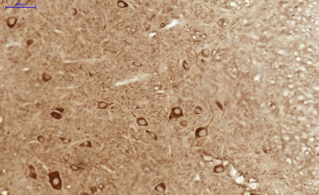
Immunohistochemistry-Paraffin: AKT3 Antibody [NBP1-80900]
Immunohistochemistry-Paraffin: AKT3 Antibody [NBP1-80900] - Staining of human cerebral cortex shows strong nuclear and cytoplasmic positivity in neuronal cells.Applications for AKT3 Antibody - BSA Free
Application
Recommended Usage
Immunohistochemistry
1:500 - 1:1000
Immunohistochemistry-Paraffin
1:50-1:200
Western Blot
0.04 - 0.4 ug/ml
Application Notes
For IHC-Paraffin, HIER pH 6 retrieval is recommended. Immunocytochemistry/Immunofluorescence Fixation Permeabilization: Use PFA/Triton X-100.
Reviewed Applications
Read 1 review rated 4 using NBP1-80900 in the following applications:
Formulation, Preparation, and Storage
Purification
Affinity purified
Formulation
PBS (pH 7.2) and 40% Glycerol
Format
BSA Free
Preservative
0.02% Sodium Azide
Concentration
Concentrations vary lot to lot. See vial label for concentration. If unlisted please contact technical services.
Shipping
The product is shipped with polar packs. Upon receipt, store it immediately at the temperature recommended below.
Stability & Storage
Store at 4C short term. Aliquot and store at -20C long term. Avoid freeze-thaw cycles.
Background: Akt3
The main function of AKT is to control inhibition of apoptosis and promote cell proliferation. Survival factors can activate AKT Ser473 and Thr308 phosphorylation sites in a transcription-independent manner, resulting in the inactivation of apoptotic signaling transduction through the tumor suppressor PTEN, an antagonist to PI3-K (5). PTEN exerts enzymatic activity as a phosphatidylinositol-3,4,5-trisphosphate (PIP3) phosphatase, opposing PI3K activity by decreasing availability of PIP3 to proliferating cells, leading to overexpression and inappropriate activation of AKT noted in many types of cancer.
AKT1 function has been linked to overall physiological growth and function (2). AKT1 has been correlated with proteus syndrome, a rare disorder characterized by overgrowth of various tissues caused by a mosaic variant in the AKT1 gene in humans.
AKT2 is strongly correlated with Type II diabetes, including phenotypes of insulin resistance, hyperglycemia and atherosclerosis (2, 6).
The function of AKT3 is specifically associated to brain development, where disruptions to AKT3 are correlated with microcephaly, hemimegalencephaly, megalencephaly and intellectual disabilities (2).
References
1. Ersahin, T., Tuncbag, N., & Cetin-Atalay, R. (2015). The PI3K/AKT/mTOR interactive pathway. Mol Biosyst, 11(7), 1946-1954. doi:10.1039/c5mb00101c
2. Cohen, M. M., Jr. (2013). The AKT genes and their roles in various disorders. Am J Med Genet A, 161a(12), 2931-2937. doi:10.1002/ajmg.a.36101
3. Georgescu, M. M. (2010). PTEN Tumor Suppressor Network in PI3K-Akt Pathway Control. Genes Cancer, 1(12), 1170-1177. doi:10.1177/1947601911407325
4. Mishra, P., Paital, B., Jena, S., Swain, S. S., Kumar, S., Yadav, M. K.,... Samanta, L. (2019). Possible activation of NRF2 by Vitamin E/Curcumin against altered thyroid hormone induced oxidative stress via NFkB/AKT/mTOR/KEAP1 signalling in rat heart. Sci Rep, 9(1), 7408. doi:10.1038/s41598-019-43320-5
5. Wedel, S., Hudak, L., Seibel, J. M., Juengel, E., Oppermann, E., Haferkamp, A., & Blaheta, R. A. (2011). Critical analysis of simultaneous blockage of histone deacetylase and multiple receptor tyrosine kinase in the treatment of prostate cancer. Prostate, 71(7), 722-735. doi:10.1002/pros.21288
6. Rotllan, N., Chamorro-Jorganes, A., Araldi, E., Wanschel, A. C., Aryal, B., Aranda, J. F.,... Fernandez-Hernando, C. (2015). Hematopoietic Akt2 deficiency attenuates the progression of atherosclerosis. Faseb j, 29(2), 597-610. doi:10.1096/fj.14-262097
Long Name
v-Akt Murine Thymoma Viral Oncogene Homolog 3
Alternate Names
PKB gamma, RAC-gamma
Entrez Gene IDs
10000 (Human)
Gene Symbol
AKT3
UniProt
Additional Akt3 Products
Product Documents for AKT3 Antibody - BSA Free
Certificate of Analysis
To download a Certificate of Analysis, please enter a lot or batch number in the search box below.
Product Specific Notices for AKT3 Antibody - BSA Free
This product is for research use only and is not approved for use in humans or in clinical diagnosis. Primary Antibodies are guaranteed for 1 year from date of receipt.
Citations for AKT3 Antibody - BSA Free
 Powered by Bioz
Customer Reviews for AKT3 Antibody - BSA Free (1)
4 out of 5
1 Customer Rating
Have you used AKT3 Antibody - BSA Free?
Submit a review and receive an Amazon gift card!
$25/€18/£15/$25CAN/¥2500 Yen for a review with an image
$10/€7/£6/$10CAN/¥1110 Yen for a review without an image
Submit a review
Customer Images
Showing
1
-
1 of
1 review
Showing All
Filter By:
-
Application: ImmunohistochemistrySample Tested: mouse spinal cordSpecies: MouseVerified Customer | Posted 05/31/2017Mouse spinal cord (DAB)High background

There are no reviews that match your criteria.
Protocols
Find general support by application which include: protocols, troubleshooting, illustrated assays, videos and webinars.
- Antigen Retrieval Protocol (PIER)
- Antigen Retrieval for Frozen Sections Protocol
- Appropriate Fixation of IHC/ICC Samples
- Cellular Response to Hypoxia Protocols
- Chromogenic IHC Staining of Formalin-Fixed Paraffin-Embedded (FFPE) Tissue Protocol
- Chromogenic Immunohistochemistry Staining of Frozen Tissue
- ClariTSA™ Fluorophore Kits
- Detection & Visualization of Antibody Binding
- Fluorescent IHC Staining of Frozen Tissue Protocol
- Graphic Protocol for Heat-induced Epitope Retrieval
- Graphic Protocol for the Preparation and Fluorescent IHC Staining of Frozen Tissue Sections
- Graphic Protocol for the Preparation and Fluorescent IHC Staining of Paraffin-embedded Tissue Sections
- Graphic Protocol for the Preparation of Gelatin-coated Slides for Histological Tissue Sections
- IHC Sample Preparation (Frozen sections vs Paraffin)
- Immunofluorescent IHC Staining of Formalin-Fixed Paraffin-Embedded (FFPE) Tissue Protocol
- Immunohistochemistry (IHC) and Immunocytochemistry (ICC) Protocols
- Immunohistochemistry Frozen Troubleshooting
- Immunohistochemistry Paraffin Troubleshooting
- Preparing Samples for IHC/ICC Experiments
- Preventing Non-Specific Staining (Non-Specific Binding)
- Primary Antibody Selection & Optimization
- Protocol for Heat-Induced Epitope Retrieval (HIER)
- Protocol for Making a 4% Formaldehyde Solution in PBS
- Protocol for VisUCyte™ HRP Polymer Detection Reagent
- Protocol for the Preparation & Fixation of Cells on Coverslips
- Protocol for the Preparation and Chromogenic IHC Staining of Frozen Tissue Sections
- Protocol for the Preparation and Chromogenic IHC Staining of Frozen Tissue Sections - Graphic
- Protocol for the Preparation and Chromogenic IHC Staining of Paraffin-embedded Tissue Sections
- Protocol for the Preparation and Chromogenic IHC Staining of Paraffin-embedded Tissue Sections - Graphic
- Protocol for the Preparation and Fluorescent IHC Staining of Frozen Tissue Sections
- Protocol for the Preparation and Fluorescent IHC Staining of Paraffin-embedded Tissue Sections
- Protocol for the Preparation of Gelatin-coated Slides for Histological Tissue Sections
- R&D Systems Quality Control Western Blot Protocol
- TUNEL and Active Caspase-3 Detection by IHC/ICC Protocol
- The Importance of IHC/ICC Controls
- Troubleshooting Guide: Immunohistochemistry
- Troubleshooting Guide: Western Blot Figures
- Western Blot Conditions
- Western Blot Protocol
- Western Blot Protocol for Cell Lysates
- Western Blot Troubleshooting
- Western Blot Troubleshooting Guide
- View all Protocols, Troubleshooting, Illustrated assays and Webinars
Loading...
Associated Pathways


IL-2 Signaling Pathways
 IL-4 Signaling Pathways
 IL-7 Signaling Pathways
 IL-9 Signaling Pathways
 IL-15 Signaling Pathways
 IL-21 Signaling Pathways
 mTOR Signaling Pathway
 Notch Signaling Pathways
 TGF-beta Signaling Pathways
 VEGF - VEGF R2 Signaling Pathways
