alpha Tubulin Antibody (DM1A) [Biotin]
Novus Biologicals | Catalog # NB100-690B
Key Product Details
Species Reactivity
Human, Mouse, Rat, Porcine, Avian, Bovine, Canine, Chicken, Chinese Hamster, Drosophila, Fungi, Goat, Guinea Pig, Hamster, Monkey, Parasite, Primate, Rabbit, Xenopus, Yeast
Applications
Immunohistochemistry, Immunohistochemistry-Paraffin, Immunohistochemistry-Frozen, Immunomicroscopy, Western Blot, Flow Cytometry, Flow (Intracellular), Immunocytochemistry/ Immunofluorescence, Immunoprecipitation, CyTOF-ready
Label
Biotin
Antibody Source
Monoclonal Mouse IgG1 kappa Clone # DM1A
Loading...
Product Specifications
Immunogen
This alpha Tubulin Antibody (DM1A) was developed against native chicken brain microtubules.
Reactivity Notes
Use in Mouse reported in scientific literature (PMID:34871568) Use in Mouse reported in scientific literature (PMID:34533563). Yeast reactivity reported in scientific literature (PMID: 25126732). Goat reactivity reported in scientific literature (PMID:31805146). Will likely react with all mammals.
Marker
Microtubule Marker
Specificity
This alpha Tubulin Antibody (DM1A) does not cross-react with beta Tubulin.
Clonality
Monoclonal
Host
Mouse
Isotype
IgG1 kappa
Scientific Data Images for alpha Tubulin Antibody (DM1A) [Biotin]
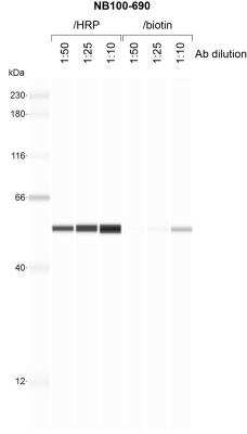
Simple Western: alpha Tubulin Antibody (DM1A) [Biotin] [NB100-690B] - 1 mg/mL human SVG-A whole cell lysate probed with the indicated concentration of each conjugated antibody. Image from verified customer review.
![alpha Tubulin Antibody (DM1A) [Biotin]](https://resources.rndsystems.com/images/products/nb100-690b_mouse-monoclonal-alpha-tubulin-antibody-dm1a-biotin-immunocytochemistry-immunofluorescence-142025111223..jpg "Alpha Tubulin (DM1A) in U-2 OS Human Cell Line.")
Alpha Tubulin (DM1A) in U-2 OS Human Cell Line.
Alpha Tubulin (DM1A) was detected in immersion fixed U-2 OS human osteosarcoma cell line using Mouse anti-Alpha Tubulin (DM1A) Protein G Purified Monoclonal Antibody conjugated to Biotin (Catalog # NB100-690B) at 2 µg/mL overnight at 4C. Cells were stained using Streptavidin conjugated to DyLight 550 (red) and counterstained with DAPI (blue). Cells were imaged using a 100X objective and digitally deconvolved.Applications for alpha Tubulin Antibody (DM1A) [Biotin]
Application
Recommended Usage
CyTOF-ready
Optimal dilutions of this antibody should be experimentally determined.
Flow (Intracellular)
Optimal dilutions of this antibody should be experimentally determined.
Flow Cytometry
Optimal dilutions of this antibody should be experimentally determined.
Immunocytochemistry/ Immunofluorescence
Optimal dilutions of this antibody should be experimentally determined.
Immunohistochemistry
Optimal dilutions of this antibody should be experimentally determined.
Immunohistochemistry-Frozen
Optimal dilutions of this antibody should be experimentally determined.
Immunohistochemistry-Paraffin
Optimal dilutions of this antibody should be experimentally determined.
Immunomicroscopy
Optimal dilutions of this antibody should be experimentally determined.
Immunoprecipitation
Optimal dilutions of this antibody should be experimentally determined.
Western Blot
Optimal dilutions of this antibody should be experimentally determined.
Application Notes
Optimal dilution of this antibody should be experimentally determined.
Reviewed Applications
Read 1 review rated 4 using NB100-690B in the following applications:
Flow Cytometry Panel Builder
Bio-Techne Knows Flow Cytometry
Save time and reduce costly mistakes by quickly finding compatible reagents using the Panel Builder Tool.
Advanced Features
- Spectra Viewer - Custom analysis of spectra from multiple fluorochromes
- Spillover Popups - Visualize the spectra of individual fluorochromes
- Antigen Density Selector - Match fluorochrome brightness with antigen density

Formulation, Preparation, and Storage
Purification
Protein G purified
Formulation
PBS
Preservative
0.05% Sodium Azide
Concentration
Please see the vial label for concentration. If unlisted please contact technical services.
Shipping
The product is shipped with polar packs. Upon receipt, store it immediately at the temperature recommended below.
Stability & Storage
Store at 4C in the dark.
Background: alpha Tubulin
Tyrosine ligase adds a C-terminal tyrosine to monomeric alpha tubulin. Assembled microtubules can again be detyrosinated by a cytoskeleton associated carboxypeptidase. Detyrosinated alpha tubulin is referred to as Glu-tubulin. Another post-translational modification of detyrosinated alpha tubulin is C-terminal polyglutamylation which is characteristic for microtubules in neuronal cells and the mitotic spindle.
Like GAPDH and beta-actin, alpha/beta tubulin is often used as a loading control in immunoblot applications (1). Alpha/beta tubulin is also good for counterstaining microtubules in immunofluorescence (2).
References
1. Hannen, R., Selmansberger, M., Hauswald, M., Pagenstecher, A., Nist, A., Stiewe, T.,... Bartsch, J. W. (2019). Comparative Transcriptomic Analysis of Temozolomide Resistant Primary GBM Stem-Like Cells and Recurrent GBM Identifies Up-Regulation of the Carbonic Anhydrase CA2 Gene as Resistance Factor. Cancers (Basel), 11(7). doi:10.3390/cancers11070921
2. Nel, M., Joubert, A. M., Dohle, W., Potter, B. V., & Theron, A. E. (2018). Modes of cell death induced by tetrahydroisoquinoline-based analogs in MDA-MB-231 breast and A549 lung cancer cell lines. Drug Des Devel Ther, 12, 1881-1904. doi:10.2147/dddt.S152718
Long Name
Tubulin Alpha 1a
Alternate Names
Alpha-Tubulin 3, B-ALPHA-1, LIS3, TUBA1A, TUBA3, Tubulin B-Alpha-1
Gene Symbol
TUBA1A
Additional alpha Tubulin Products
Product Documents for alpha Tubulin Antibody (DM1A) [Biotin]
Certificate of Analysis
To download a Certificate of Analysis, please enter a lot or batch number in the search box below.
Product Specific Notices for alpha Tubulin Antibody (DM1A) [Biotin]
This product is for research use only and is not approved for use in humans or in clinical diagnosis. Primary Antibodies are guaranteed for 1 year from date of receipt.
Related Research Areas
Customer Reviews for alpha Tubulin Antibody (DM1A) [Biotin] (1)
4 out of 5
1 Customer Rating
Have you used alpha Tubulin Antibody (DM1A) [Biotin]?
Submit a review and receive an Amazon gift card!
$25/€18/£15/$25CAN/¥2500 Yen for a review with an image
$10/€7/£6/$10CAN/¥1110 Yen for a review without an image
Submit a review
Customer Images
![alpha Tubulin Antibody (DM1A) [Biotin] NB100-690B](https://resources.rndsystems.com/images/reviews/review_nb100-690b_47946_0_0.jpg)
Showing
1
-
1 of
1 review
Showing All
Filter By:
-
Application: Simple WesternSample Tested: SVG-A whole cell lysateSpecies: HumanVerified Customer | Posted 05/15/20191 mg/mL SVG-A whole cell lysate probed with the indicated concentration of each conjugated antibody.
![alpha Tubulin Antibody (DM1A) [Biotin] NB100-690B](data:image/png;base64,R0lGODlhAQABAAD/ACwAAAAAAQABAAACADs=)
There are no reviews that match your criteria.
Protocols
Find general support by application which include: protocols, troubleshooting, illustrated assays, videos and webinars.
- 7-Amino Actinomycin D (7-AAD) Cell Viability Flow Cytometry Protocol
- Antigen Retrieval Protocol (PIER)
- Antigen Retrieval for Frozen Sections Protocol
- Appropriate Fixation of IHC/ICC Samples
- Cellular Response to Hypoxia Protocols
- Chromogenic IHC Staining of Formalin-Fixed Paraffin-Embedded (FFPE) Tissue Protocol
- Chromogenic Immunohistochemistry Staining of Frozen Tissue
- ClariTSA™ Fluorophore Kits
- Detection & Visualization of Antibody Binding
- Extracellular Membrane Flow Cytometry Protocol
- Flow Cytometry Protocol for Cell Surface Markers
- Flow Cytometry Protocol for Staining Membrane Associated Proteins
- Flow Cytometry Staining Protocols
- Flow Cytometry Troubleshooting Guide
- Fluorescent IHC Staining of Frozen Tissue Protocol
- Graphic Protocol for Heat-induced Epitope Retrieval
- Graphic Protocol for the Preparation and Fluorescent IHC Staining of Frozen Tissue Sections
- Graphic Protocol for the Preparation and Fluorescent IHC Staining of Paraffin-embedded Tissue Sections
- Graphic Protocol for the Preparation of Gelatin-coated Slides for Histological Tissue Sections
- ICC Cell Smear Protocol for Suspension Cells
- ICC Immunocytochemistry Protocol Videos
- ICC for Adherent Cells
- IHC Sample Preparation (Frozen sections vs Paraffin)
- Immunocytochemistry (ICC) Protocol
- Immunocytochemistry Troubleshooting
- Immunofluorescence of Organoids Embedded in Cultrex Basement Membrane Extract
- Immunofluorescent IHC Staining of Formalin-Fixed Paraffin-Embedded (FFPE) Tissue Protocol
- Immunohistochemistry (IHC) and Immunocytochemistry (ICC) Protocols
- Immunohistochemistry Frozen Troubleshooting
- Immunohistochemistry Paraffin Troubleshooting
- Immunoprecipitation Protocol
- Intracellular Flow Cytometry Protocol Using Alcohol (Methanol)
- Intracellular Flow Cytometry Protocol Using Detergents
- Intracellular Nuclear Staining Flow Cytometry Protocol Using Detergents
- Intracellular Staining Flow Cytometry Protocol Using Alcohol Permeabilization
- Intracellular Staining Flow Cytometry Protocol Using Detergents to Permeabilize Cells
- Preparing Samples for IHC/ICC Experiments
- Preventing Non-Specific Staining (Non-Specific Binding)
- Primary Antibody Selection & Optimization
- Propidium Iodide Cell Viability Flow Cytometry Protocol
- Protocol for Heat-Induced Epitope Retrieval (HIER)
- Protocol for Liperfluo
- Protocol for Making a 4% Formaldehyde Solution in PBS
- Protocol for VisUCyte™ HRP Polymer Detection Reagent
- Protocol for the Characterization of Human Th22 Cells
- Protocol for the Characterization of Human Th9 Cells
- Protocol for the Fluorescent ICC Staining of Cell Smears - Graphic
- Protocol for the Fluorescent ICC Staining of Cultured Cells on Coverslips - Graphic
- Protocol for the Preparation & Fixation of Cells on Coverslips
- Protocol for the Preparation and Chromogenic IHC Staining of Frozen Tissue Sections
- Protocol for the Preparation and Chromogenic IHC Staining of Frozen Tissue Sections - Graphic
- Protocol for the Preparation and Chromogenic IHC Staining of Paraffin-embedded Tissue Sections
- Protocol for the Preparation and Chromogenic IHC Staining of Paraffin-embedded Tissue Sections - Graphic
- Protocol for the Preparation and Fluorescent ICC Staining of Cells on Coverslips
- Protocol for the Preparation and Fluorescent ICC Staining of Non-adherent Cells
- Protocol for the Preparation and Fluorescent ICC Staining of Stem Cells on Coverslips
- Protocol for the Preparation and Fluorescent IHC Staining of Frozen Tissue Sections
- Protocol for the Preparation and Fluorescent IHC Staining of Paraffin-embedded Tissue Sections
- Protocol for the Preparation of Gelatin-coated Slides for Histological Tissue Sections
- Protocol for the Preparation of a Cell Smear for Non-adherent Cell ICC - Graphic
- Protocol: Annexin V and PI Staining by Flow Cytometry
- Protocol: Annexin V and PI Staining for Apoptosis by Flow Cytometry
- R&D Systems Quality Control Western Blot Protocol
- TUNEL and Active Caspase-3 Detection by IHC/ICC Protocol
- The Importance of IHC/ICC Controls
- Troubleshooting Guide: Fluorokine Flow Cytometry Kits
- Troubleshooting Guide: Immunohistochemistry
- Troubleshooting Guide: Western Blot Figures
- Western Blot Conditions
- Western Blot Protocol
- Western Blot Protocol for Cell Lysates
- Western Blot Troubleshooting
- Western Blot Troubleshooting Guide
- View all Protocols, Troubleshooting, Illustrated assays and Webinars
Loading...