B7-2/CD86 Antibody (BU63)
Novus Biologicals | Catalog # NBP2-25208
Clone BU63 was used by HLDA to establish CD designation.
Key Product Details
Validated by
Knockout/Knockdown
Species Reactivity
Validated:
Human, Mouse, Rat
Cited:
Human, Mouse, Rat
Applications
Validated:
Knockout Validated, Immunohistochemistry, Immunohistochemistry-Paraffin, Immunohistochemistry-Frozen, Western Blot, Flow Cytometry, Immunocytochemistry/ Immunofluorescence, Simple Western
Cited:
Immunohistochemistry-Paraffin, Immunohistochemistry-Frozen, Western Blot, Block/Neutralize, Immunofluorescence, Immunocytochemistry/ Immunofluorescence, IF/IHC
Label
Unconjugated
Antibody Source
Monoclonal Mouse IgG1 kappa Clone # BU63
Loading...
Product Specifications
Immunogen
ARH-77 (B-lymphoblastoid cell line)
Localization
Cell Surface
Marker
Dendritic Cells Maturation Marker
Clonality
Monoclonal
Host
Mouse
Isotype
IgG1 kappa
Theoretical MW
70 kDa.
Disclaimer note: The observed molecular weight of the protein may vary from the listed predicted molecular weight due to post translational modifications, post translation cleavages, relative charges, and other experimental factors.
Disclaimer note: The observed molecular weight of the protein may vary from the listed predicted molecular weight due to post translational modifications, post translation cleavages, relative charges, and other experimental factors.
Description
200ug/ml of antibody purified from Bioreactor Concentrate by Protein A or G. Prepared in 10 mM PBS with 0.05% BSA & 0.05% azide. Also available WITHOUT BSA & azide at 1.0 mg/ml. (NBP2-34569)
Antibody with azide - store at 2 to 8C. Antibody without azide - store at -20 to -80C.
Antibody with azide - store at 2 to 8C. Antibody without azide - store at -20 to -80C.
Scientific Data Images for B7-2/CD86 Antibody (BU63)
![Knockout Validated: B7-2/CD86 Antibody (BU63) [NBP2-25208]](https://resources.rndsystems.com/images/products/B7-2-CD86-Antibody-BU63-Knockout-Validated-NBP2-25208-img0005.jpg "Western Blot: B7-2/CD86 Antibody (BU63) [NBP2-25208]")
Western Blot: B7-2/CD86 Antibody (BU63) [NBP2-25208]
Western Blot: B7-2/CD86 Antibody (BU63) [NBP2-25208] - Western blot shows lysates of Ramos human Burkitt's lymphoma parental cell line and B7-2 knockout (KO) Ramos cell line. PVDF membrane was probed with 1.0 ug/mL of Mouse Anti-Human B7-2 Monoclonal Antibody (Catalog # NBP2-25208) followed by HRP-conjugated Anti-Mouse IgG Secondary Antibody (Catalog #HAF018). Specific band was detected for B7-2 at approximately 90 kDa (as indicated) in the parental Ramos cell line, but is not detectable in the knockout Ramos cell line. This experiment was conducted under reducing conditions.![Immunohistochemistry: B7-2/CD86 Antibody (BU63) [NBP2-25208]](https://resources.rndsystems.com/images/products/B7-2-CD86-Antibody-BU63-Immunohistochemistry-NBP2-25208-img0009.jpg "Immunohistochemistry: B7-2/CD86 Antibody (BU63) [NBP2-25208]")
Immunohistochemistry: B7-2/CD86 Antibody (BU63) [NBP2-25208]
B7-2-CD86-Antibody-BU63-Immunohistochemistry-NBP2-25208-img0009.jpg![Immunocytochemistry/ Immunofluorescence: B7-2/CD86 Antibody (BU63) [NBP2-25208]](https://resources.rndsystems.com/images/products/B7-2-CD86-Antibody-BU63-Immunocytochemistry-Immunofluorescence-NBP2-25208-img0006.jpg "Immunocytochemistry/ Immunofluorescence: B7-2/CD86 Antibody (BU63) [NBP2-25208]")
Immunocytochemistry/ Immunofluorescence: B7-2/CD86 Antibody (BU63) [NBP2-25208]
Immunocytochemistry/Immunofluorescence: B7-2/CD86 Antibody (BU63) [NBP2-25208] - Immunofluorescence staining of PFA-fixed Ramos cells using followed by goat anti-Mouse IgG conjugated to CF488 (green). Nuclei are stained with Red Dot.![Immunohistochemistry: B7-2/CD86 Antibody (BU63) [NBP2-25208]](https://resources.rndsystems.com/images/products/B7-2-CD86-Antibody-BU63-Immunohistochemistry-NBP2-25208-img0010.jpg "Immunohistochemistry: B7-2/CD86 Antibody (BU63) [NBP2-25208]")
Immunohistochemistry: B7-2/CD86 Antibody (BU63) [NBP2-25208]
B7-2-CD86-Antibody-BU63-Immunohistochemistry-NBP2-25208-img0010.jpg![Flow Cytometry: B7-2/CD86 Antibody (BU63) [NBP2-25208]](https://resources.rndsystems.com/images/products/B7-2-CD86-Antibody-BU63-Flow-Cytometry-NBP2-25208-img0008.jpg "Flow Cytometry: B7-2/CD86 Antibody (BU63) [NBP2-25208]")
Flow Cytometry: B7-2/CD86 Antibody (BU63) [NBP2-25208]
Flow Cytometry: B7-2/CD86 Antibody (BU63) [NBP2-25208] - Flow Cytometric Analysis of PFA-fixed Ramos cells. B7-2/CD86 Antibody (BU63) followed by goat anti-Mouse IgG-CF488 (Blue); Isotype Control (Red).![Immunohistochemistry-Paraffin: B7-2/CD86 Antibody (BU63) [NBP2-25208]](https://resources.rndsystems.com/images/products/B7-2-CD86-Antibody-BU63-Immunohistochemistry-Paraffin-NBP2-25208-img0004.jpg "Immunohistochemistry-Paraffin: B7-2/CD86 Antibody (BU63) [NBP2-25208]")
Immunohistochemistry-Paraffin: B7-2/CD86 Antibody (BU63) [NBP2-25208]
Immunohistochemistry-Paraffin: B7-2/CD86 Antibody (BU63) [NBP2-25208] - Formalin-fixed, paraffin-embedded esophagus tumor tissue stained with CD86 antibody (5 ug/ml), peroxidase-conjugate and DAB chromogen. Note strong staining of differentiated squamous cells. TMA was used for this test.![Flow Cytometry: B7-2/CD86 Antibody (BU63) [NBP2-25208]](https://resources.rndsystems.com/images/products/B7-2-CD86-Antibody-BU63-Flow-Cytometry-NBP2-25208-img0007.jpg "Flow Cytometry: B7-2/CD86 Antibody (BU63) [NBP2-25208]")
Flow Cytometry: B7-2/CD86 Antibody (BU63) [NBP2-25208]
Flow Cytometry: B7-2/CD86 Antibody (BU63) [NBP2-25208] - Flow Cytometric Analysis of human PBMCs using B7-2/CD86 Antibody (BU63); Goat anti-Mouse IgG-CF488 (red); Isotype Control (green). [NBP2-25208] -")
Immunohistochemistry: B7-2/CD86 Antibody (BU63) [NBP2-25208] -
Immunohistochemistry: B7-2/CD86 Antibody (BU63) [NBP2-25208] - Macrophage polarization states. Distribution & proportion of CD68+ (a), CD86+ (c) & CD163+ (e) macrophages (brown) were identified using IHC procedures. Intensity for the color response to antibodies was visible in lung tissues from all groups. Population proportions for the stained cells as shown by red arrows were calculated as a fold change of the control. (b, d, f). The results were expressed as Mean ± SD (n = 3). **: a p-value of < 0.01 vs NS, OVA/BUD & OVA/PCI except the number of CD86+ cells in OVA/PCI group. #: P < 0.05 vs NS & ##; P < 0.01 vs either NS or OVA/BUD group Image collected & cropped by CiteAb from the following publication (https://pubmed.ncbi.nlm.nih.gov/32111211), licensed under a CC-BY license. Not internally tested by Novus Biologicals. [NBP2-25208] -")
Immunocytochemistry/ Immunofluorescence: B7-2/CD86 Antibody (BU63) [NBP2-25208] -
Immunocytochemistry/ Immunofluorescence: B7-2/CD86 Antibody (BU63) [NBP2-25208] - Effects of 20(S)-Rg3 on macrophage polarization in atherosclerotic lesions of diabetic ApoE–/– mice. (A,B) Co-immunofluorescence staining of the aortic root for macrophage (anti-Moma-2 antibody, green) & M1 marker iNos (red), & a bar graph summarizing the results (n = 5, respectively). (C,D) Co-immunofluorescence staining for macrophage (anti-Moma-2 antibody, green) & M1 marker CD86 (red), & a bar graph summarizing the results (n = 5, respectively). Scale bar: 20 μm. Data are mean ± SEM. ∗p < 0.05, ∗∗p < 0.01, ∗∗∗p < 0.001. Image collected & cropped by CiteAb from the following publication (https://pubmed.ncbi.nlm.nih.gov/29867472), licensed under a CC-BY license. Not internally tested by Novus Biologicals. [NBP2-25208] -")
Western Blot: B7-2/CD86 Antibody (BU63) [NBP2-25208] -
M1 macrophages were induced in the in vivo mouse intervertebral disc (mIVD) degeneration model (puncture model). (a) Immunohistological analysis of Iba-1, iNOS, and arginase (brown staining) expression in nucleus pulposus (NP), annulus fibrosis (AF), and cartilage endplate (CEP) [right] at high magnification and in whole mIVDs (left) at low magnification. Images using an IgG isotype control antibody are included to demonstrate the specificity of the IHC staining. (b) Protein was extracted from punctured mIVDs in the puncture model, and western blotting was performed using antibodies against CD86, CD163, and beta -actin. (c) Images of the panel in (b), captured using a ChemiDoc Touch system and quantified using ImageJ. Biological and technical replicates were performed three times. Values represent the mean +/- SD. *p < 0.05, **p < 0.01, relative to the corresponding control. Image collected and cropped by CiteAb from the following open publication (https://pubmed.ncbi.nlm.nih.gov/41053148), licensed under a CC-BY license. Not internally tested by Novus Biologicals. [NBP2-25208] -")
Western Blot: B7-2/CD86 Antibody (BU63) [NBP2-25208] -
Thrombin promotes polarization into M1 macrophages. (a) Mouse bone marrow was stimulated with thrombin, and quantitative PCR was performed using specific primers for CD86 and CD163. Biological and technical replicates were performed three times. (b) Mouse bone marrow was stimulated with thrombin, and western blotting was performed using antibodies for the M1 markers iNOS and CD86, and the M2 markers CD163 and Arginase-1. beta -actin was used as an internal control. n = 3. Values represent the mean +/- SD. *p < 0.05, **p < 0.01, relative to the corresponding control. Image collected and cropped by CiteAb from the following open publication (https://pubmed.ncbi.nlm.nih.gov/41053148), licensed under a CC-BY license. Not internally tested by Novus Biologicals. [NBP2-25208] -")
Western Blot: B7-2/CD86 Antibody (BU63) [NBP2-25208] -
IGU alleviates the inflammation of type II alveolar epithelial cells caused by M1 macrophages. (A–C) Representative western blot and quantitation for iNOS and CD86 in Raw264.7 cells. beta -actin was used as an internal control. (D) QRT-PCR for the mRNA levels of M1 macrophage polarization-related inflammatory cytokines TNF-alpha, IL-6 and IL-1 beta in Raw264.7 cells. (E, F) ELISA was used to measure the level of TNF-alpha and IL-6 in Raw264.7 cell culture supernatant. (G, H) QRT-PCR for the mRNA levels of MMP2 and MMP9. (I) Representative gelatin zymogram of MMPs in Raw264.7 cell culture supernatants. (J) Schematic representation of Raw264.7 cells co-cultured with type II alveolar epithelial cells. (K–M) QRT-PCR for the mRNA levels of TNF-alpha, IL-6 and IL-1 beta in type II alveolar epithelial cells (n=3). *P<0.05, **P<0.01, ***P<0.001, ns, No significance. Image collected and cropped by CiteAb from the following open publication (https://pubmed.ncbi.nlm.nih.gov/40181990), licensed under a CC-BY license. Not internally tested by Novus Biologicals. [NBP2-25208] -")
Western Blot: B7-2/CD86 Antibody (BU63) [NBP2-25208] -
Rescue experiments of Raw264.7 cells after overexpression of TLR4. (A, B) Representative western blot and quantitation of TLR4 in TLR4-overexpressing Raw264.7 cells. beta -actin was used as an internal control. (C–E) QRT-PCR for the mRNA levels of M1 macrophage polarization-related inflammatory cytokines TNF-alpha, IL-6 and IL-1 beta in Raw264.7 cells. (F–J) Representative western blot and quantitation of protein levels of TLR4, NF-kappa B, iNOS and CD86 in Raw264.7 cells beta -actin was used as an internal control. (n=3). *P<0.05, **P<0.01, ***P<0.001, ns, No significance. Image collected and cropped by CiteAb from the following open publication (https://pubmed.ncbi.nlm.nih.gov/40181990), licensed under a CC-BY license. Not internally tested by Novus Biologicals.Applications for B7-2/CD86 Antibody (BU63)
Application
Recommended Usage
Flow Cytometry
1-2 ug/million cells
Immunocytochemistry/ Immunofluorescence
1-2 ug/ml
Immunohistochemistry
0.5-1ug/ml
Immunohistochemistry-Paraffin
2-4 ug/ml
Simple Western
1:25
Western Blot
0.5-1ug/ml
Application Notes
Immunohistochemistry (Formalin-fixed): 2-4ug/ml for 30 minutes at RT. Staining of formalin-fixed tissues requires heating tissue sections in 10mM Tris buffer with 1mM EDTA, pH 9.0, for 45 min at 95C followed by cooling at RT for 20 minutes.
Optimal dilution for a specific application should be determined.
IHC-fr reported in the literature (PMID: 29867472, 31629891)
See Simple Western Antibody Database for Simple Western validation: antibody dilution of 1:25
Optimal dilution for a specific application should be determined.
IHC-fr reported in the literature (PMID: 29867472, 31629891)
See Simple Western Antibody Database for Simple Western validation: antibody dilution of 1:25
Reviewed Applications
Read 1 review rated 3 using NBP2-25208 in the following applications:
Flow Cytometry Panel Builder
Bio-Techne Knows Flow Cytometry
Save time and reduce costly mistakes by quickly finding compatible reagents using the Panel Builder Tool.
Advanced Features
- Spectra Viewer - Custom analysis of spectra from multiple fluorochromes
- Spillover Popups - Visualize the spectra of individual fluorochromes
- Antigen Density Selector - Match fluorochrome brightness with antigen density

Formulation, Preparation, and Storage
Purification
Protein A or G purified
Formulation
10 mM PBS with 0.05% BSA
Preservative
0.05% Sodium Azide
Concentration
0.2 mg/ml
Shipping
The product is shipped with polar packs. Upon receipt, store it immediately at the temperature recommended below.
Stability & Storage
Store at 4C.
Background: B7-2/CD86
References
1. Collins M, Ling V, Carreno BM. The B7 family of immune-regulatory ligands. Genome Biol. 2005;6(6):223. https://doi.org/10.1186/gb-2005-6-6-223
2. Greaves P, Gribben JG. The role of B7 family molecules in hematologic malignancy. Blood. 2013;121(5):734-744. https://doi.org/10.1182/blood-2012-10-385591
3. Bolandi N, Derakhshani A, Hemmat N, et al. The Positive and Negative Immunoregulatory Role of B7 Family: Promising Novel Targets in Gastric Cancer Treatment. Int J Mol Sci. 2021;22(19):10719. https://doi.org/10.3390/ijms221910719
4. Uniprot (P42081)
5. Bhatia S, Edidin M, Almo SC, Nathenson SG. B7-1 and B7-2: similar costimulatory ligands with different biochemical, oligomeric and signaling properties. Immunol Lett. 2006;104(1-2):70-75. https://doi.org/10.1016/j.imlet.2005.11.019
6. Ohue Y, Nishikawa H. Regulatory T (Treg) cells in cancer: Can Treg cells be a new therapeutic target?. Cancer Sci. 2019;110(7):2080-2089. https://doi.org/10.1111/cas.14069
7. Chen L, Flies DB. Molecular mechanisms of T cell co-stimulation and co-inhibition [published correction appears in Nat Rev Immunol. 2013 Jul;13(7):542]. Nat Rev Immunol. 2013;13(4):227-242. https://doi.org/1010.1038/nri3405
8. Karimi A, Alilou S, Mirzaei HR. Adverse Events Following Administration of Anti-CTLA4 Antibody Ipilimumab. Front Oncol. 2021;11:624780. https://doi.org/101010.3389/fonc.2021.624780
Additional B7-2/CD86 Products
Product Documents for B7-2/CD86 Antibody (BU63)
Certificate of Analysis
To download a Certificate of Analysis, please enter a lot or batch number in the search box below.
Product Specific Notices for B7-2/CD86 Antibody (BU63)
This product is for research use only and is not approved for use in humans or in clinical diagnosis. Primary Antibodies are guaranteed for 1 year from date of receipt.
Citations for B7-2/CD86 Antibody (BU63)
 Powered by Bioz
Customer Reviews for B7-2/CD86 Antibody (BU63) (1)
3 out of 5
1 Customer Rating
Have you used B7-2/CD86 Antibody (BU63)?
Submit a review and receive an Amazon gift card!
$25/€18/£15/$25CAN/¥2500 Yen for a review with an image
$10/€7/£6/$10CAN/¥1110 Yen for a review without an image
Submit a review
Customer Images
Showing
1
-
1 of
1 review
Showing All
Filter By:
-
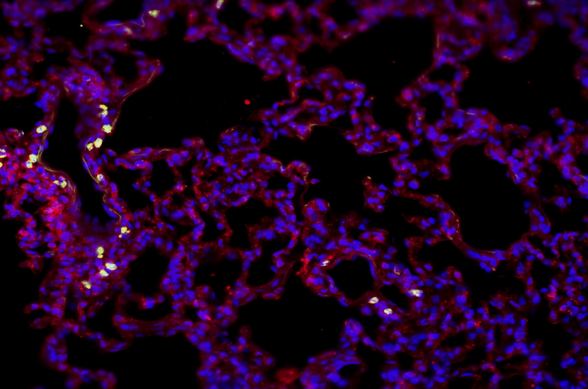
Application: Immunohistochemistry-ParaffinSample Tested: Rat Lung TissueSpecies: RatVerified Customer | Posted 07/03/2025Native lung tissue (Rat) stained with CD86 (green) and Elastin (Eln) (red)Native lung tissue (Rat) stained with CD86 (green) and Elastin (Eln) (red). Fixed in 4% PFA then paraffin-embedded and sectioned.

There are no reviews that match your criteria.
Protocols
Find general support by application which include: protocols, troubleshooting, illustrated assays, videos and webinars.
- 7-Amino Actinomycin D (7-AAD) Cell Viability Flow Cytometry Protocol
- Antigen Retrieval Protocol (PIER)
- Antigen Retrieval for Frozen Sections Protocol
- Appropriate Fixation of IHC/ICC Samples
- Cellular Response to Hypoxia Protocols
- Chromogenic IHC Staining of Formalin-Fixed Paraffin-Embedded (FFPE) Tissue Protocol
- Chromogenic Immunohistochemistry Staining of Frozen Tissue
- ClariTSA™ Fluorophore Kits
- Detection & Visualization of Antibody Binding
- Extracellular Membrane Flow Cytometry Protocol
- Flow Cytometry Protocol for Cell Surface Markers
- Flow Cytometry Protocol for Staining Membrane Associated Proteins
- Flow Cytometry Staining Protocols
- Flow Cytometry Troubleshooting Guide
- Fluorescent IHC Staining of Frozen Tissue Protocol
- Graphic Protocol for Heat-induced Epitope Retrieval
- Graphic Protocol for the Preparation and Fluorescent IHC Staining of Frozen Tissue Sections
- Graphic Protocol for the Preparation and Fluorescent IHC Staining of Paraffin-embedded Tissue Sections
- Graphic Protocol for the Preparation of Gelatin-coated Slides for Histological Tissue Sections
- ICC Cell Smear Protocol for Suspension Cells
- ICC Immunocytochemistry Protocol Videos
- ICC for Adherent Cells
- IHC Sample Preparation (Frozen sections vs Paraffin)
- Immunocytochemistry (ICC) Protocol
- Immunocytochemistry Troubleshooting
- Immunofluorescence of Organoids Embedded in Cultrex Basement Membrane Extract
- Immunofluorescent IHC Staining of Formalin-Fixed Paraffin-Embedded (FFPE) Tissue Protocol
- Immunohistochemistry (IHC) and Immunocytochemistry (ICC) Protocols
- Immunohistochemistry Frozen Troubleshooting
- Immunohistochemistry Paraffin Troubleshooting
- Intracellular Flow Cytometry Protocol Using Alcohol (Methanol)
- Intracellular Flow Cytometry Protocol Using Detergents
- Intracellular Nuclear Staining Flow Cytometry Protocol Using Detergents
- Intracellular Staining Flow Cytometry Protocol Using Alcohol Permeabilization
- Intracellular Staining Flow Cytometry Protocol Using Detergents to Permeabilize Cells
- Preparing Samples for IHC/ICC Experiments
- Preventing Non-Specific Staining (Non-Specific Binding)
- Primary Antibody Selection & Optimization
- Propidium Iodide Cell Viability Flow Cytometry Protocol
- Protocol for Heat-Induced Epitope Retrieval (HIER)
- Protocol for Liperfluo
- Protocol for Making a 4% Formaldehyde Solution in PBS
- Protocol for VisUCyte™ HRP Polymer Detection Reagent
- Protocol for the Characterization of Human Th22 Cells
- Protocol for the Characterization of Human Th9 Cells
- Protocol for the Fluorescent ICC Staining of Cell Smears - Graphic
- Protocol for the Fluorescent ICC Staining of Cultured Cells on Coverslips - Graphic
- Protocol for the Preparation & Fixation of Cells on Coverslips
- Protocol for the Preparation and Chromogenic IHC Staining of Frozen Tissue Sections
- Protocol for the Preparation and Chromogenic IHC Staining of Frozen Tissue Sections - Graphic
- Protocol for the Preparation and Chromogenic IHC Staining of Paraffin-embedded Tissue Sections
- Protocol for the Preparation and Chromogenic IHC Staining of Paraffin-embedded Tissue Sections - Graphic
- Protocol for the Preparation and Fluorescent ICC Staining of Cells on Coverslips
- Protocol for the Preparation and Fluorescent ICC Staining of Non-adherent Cells
- Protocol for the Preparation and Fluorescent ICC Staining of Stem Cells on Coverslips
- Protocol for the Preparation and Fluorescent IHC Staining of Frozen Tissue Sections
- Protocol for the Preparation and Fluorescent IHC Staining of Paraffin-embedded Tissue Sections
- Protocol for the Preparation of Gelatin-coated Slides for Histological Tissue Sections
- Protocol for the Preparation of a Cell Smear for Non-adherent Cell ICC - Graphic
- Protocol: Annexin V and PI Staining by Flow Cytometry
- Protocol: Annexin V and PI Staining for Apoptosis by Flow Cytometry
- R&D Systems Quality Control Western Blot Protocol
- TUNEL and Active Caspase-3 Detection by IHC/ICC Protocol
- The Importance of IHC/ICC Controls
- Troubleshooting Guide: Fluorokine Flow Cytometry Kits
- Troubleshooting Guide: Immunohistochemistry
- Troubleshooting Guide: Western Blot Figures
- Western Blot Conditions
- Western Blot Protocol
- Western Blot Protocol for Cell Lysates
- Western Blot Troubleshooting
- Western Blot Troubleshooting Guide
- View all Protocols, Troubleshooting, Illustrated assays and Webinars


