beta-Catenin Antibody (12F7) - BSA Free
Novus Biologicals | Catalog # NBP1-54467
Key Product Details
Validated by
Species Reactivity
Validated:
Cited:
Applications
Validated:
Cited:
Label
Antibody Source
Format
Product Specifications
Immunogen
Reactivity Notes
Localization
Marker
Clonality
Host
Isotype
Theoretical MW
Disclaimer note: The observed molecular weight of the protein may vary from the listed predicted molecular weight due to post translational modifications, post translation cleavages, relative charges, and other experimental factors.
Scientific Data Images for beta-Catenin Antibody (12F7) - BSA Free
![Western Blot: beta-Catenin Antibody (12F7)BSA Free [NBP1-54467]](https://resources.rndsystems.com/images/products/beta-Catenin-Antibody-12F7-Western-Blot-NBP1-54467-img0008.jpg "Western Blot: beta-Catenin Antibody (12F7)BSA Free [NBP1-54467]")
Western Blot: beta-Catenin Antibody (12F7)BSA Free [NBP1-54467]
Western Blot: beta-Catenin Antibody (12F7) [NBP1-54467] - Total protein from human HepG2 and HeLa cells, rat PC12 cells and mouse 3T3 cells was separated on a 7.5% gel by SDS-PAGE, transferred to PVDF membrane and blocked in 5% non-fat milk in TBST. The membrane was probed with 1.0 ug/ml anti-Beta catenin in 1% non-fat milk in TBST and detected with an anti-mouse HRP secondary antibody using chemiluminescence.![Western Blot: beta-Catenin Antibody (12F7)BSA Free [NBP1-54467]](https://resources.rndsystems.com/images/products/beta-Catenin-Antibody-12F7-Western-Blot-NBP1-54467-img0014.jpg "Western Blot: beta-Catenin Antibody (12F7)BSA Free [NBP1-54467]")
Western Blot: beta-Catenin Antibody (12F7)BSA Free [NBP1-54467]
beta-Catenin-Antibody-12F7-Western-Blot-NBP1-54467-img0014.jpg![Immunocytochemistry/ Immunofluorescence: beta-Catenin Antibody (12F7) - BSA Free [NBP1-54467]](https://resources.rndsystems.com/images/products/beta-Catenin-Antibody-12F7-Immunocytochemistry-Immunofluorescence-NBP1-54467-img0004.jpg "Immunocytochemistry/ Immunofluorescence: beta-Catenin Antibody (12F7) - BSA Free [NBP1-54467]")
Immunocytochemistry/ Immunofluorescence: beta-Catenin Antibody (12F7) - BSA Free [NBP1-54467]
Immunocytochemistry/Immunofluorescence: beta-Catenin Antibody (12F7) [NBP1-54467] - The beta- Catenin antibody was tested in MCF-7 cells against Dylight 488 (Green). Actin and nuclei were counterstained against Phalloidin 550 (Red) and DAPI (Blue).![Immunohistochemistry: beta-Catenin Antibody (12F7) - BSA Free [NBP1-54467]](https://resources.rndsystems.com/images/products/beta-Catenin-Antibody-12F7-Immunohistochemistry-NBP1-54467-img0013.jpg "Immunohistochemistry: beta-Catenin Antibody (12F7) - BSA Free [NBP1-54467]")
Immunohistochemistry: beta-Catenin Antibody (12F7) - BSA Free [NBP1-54467]
Immunohistochemistry: beta-Catenin Antibody (12F7) [NBP1-54467] - Beta-catening (green) was detected in human skin (psoriasis) using beta-catenin-FITC antibody (1:40) in PBS for 1 hour. Nuclei were stained with Dapi (blue). Image from verified customer review. Image using the FITC format of this antibody.![Flow Cytometry: beta-Catenin Antibody (12F7)BSA Free [NBP1-54467]](https://resources.rndsystems.com/images/products/beta-Catenin-Antibody-12F7-Flow-Cytometry-NBP1-54467-img0012.jpg "Flow Cytometry: beta-Catenin Antibody (12F7)BSA Free [NBP1-54467]")
Flow Cytometry: beta-Catenin Antibody (12F7)BSA Free [NBP1-54467]
Flow Cytometry: beta-Catenin Antibody (12F7) [NBP1-54467] - An intracellular stain was performed on SK-MEL-28 cells with beta-Catenin Antibody [12F7] NBP1-54467B (blue) and a matched isotype control (orange). Both antibodies were conjugated to Biotin. Cells were fixed with 4% PFA and then permeabilized with 0.1% saponin. Cells were incubated in an antibody dilution of 2.5 ug/mL for 30 minutes at room temperature, followed by Streptavidin - R-Phycoerythrin Protein (2012-1000, Novus Biologicals).![Western Blot: beta-Catenin Antibody (12F7)BSA Free [NBP1-54467]](https://resources.rndsystems.com/images/products/beta-Catenin-Antibody-12F7-Western-Blot-NBP1-54467-img0001.jpg "Western Blot: beta-Catenin Antibody (12F7)BSA Free [NBP1-54467]")
Western Blot: beta-Catenin Antibody (12F7)BSA Free [NBP1-54467]
Western Blot: beta-Catenin Antibody (12F7) [NBP1-54467] - Analysis of beta Catenin expression in 1) HepG2, 2) MCF7, and 3) Cos7 whole cell lysates using NBP1-54467.![Western Blot: beta-Catenin Antibody (12F7)BSA Free [NBP1-54467]](https://resources.rndsystems.com/images/products/beta-Catenin-Antibody-12F7-Western-Blot-NBP1-54467-img0005.jpg "Western Blot: beta-Catenin Antibody (12F7)BSA Free [NBP1-54467]")
Western Blot: beta-Catenin Antibody (12F7)BSA Free [NBP1-54467]
Western Blot: beta-Catenin Antibody (12F7) [NBP1-54467] - Analysis of beta- Catenin in embryonic lung (lane 1) and embryonic limb (lane 2) lysates using anti-beta- Catenin antibody. Each lane was loaded with 5ug of protein sample. Image from verified customer review.![Immunohistochemistry-Paraffin: beta-Catenin Antibody (12F7) - BSA Free [NBP1-54467]](https://resources.rndsystems.com/images/products/beta-Catenin-Antibody-12F7-Immunohistochemistry-Paraffin-NBP1-54467-img0002.jpg "Immunohistochemistry-Paraffin: beta-Catenin Antibody (12F7) - BSA Free [NBP1-54467]")
Immunohistochemistry-Paraffin: beta-Catenin Antibody (12F7) - BSA Free [NBP1-54467]
Immunohistochemistry-Paraffin: beta-Catenin Antibody (12F7) [NBP1-54467] - Analysis of beta Catenin in mouse intestine using DAB with hematoxylin counterstain.![Immunohistochemistry-Paraffin: beta-Catenin Antibody (12F7) - BSA Free [NBP1-54467]](https://resources.rndsystems.com/images/products/beta-Catenin-Antibody-12F7-Immunohistochemistry-Paraffin-NBP1-54467-img0003.jpg "Immunohistochemistry-Paraffin: beta-Catenin Antibody (12F7) - BSA Free [NBP1-54467]")
Immunohistochemistry-Paraffin: beta-Catenin Antibody (12F7) - BSA Free [NBP1-54467]
Immunohistochemistry-Paraffin: beta-Catenin Antibody (12F7) [NBP1-54467] - Analysis of beta- Catenin in mouse intestine using DAB with hematoxylin counterstain.![Flow (Intracellular): beta-Catenin Antibody (12F7) - BSA Free [NBP1-54467]](https://resources.rndsystems.com/images/products/beta-Catenin-Antibody-12F7-Flow-Intracellular-NBP1-54467-img0009.jpg "Flow (Intracellular): beta-Catenin Antibody (12F7) - BSA Free [NBP1-54467]")
Flow (Intracellular): beta-Catenin Antibody (12F7) - BSA Free [NBP1-54467]
Flow (Intracellular): beta-Catenin Antibody (12F7) [NBP1-54467] - An intracellular stain was performed on HeLa cells with beta-Catenin Antibody (12F7) NBP1-54467AF647 (blue) and a matched isotype control (orange). Cells were fixed with 4% PFA and then permeabilized with 0.1% saponin. Cells were incubated in an antibody dilution of 5 ug/mL for 30 minutes at room temperature. Both antibodies were conjugated to Alexa Fluor 647.![Simple Western: beta-Catenin Antibody (12F7)BSA Free [NBP1-54467]](https://resources.rndsystems.com/images/products/beta-Catenin-Antibody-12F7-Simple-Western-NBP1-54467-img0006.jpg "Simple Western: beta-Catenin Antibody (12F7)BSA Free [NBP1-54467]")
Simple Western: beta-Catenin Antibody (12F7)BSA Free [NBP1-54467]
Simple Western: beta-Catenin Antibody (12F7) [NBP1-54467] - Simple Western lane view shows a specific band for Beta- Catenin in 0.5 mg/ml of HepG2 lysate. This experiment was performed under reducing conditions using the 12-230 kDa separation system. - BSA Free [NBP1-54467] -")
Western Blot: beta-Catenin Antibody (12F7) - BSA Free [NBP1-54467] -
Activity of Wnt/ beta -catenin pathway in the embryonic chick lung.(A) Western blot analysis of active and total beta -catenin in stage b1, b2 and b3 lungs, and stage 24 limb (as positive control). Control loading was performed using beta -tubulin (55 kDa). Total and active beta -catenin correspond to 92 kDa. (B) Semi-quantitative analysis for active and total beta -catenin. Results are presented as arbitrary units normalized for beta -tubulin. p<0.05: * vs limb. - BSA Free [NBP1-54467] -")
Immunohistochemistry: beta-Catenin Antibody (12F7) - BSA Free [NBP1-54467] -
Downregulation of CREBRF and NR3C2 increase poor prognosis in HNSCC. (A) Histological analysis of miR124-3p and miR766-3p target gene expression (CREBRF-ATG5/CREB3, NR3C2-beta -catenin/c-Myc) in Responder vs. Non-Responder HNSCC clinical samples. Magnification, x100. Scale bar, 210 um. The quantitative data from all specimens are shown in the bar chart. Each dot in the graph represents an individual clinical sample. Two-sided unpaired Student t test was used to analyze comparisons, and data are presented as means +/- SEM. * p < 0.05 and *** p < 0.001. (B) Histological analysis of tumor morphology in relation to miR124-3p and miR766-3p target gene expression. Representative images of CREBRF, ATG5, CREB3, NR3C2, beta -catenin, and c-Myc expression in the serial section of responder and non-responder HNSCC specimens. BV: Blood Vessel. T: Tumor. N: Normal tissue. The invasive cancer cells are indicated by red arrowhead. Magnification, ×200. Scale bar, 100 um. (C) Summary of resistance mechanisms regulated by miR124-3p and miR766-3p. Our data indicated that upon acquired resistance in HNSCC cells or in non-responder HNSCC tumors, the levels of miR124-3p and miR766-3p go up, which in turn down-regulate its direct target genes: CREBRF and NR3C2, and consequently the expression of downstream targets of CREBRF (ATG5/CREB3) and NR3C2 ( beta -catenin/c-Myc) increased in resistant tumors, which are positively correlated with poor prognosis. Thus, by enhancing the CREBRF-ATG5/CREB3 and NR3C2-beta -catenin/c-Myc axis, miR124-3p and miR766-3p support aggressive HNSCC progression. Image collected and cropped by CiteAb from the following open publication (https://pubmed.ncbi.nlm.nih.gov/36358691), licensed under a CC-BY license. Not internally tested by Novus Biologicals. - BSA Free [NBP1-54467] -")
Immunohistochemistry: beta-Catenin Antibody (12F7) - BSA Free [NBP1-54467] -
Downregulation of CREBRF and NR3C2 increase poor prognosis in HNSCC. (A) Histological analysis of miR124-3p and miR766-3p target gene expression (CREBRF-ATG5/CREB3, NR3C2-beta -catenin/c-Myc) in Responder vs. Non-Responder HNSCC clinical samples. Magnification, x100. Scale bar, 210 um. The quantitative data from all specimens are shown in the bar chart. Each dot in the graph represents an individual clinical sample. Two-sided unpaired Student t test was used to analyze comparisons, and data are presented as means +/- SEM. * p < 0.05 and *** p < 0.001. (B) Histological analysis of tumor morphology in relation to miR124-3p and miR766-3p target gene expression. Representative images of CREBRF, ATG5, CREB3, NR3C2, beta -catenin, and c-Myc expression in the serial section of responder and non-responder HNSCC specimens. BV: Blood Vessel. T: Tumor. N: Normal tissue. The invasive cancer cells are indicated by red arrowhead. Magnification, ×200. Scale bar, 100 um. (C) Summary of resistance mechanisms regulated by miR124-3p and miR766-3p. Our data indicated that upon acquired resistance in HNSCC cells or in non-responder HNSCC tumors, the levels of miR124-3p and miR766-3p go up, which in turn down-regulate its direct target genes: CREBRF and NR3C2, and consequently the expression of downstream targets of CREBRF (ATG5/CREB3) and NR3C2 ( beta -catenin/c-Myc) increased in resistant tumors, which are positively correlated with poor prognosis. Thus, by enhancing the CREBRF-ATG5/CREB3 and NR3C2-beta -catenin/c-Myc axis, miR124-3p and miR766-3p support aggressive HNSCC progression. Image collected and cropped by CiteAb from the following open publication (https://pubmed.ncbi.nlm.nih.gov/36358691), licensed under a CC-BY license. Not internally tested by Novus Biologicals.Applications for beta-Catenin Antibody (12F7) - BSA Free
Immunocytochemistry/ Immunofluorescence
Immunohistochemistry
Immunohistochemistry-Paraffin
Immunoprecipitation
Simple Western
Western Blot
In Simple Western only 10 - 15 uL of the recommended dilution is used per data point.
See Simple Western Antibody Database for Simple Western validation: Tested in HepG2 lysate 0.5 mg/mL, separated by Size, antibody dilution of 1:100, apparent MW was 90 kDa. Separated by Size-Wes, Sally Sue/Peggy Sue.
Reviewed Applications
Read 3 reviews rated 4.3 using NBP1-54467 in the following applications:
Flow Cytometry Panel Builder
Bio-Techne Knows Flow Cytometry
Save time and reduce costly mistakes by quickly finding compatible reagents using the Panel Builder Tool.
Advanced Features
- Spectra Viewer - Custom analysis of spectra from multiple fluorochromes
- Spillover Popups - Visualize the spectra of individual fluorochromes
- Antigen Density Selector - Match fluorochrome brightness with antigen density

Formulation, Preparation, and Storage
Purification
Formulation
Format
Preservative
Concentration
Shipping
Stability & Storage
Background: beta-Catenin
Alternate Names
Gene Symbol
UniProt
Additional beta-Catenin Products
Product Documents for beta-Catenin Antibody (12F7) - BSA Free
Certificate of Analysis
To download a Certificate of Analysis, please enter a lot or batch number in the search box below.
Product Specific Notices for beta-Catenin Antibody (12F7) - BSA Free
This product is for research use only and is not approved for use in humans or in clinical diagnosis. Primary Antibodies are guaranteed for 1 year from date of receipt.
Citations for beta-Catenin Antibody (12F7) - BSA Free
 Powered by Bioz
Customer Reviews for beta-Catenin Antibody (12F7) - BSA Free (3)
Have you used beta-Catenin Antibody (12F7) - BSA Free?
Submit a review and receive an Amazon gift card!
$25/€18/£15/$25CAN/¥2500 Yen for a review with an image
$10/€7/£6/$10CAN/¥1110 Yen for a review without an image
Submit a review
Customer Images
-(01-ml)_NBP1-54467_11486.tif)
-
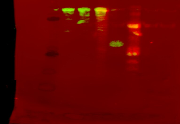
Application: Western BlotSample Tested: recombinant beta-catenin, beta catenin truncate (novus bio) and random antibody (- control) with Alexa 647Species: recombinant beta-catenin and N/AVerified Customer | Posted 05/08/2020from left to right: B-catenin biotin (8-1) B-catenin biotin (10-15) B-catenin 647 (8-15) Pure b-catenin truncate (Novus Biologics) Irrelevant antibody 6471:1000 antibody dilution for incubation (overnight, 4C) blocked with 2% TBST

-
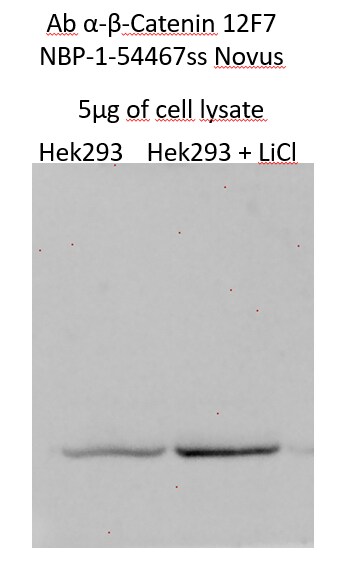
Application: Western BlotSample Tested: embryonic kidney cells and HEK293 CellsSpecies: HumanVerified Customer | Posted 07/12/2017
-
Application: Western BlotSample Tested: 1 - Embryonic Lung; 2 - Embryonic limbSpecies: OtherVerified Customer | Posted 10/24/2014Western blot analysis of chick proteins
There are no reviews that match your criteria.
Protocols
View specific protocols for beta-Catenin Antibody (12F7) - BSA Free (NBP1-54467):
Sample Preparation.
1. Grow cells to 60-85% confluency. Flow cytometry requires between 2 x 10⁵ and 1 x 10⁶ cells for optimal performance.
2. If cells are adherent, harvest gently by washing once with staining buffer and then scraping. Avoid using trypsin as this can disrupt certain epitopes of interest. If enzymatic harvest is required, use Accutase, Collagenase, or TrypLE Express for a less damaging option.
3. Reserve 100 uL for counting, then transfer cell volume into a 50 mL conical tube and centrifuge for 8 minutes at 400 RCF.
a. Count cells using a hemocytometer and a 1:1 trypan blue exclusion stain to determine cell viability before starting the flow protocol. If cells appear blue, do not proceed.
4. Re-suspend cells to a concentration of 1 x 10⁶ cells/mL in staining buffer (NBP2-26247).
5. Aliquot out 1 mL samples in accordance with your experimental samples.
Tip: When cell surface and intracellular staining are required in the same sample, it is advisable that the cell surface staining be performed first since the fixation and permeablization steps might reduce the availability of surface antigens.
Intracellular Staining.
Tip: When performing intracellular staining, it is important to use appropriate fixation and permeabilization reagents based upon the target and its subcellular location. Generally, our Intracellular Flow Assay Kit (NBP2-29450) is a good place to start as it contains an optimized combination of reagents for intracellular staining as well as an inhibitor of intracellular protein transport (necessary if staining secreted proteins). Certain targets may require more gentle or transient permeabilization protocols such as the commonly employed methanol or saponin-based methods.
Protocol for Cytoplasmic Targets:
Optional: Perform cell surface staining as described in the previous section.
1. Fix the cells by adding 100 uL fixation solution (such as 4% PFA) to each sample for 10-15 minutes.
2. Permeabilize cells by adding 100 uL of a permeabization buffer to every 1 x 10⁶ cells present in the sample. Mix well and incubate at room temperature for 15 minutes.
a. For cytoplasmic targets, use a gentle permeabilization solution such as 1X PBS + 0.5% Saponin or 1X PBS + 0.5% Tween-20.
b. To maintain the permeabilized state throughout your experiment, use staining buffer + 0.1% of the permeabilization reagent (i.e. 0.1% Tween-20 or 0.1% Saponin).
3. Following the 15 minute incubation, add 2 mL of the staining buffer + 0.1% permeabilizer to each sample.
4. Centrifuge for 5 minutes at 400 RCF.
5. Discard supernatant and re-suspend in 1 mL of staining buffer + 0.1% permeabilizer.
6. Stain each sample at 1 uL/ 1 x 10⁶ cells of primary antibody or 1-3 uL/ 1 x 10⁶ cells for directly conjugated antibodies. Mix well and incubate at room temperature for 30 minutes- 1 hour. Gently mix samples every 10-15 minutes.
7. Following the primary/conjugate incubation, add 2 mL/sample of staining buffer +0.1% permeabilizer and centrifuge for 5 minutes at 400 RCF.
8. Remove supernatant and re-suspend each sample in 2 mL staining buffer + 0.1% permeabilizer, repeat wash for 5 minutes at 400 RCF.
9. If using a directly conjugated antibody, after the second wash, re-suspend cell pellet to a final volume of 500 uL per sample and proceed with flow analysis.
Immunocytochemistry Protocol
Culture cells to appropriate density in 35 mm culture dishes or 6-well plates.
1. Remove culture medium and add 10% formalin to the dish. Fix at room temperature for 30 minutes.
2. Remove the formalin and add ice cold methanol. Incubate for 5-10 minutes.
3. Remove methanol and add washing solution (i.e. PBS). Be sure to not let the specimen dry out. Wash three times for 10 minutes.
4. To block nonspecific antibody binding incubate in 10% normal goat serum from 1 hour to overnight at room temperature.
5. Add primary antibody at appropriate dilution and incubate at room temperature from 2 hours to overnight at room temperature.
6. Remove primary antibody and replace with washing solution. Wash three times for 10 minutes.
7. Add secondary antibody at appropriate dilution. Incubate for 1 hour at room temperature.
8. Remove antibody and replace with wash solution, then wash for 10 minutes. Add Hoechst 33258 to wash solution at 1:25,0000 and incubate for 10 minutes. Wash a third time for 10 minutes.
9. Cells can be viewed directly after washing. The plates can also be stored in PBS containing Azide covered in Parafilm (TM). Cells can also be cover-slipped using Fluoromount, with appropriate sealing.
*The above information is only intended as a guide. The researcher should determine what protocol best meets their needs. Please follow safe laboratory procedures.
Immunohistochemistry-Paraffin Embedded Sections
Antigen Unmasking:
Bring slides to a boil in 10 mM sodium citrate buffer (pH 6.0) then maintain at a sub-boiling temperature for 10 minutes. Cool slides on bench-top for 30 minutes.
Staining:
1. Wash sections in deionized water three times for 5 minutes each.
2. Wash sections in wash buffer for 5 minutes.
3. Block each section with 100-400 ul blocking solution for 1 hour at room temperature.
4. Remove blocking solution and add 100-400 ul diluted primary antibody. Incubate overnight at 4 C.
5. Remove antibody solution and wash sections in wash buffer three times for 5 minutes each.
6. Add 100-400 ul biotinylated diluted secondary antibody. Incubate 30 minutes at room temperature.
7. Remove secondary antibody solution and wash sections three times with wash buffer for 5 minutes each.
8. Add 100-400 ul Streptavidin-HRP reagent to each section and incubate for 30 minutes at room temperature.
9. Wash sections three times in wash buffer for 5 minutes each.
10. Add 100-400 ul DAB substrate to each section and monitor staining closely.
11. As soon as the sections develop, immerse slides in deionized water.
12. Counterstain sections in hematoxylin.
13. Wash sections in deionized water two times for 5 minutes each.
14. Dehydrate sections.
15. Mount coverslips.
*The above information is only intended as a guide. The researcher should determine what protocol best meets their needs. Please follow safe laboratory procedures.
Western Blot Protocol
1. Perform SDS-PAGE on samples to be analyzed, loading 40 ug of total protein per lane.
2. Transfer proteins to membrane according to the instructions provided by the manufacturer of the membrane and transfer apparatus.
3. Stain according to standard Ponceau S procedure (or similar product) to assess transfer success, and mark molecular weight standards where appropriate.
4. Rinse the blot.
5. Block the membrane using standard blocking buffer for at least 1 hour.
6. Wash the membrane in wash buffer three times for 10 minutes each.
7. Dilute primary antibody in blocking buffer and incubate 1 hour at room temperature.
8. Wash the membrane in wash buffer three times for 10 minutes each.
9. Apply the diluted HRP conjugated secondary antibody in blocking buffer (as per manufacturers instructions) and incubate 1 hour at room temperature.
10. Wash the blot in wash buffer three times for 10 minutes each (this step can be repeated as required to reduce background).
11. Apply the detection reagent of choice in accordance with the manufacturers instructions.
Note: Tween-20 can be added to the blocking or antibody dilution buffer at a final concentration of 0.05-0.2%.
*The above information is only intended as a guide. The researcher should determine what protocol best meets their needs. Please follow safe laboratory procedures.
Find general support by application which include: protocols, troubleshooting, illustrated assays, videos and webinars.
- 7-Amino Actinomycin D (7-AAD) Cell Viability Flow Cytometry Protocol
- Antigen Retrieval Protocol (PIER)
- Antigen Retrieval for Frozen Sections Protocol
- Appropriate Fixation of IHC/ICC Samples
- Cellular Response to Hypoxia Protocols
- Chromogenic IHC Staining of Formalin-Fixed Paraffin-Embedded (FFPE) Tissue Protocol
- Chromogenic Immunohistochemistry Staining of Frozen Tissue
- ClariTSA™ Fluorophore Kits
- Detection & Visualization of Antibody Binding
- Extracellular Membrane Flow Cytometry Protocol
- Flow Cytometry Protocol for Cell Surface Markers
- Flow Cytometry Protocol for Staining Membrane Associated Proteins
- Flow Cytometry Staining Protocols
- Flow Cytometry Troubleshooting Guide
- Fluorescent IHC Staining of Frozen Tissue Protocol
- Graphic Protocol for Heat-induced Epitope Retrieval
- Graphic Protocol for the Preparation and Fluorescent IHC Staining of Frozen Tissue Sections
- Graphic Protocol for the Preparation and Fluorescent IHC Staining of Paraffin-embedded Tissue Sections
- Graphic Protocol for the Preparation of Gelatin-coated Slides for Histological Tissue Sections
- ICC Cell Smear Protocol for Suspension Cells
- ICC Immunocytochemistry Protocol Videos
- ICC for Adherent Cells
- IHC Sample Preparation (Frozen sections vs Paraffin)
- Immunocytochemistry (ICC) Protocol
- Immunocytochemistry Troubleshooting
- Immunofluorescence of Organoids Embedded in Cultrex Basement Membrane Extract
- Immunofluorescent IHC Staining of Formalin-Fixed Paraffin-Embedded (FFPE) Tissue Protocol
- Immunohistochemistry (IHC) and Immunocytochemistry (ICC) Protocols
- Immunohistochemistry Frozen Troubleshooting
- Immunohistochemistry Paraffin Troubleshooting
- Immunoprecipitation Protocol
- Intracellular Flow Cytometry Protocol Using Alcohol (Methanol)
- Intracellular Flow Cytometry Protocol Using Detergents
- Intracellular Nuclear Staining Flow Cytometry Protocol Using Detergents
- Intracellular Staining Flow Cytometry Protocol Using Alcohol Permeabilization
- Intracellular Staining Flow Cytometry Protocol Using Detergents to Permeabilize Cells
- Preparing Samples for IHC/ICC Experiments
- Preventing Non-Specific Staining (Non-Specific Binding)
- Primary Antibody Selection & Optimization
- Propidium Iodide Cell Viability Flow Cytometry Protocol
- Protocol for Heat-Induced Epitope Retrieval (HIER)
- Protocol for Liperfluo
- Protocol for Making a 4% Formaldehyde Solution in PBS
- Protocol for VisUCyte™ HRP Polymer Detection Reagent
- Protocol for the Characterization of Human Th22 Cells
- Protocol for the Characterization of Human Th9 Cells
- Protocol for the Fluorescent ICC Staining of Cell Smears - Graphic
- Protocol for the Fluorescent ICC Staining of Cultured Cells on Coverslips - Graphic
- Protocol for the Preparation & Fixation of Cells on Coverslips
- Protocol for the Preparation and Chromogenic IHC Staining of Frozen Tissue Sections
- Protocol for the Preparation and Chromogenic IHC Staining of Frozen Tissue Sections - Graphic
- Protocol for the Preparation and Chromogenic IHC Staining of Paraffin-embedded Tissue Sections
- Protocol for the Preparation and Chromogenic IHC Staining of Paraffin-embedded Tissue Sections - Graphic
- Protocol for the Preparation and Fluorescent ICC Staining of Cells on Coverslips
- Protocol for the Preparation and Fluorescent ICC Staining of Non-adherent Cells
- Protocol for the Preparation and Fluorescent ICC Staining of Stem Cells on Coverslips
- Protocol for the Preparation and Fluorescent IHC Staining of Frozen Tissue Sections
- Protocol for the Preparation and Fluorescent IHC Staining of Paraffin-embedded Tissue Sections
- Protocol for the Preparation of Gelatin-coated Slides for Histological Tissue Sections
- Protocol for the Preparation of a Cell Smear for Non-adherent Cell ICC - Graphic
- Protocol: Annexin V and PI Staining by Flow Cytometry
- Protocol: Annexin V and PI Staining for Apoptosis by Flow Cytometry
- R&D Systems Quality Control Western Blot Protocol
- TUNEL and Active Caspase-3 Detection by IHC/ICC Protocol
- The Importance of IHC/ICC Controls
- Troubleshooting Guide: Fluorokine Flow Cytometry Kits
- Troubleshooting Guide: Immunohistochemistry
- Troubleshooting Guide: Western Blot Figures
- Western Blot Conditions
- Western Blot Protocol
- Western Blot Protocol for Cell Lysates
- Western Blot Troubleshooting
- Western Blot Troubleshooting Guide
- View all Protocols, Troubleshooting, Illustrated assays and Webinars
FAQs for beta-Catenin Antibody (12F7) - BSA Free
-
Q: Hello, I was wondering if the NBP1-54467SS antibody is for global B-catenin, or only for phosphorylated version
A: This monoclonal Ab should detect both phosphorylated and unphosphorylated beta-Catenin.
Associated Pathways


