![Western Blot: beta-Catenin Antibody [NBP1-32239]](https://resources.rndsystems.com/images/products/beta-Catenin-Antibody-Western-Blot-NBP1-32239-img0033.jpg "Western Blot: beta-Catenin Antibody [NBP1-32239]")
Loading...
Key Product Details
Validated by
Knockout/Knockdown, Biological Validation
Species Reactivity
Validated:
Human, Mouse, Rat, Canine, Feline, Rabbit, Zebrafish
Cited:
Human, Mouse, Rat
Predicted:
Bovine (99%), Chicken (97%), Porcine (99%), Rhesus Macaque (100%), Sheep (99%), Xenopus (97%). Backed by our 100% Guarantee.
Applications
Validated:
Immunohistochemistry, Immunohistochemistry-Paraffin, Immunohistochemistry-Frozen, Immunohistochemistry Whole-Mount, Western Blot, Flow Cytometry, Immunocytochemistry/ Immunofluorescence, Immunoprecipitation, Chromatin Immunoprecipitation (ChIP)
Cited:
Western Blot, Immunocytochemistry/ Immunofluorescence, Immunoprecipitation, Chemotaxis, Proximity Ligation Assay, IF/IHC
Label
Unconjugated
Antibody Source
Polyclonal Rabbit IgG
Loading...
Product Specifications
Immunogen
Recombinant protein encompassing a sequence within the N-terminus region of human beta-Catenin. The exact sequence is proprietary.
Reactivity Notes
Xenopus laevis (97%), Cat (100%).
Localization
Cytoplasm, Nucleus, cytoskeleton
Marker
Epithelial Cell Marker, Adherens Junction Marker
Clonality
Polyclonal
Host
Rabbit
Isotype
IgG
Theoretical MW
85 kDa.
Disclaimer note: The observed molecular weight of the protein may vary from the listed predicted molecular weight due to post translational modifications, post translation cleavages, relative charges, and other experimental factors.
Disclaimer note: The observed molecular weight of the protein may vary from the listed predicted molecular weight due to post translational modifications, post translation cleavages, relative charges, and other experimental factors.
Scientific Data Images for beta-Catenin Antibody
Western Blot: beta-Catenin Antibody [NBP1-32239]
Western Blot: beta-Catenin Antibody [NBP1-32239] - Various whole cell extracts (30 ug) were separated by 7.5% SDS-PAGE, and the membrane was blotted with beta Catenin antibody [N1N2-2], N-term diluted at 1:3000.![Immunocytochemistry/ Immunofluorescence: beta-Catenin Antibody [NBP1-32239]](https://resources.rndsystems.com/images/products/beta-Catenin-Antibody-Immunocytochemistry-Immunofluorescence-NBP1-32239-img0017.jpg "Immunocytochemistry/ Immunofluorescence: beta-Catenin Antibody [NBP1-32239]")
Immunocytochemistry/ Immunofluorescence: beta-Catenin Antibody [NBP1-32239]
Immunocytochemistry/Immunofluorescence: beta-Catenin Antibody [NBP1-32239] - Paraformaldehyde-fixed A431, using beta- Catenin antibody (Green) at 1:200 dilution. Alpha-tubulin filaments were labeled with an alpha Tubulin antibody (Red) at 1:2000.![Immunohistochemistry-Paraffin: beta-Catenin Antibody [NBP1-32239]](https://resources.rndsystems.com/images/products/beta-Catenin-Antibody-Immunohistochemistry-Paraffin-NBP1-32239-img0043.jpg "Immunohistochemistry-Paraffin: beta-Catenin Antibody [NBP1-32239]")
Immunohistochemistry-Paraffin: beta-Catenin Antibody [NBP1-32239]
beta-Catenin-Antibody-Immunohistochemistry-Paraffin-NBP1-32239-img0043.jpg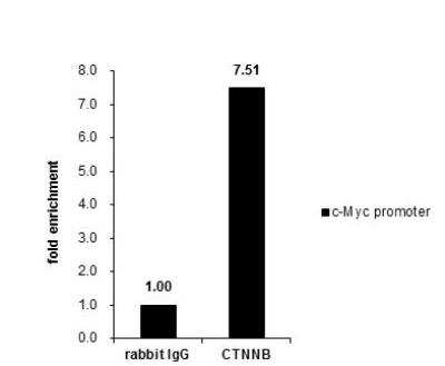
Chromatin Immunoprecipitation (ChIP): beta-Catenin Antibody [NBP1-32239] - Cross-linked ChIP was performed with HCT116 chromatin extract and 5 ug of either control rabbit IgG or anti-beta Catenin antibody. The precipitated DNA was detected by PCR with primer set targeting to c-Myc promoter.
![Western Blot: beta-Catenin Antibody [NBP1-32239]](https://resources.rndsystems.com/images/products/beta-Catenin-Antibody-Western-Blot-NBP1-32239-img0008.jpg "Western Blot: beta-Catenin Antibody [NBP1-32239]")
![Western Blot: beta-Catenin Antibody [NBP1-32239]](https://resources.rndsystems.com/images/products/beta-Catenin-Antibody-Western-Blot-NBP1-32239-img0013.jpg "Western Blot: beta-Catenin Antibody [NBP1-32239]")
Western Blot: beta-Catenin Antibody [NBP1-32239]
Western Blot: beta-Catenin Antibody [NBP1-32239] - Sample (50 ug of whole cell lysate) A: mouse brain 7.5% SDS PAGE diluted at 1:1000![Western Blot: beta-Catenin Antibody [NBP1-32239]](https://resources.rndsystems.com/images/products/beta-Catenin-Antibody-Western-Blot-NBP1-32239-img0014.jpg "Western Blot: beta-Catenin Antibody [NBP1-32239]")
Western Blot: beta-Catenin Antibody [NBP1-32239]
Western Blot: beta-Catenin Antibody [NBP1-32239] - A. 30 ug PC-12 whole cell lysate/extract 7.5 % SDS-PAGEbeta Catenin antibody [N1N2-2], N-term dilution: 1:1000![Western Blot: beta-Catenin Antibody [NBP1-32239]](https://resources.rndsystems.com/images/products/beta-Catenin-Antibody-Western-Blot-NBP1-32239-img0026.jpg "Western Blot: beta-Catenin Antibody [NBP1-32239]")
![Immunocytochemistry/ Immunofluorescence: beta-Catenin Antibody [NBP1-32239]](https://resources.rndsystems.com/images/products/beta-Catenin-Antibody-Immunocytochemistry-Immunofluorescence-NBP1-32239-img0032.jpg "Immunocytochemistry/ Immunofluorescence: beta-Catenin Antibody [NBP1-32239]")
Immunocytochemistry/ Immunofluorescence: beta-Catenin Antibody [NBP1-32239]
Immunocytochemistry/Immunofluorescence: beta-Catenin Antibody [NBP1-32239] - HeLa cells were fixed in 4% paraformaldehyde at RT for 15 min. Green: beta Catenin protein stained by beta Catenin antibody [N1N2-2], N-term diluted at 1:500. Blue: Hoechst 33342 staining.![Immunohistochemistry-Paraffin: beta-Catenin Antibody [NBP1-32239]](https://resources.rndsystems.com/images/products/beta-Catenin-Antibody-Immunohistochemistry-Paraffin-NBP1-32239-img0019.jpg "Immunohistochemistry-Paraffin: beta-Catenin Antibody [NBP1-32239]")
Immunohistochemistry-Paraffin: beta-Catenin Antibody [NBP1-32239]
Immunohistochemistry-Paraffin: beta-Catenin Antibody [NBP1-32239] - Paraffin-embedded mouse urinary bladder diluted at 1:500.![Immunohistochemistry-Paraffin: beta-Catenin Antibody [NBP1-32239]](https://resources.rndsystems.com/images/products/beta-Catenin-Antibody-Immunohistochemistry-Paraffin-NBP1-32239-img0020.jpg "Immunohistochemistry-Paraffin: beta-Catenin Antibody [NBP1-32239]")
Immunohistochemistry-Paraffin: beta-Catenin Antibody [NBP1-32239]
Immunohistochemistry-Paraffin: beta-Catenin Antibody [NBP1-32239] - Paraffin-embedded mouse skin dilution: 1:500.![Immunohistochemistry-Paraffin: beta-Catenin Antibody [NBP1-32239]](https://resources.rndsystems.com/images/products/beta-Catenin-Antibody-Immunohistochemistry-Paraffin-NBP1-32239-img0021.jpg "Immunohistochemistry-Paraffin: beta-Catenin Antibody [NBP1-32239]")
Immunohistochemistry-Paraffin: beta-Catenin Antibody [NBP1-32239]
Immunohistochemistry-Paraffin: beta-Catenin Antibody [NBP1-32239] - Paraffin-embedded mouse colon dilution: 1:500.![Immunohistochemistry-Paraffin: beta-Catenin Antibody [NBP1-32239]](https://resources.rndsystems.com/images/products/beta-Catenin-Antibody-Immunohistochemistry-Paraffin-NBP1-32239-img0036.jpg "Immunohistochemistry-Paraffin: beta-Catenin Antibody [NBP1-32239]")
Immunohistochemistry-Paraffin: beta-Catenin Antibody [NBP1-32239]
Immunohistochemistry-Paraffin: beta-Catenin Antibody [NBP1-32239] - Paraffin-embedded mouse duodenum. beta Catenin antibody [N1N2-2], N-term diluted at 1:500.![Immunohistochemistry-Paraffin: beta-Catenin Antibody [NBP1-32239]](https://resources.rndsystems.com/images/products/beta-Catenin-Antibody-Immunohistochemistry-Paraffin-NBP1-32239-img0037.jpg "Immunohistochemistry-Paraffin: beta-Catenin Antibody [NBP1-32239]")
Immunohistochemistry-Paraffin: beta-Catenin Antibody [NBP1-32239]
Immunohistochemistry-Paraffin: beta-Catenin Antibody [NBP1-32239] - Paraffin-embedded human esophagus. beta Catenin antibody [N1N2-2], N-term diluted at 1:500.![Immunohistochemistry-Paraffin: beta-Catenin Antibody [NBP1-32239]](https://resources.rndsystems.com/images/products/beta-Catenin-Antibody-Immunohistochemistry-Paraffin-NBP1-32239-img0038.jpg "Immunohistochemistry-Paraffin: beta-Catenin Antibody [NBP1-32239]")
Immunohistochemistry-Paraffin: beta-Catenin Antibody [NBP1-32239]
Immunohistochemistry-Paraffin: beta-Catenin Antibody [NBP1-32239] - Paraffin-embedded human cervix. beta Catenin antibody [N1N2-2], N-term diluted at 1:500.![Immunohistochemistry-Paraffin: beta-Catenin Antibody [NBP1-32239]](https://resources.rndsystems.com/images/products/beta-Catenin-Antibody-Immunohistochemistry-Paraffin-NBP1-32239-img0039.jpg "Immunohistochemistry-Paraffin: beta-Catenin Antibody [NBP1-32239]")
Immunohistochemistry-Paraffin: beta-Catenin Antibody [NBP1-32239]
Immunohistochemistry-Paraffin: beta-Catenin Antibody [NBP1-32239] - Paraffin-embedded mouse duodenum. beta Catenin antibody [N1N2-2], N-term diluted at 1:500.![Immunohistochemistry-Paraffin: beta-Catenin Antibody [NBP1-32239]](https://resources.rndsystems.com/images/products/beta-Catenin-Antibody-Immunohistochemistry-Paraffin-NBP1-32239-img0040.jpg "Immunohistochemistry-Paraffin: beta-Catenin Antibody [NBP1-32239]")
Immunohistochemistry-Paraffin: beta-Catenin Antibody [NBP1-32239]
Immunohistochemistry-Paraffin: beta-Catenin Antibody [NBP1-32239] - Paraffin-embedded rat colon. beta Catenin antibody [N1N2-2], N-term diluted at 1:500.![Immunohistochemistry-Paraffin: beta-Catenin Antibody [NBP1-32239]](https://resources.rndsystems.com/images/products/beta-Catenin-Antibody-Immunohistochemistry-Paraffin-NBP1-32239-img0041.jpg "Immunohistochemistry-Paraffin: beta-Catenin Antibody [NBP1-32239]")
Immunohistochemistry-Paraffin: beta-Catenin Antibody [NBP1-32239]
Immunohistochemistry-Paraffin: beta-Catenin Antibody [NBP1-32239] - Paraffin-embedded rat duodenum. beta Catenin antibody [N1N2-2], N-term diluted at 1:500.![Immunohistochemistry-Paraffin: beta-Catenin Antibody [NBP1-32239]](https://resources.rndsystems.com/images/products/beta-Catenin-Antibody-Immunohistochemistry-Paraffin-NBP1-32239-img0042.jpg "Immunohistochemistry-Paraffin: beta-Catenin Antibody [NBP1-32239]")
Immunohistochemistry-Paraffin: beta-Catenin Antibody [NBP1-32239]
Immunohistochemistry-Paraffin: beta-Catenin Antibody [NBP1-32239] - Paraffin-embedded mouse intestine. beta Catenin antibody [N1N2-2], N-term diluted at 1:500.
Immunocytochemistry/ Immunofluorescence: beta-Catenin Antibody [NBP1-32239] -
Immunocytochemistry/ Immunofluorescence: beta-Catenin Antibody [NBP1-32239] - P-cadherin is co-localized with other junctional proteins at the RPE cell border in mice.Immunofluorescence of mouse RPE flat-mounts. Double staining: P-cadherin (red; A, E, I) & either ZO-1 (green; B), beta -catenin (green; F), or F-actin (green; J), with nuclear stain by DAPI (blue; C, G, K). Merged images (D, H, L) show the co-localization of P-cadherin with ZO-1 (tight junction), beta -catenin (adherens junction), & F-actin (adherens junction) at the cell-cell border. Image collected & cropped by CiteAb from the following publication (https://pubmed.ncbi.nlm.nih.gov/29338041), licensed under a CC-BY license. Not internally tested by Novus Biologicals.
Immunocytochemistry/ Immunofluorescence: beta-Catenin Antibody [NBP1-32239] -
Immunocytochemistry/ Immunofluorescence: beta-Catenin Antibody [NBP1-32239] - WNT10A/ beta -catenin signalling is required for region-specific differentiation.(a–d) Filiform papillae are present in Wnt10a−/− & inducible beta -catenin mutant dorsal tongue (yellow arrows), but horny structures & expression of hard keratins (in situ hybridization, purple signals) are decreased (red arrows). (e–l) Epithelial deletion of Wnt10a (e–f″,i,j) or beta -catenin (g–h″,k,l) induced from P25, P110 or P15 as indicated causes decreased expression of nuclear beta -catenin, LEF1 & HOXC13 (white arrows, LEF1+ proliferating cells; yellow arrows, HOXC13+ differentiating cells). (m) qPCR shows significantly decreased Hoxc13 levels in Wnt10a & beta -catenin mutant tongue epithelium. (n–r) IF & qPCR reveal reduced levels of KRT9 protein (n–q) & mRNA (r) in Wnt10a−/− & inducible beta -catenin mutant footpad epidermis. (s–v″) Co-IF for KRT9 & KRT10 in plantar epidermis from patients homozygous for WNT10A c.756+1G>A (s–t″) or WNT10A c.391G>A (u–v″) compared with similarly aged sex-matched controls. For qPCR, RNA levels were quantified in six control & six mutant (P40) or four control & four mutant (P20-100) samples with three technical replicates for each, & normalized to beta -actin mRNA. Significance was calculated with two-tailed Student's t-test. Error bars indicate s.e.m. Scale bar, 25 μm (e–l) or 50 μm (a–d,n–q,s–v″). Image collected & cropped by CiteAb from the following publication (https://www.nature.com/articles/ncomms15397), licensed under a CC-BY license. Not internally tested by Novus Biologicals.
Immunocytochemistry/ Immunofluorescence: beta-Catenin Antibody [NBP1-32239] -
Immunocytochemistry/ Immunofluorescence: beta-Catenin Antibody [NBP1-32239] - Oxidative stress-induced dissociation of adherens junctions results in nuclear translocation of beta -catenin & an increase of EMT-related factors in mouse RPE.(A) Immunofluorescence of mouse RPE flat-mounts. Mice were injected with NaIO3 (15 mg/kg body weight) on Day 0, & the localization of beta -catenin (green) & P-cadherin (red) was analyzed along with nuclear stain by DAPI (blue) on Days 0 (a-c), 1 (d-f), 3 (g-i) & 7 (j-l). Double staining: beta -catenin (a, d, g, j), P-cadherin (b, e, h, k), & merged images with DAPI (c, f, i, l). The localization of beta -catenin & P-cadherin at the cell-cell border was significantly disrupted, & instead prominently detected on/in the nucleus on Day 3. (B) Immunofluorescence of mouse retinal sections with a focus on the RPE nuclei. Mice were injected with NaIO3 (15 mg/kg body weight) on Day 0, & the localization of beta -catenin (green) & P-cadherin (red) was analyzed along with nuclear stain by DAPI (blue) on Days 0 (m-o) & 3 (two representative nuclei; p-r & s-u). Double staining: beta -catenin (m, p, s), P-cadherin (n, q, t), & merged images with DAPI (o, r, u). On Day 3, beta -catenin was detected in the nuclei of mouse RPE. (C) Western blot analyses of mouse RPE proteins. Mice were injected with NaIO3 (15 mg/kg body weight) on Day 0, & RPE protein lysates were prepared on Days 0, 1, 3, & 7. The protein levels were analyzed using Western blotting with antibodies against P-cadherin, beta -catenin, SNAI1 (Snail), vimentin, & control beta -actin. The protein levels of beta -catenin & SNAI1 increased similarly on Day 1 following oxidative stress. Image collected & cropped by CiteAb from the following publication (https://pubmed.ncbi.nlm.nih.gov/29338041), licensed under a CC-BY license. Not internally tested by Novus Biologicals.
Western Blot: beta-Catenin Antibody [NBP1-32239] -
Western Blot: beta-Catenin Antibody [NBP1-32239] - Whole cell extract (30 ug) was separated by 7.5% SDS-PAGE, and the membrane was blotted with beta-Catenin antibody (NBP1-32239) diluted at 1:1000. The HRP-conjugated anti-rabbit IgG antibody was used to detect the primary antibody, and the signal was developed with Trident ECL plus-Enhanced.
Immunohistochemistry-Paraffin: beta-Catenin Antibody [NBP1-32239] -
Immunohistochemistry-Paraffin: beta-Catenin Antibody [NBP1-32239] - beta-Catenin antibody detects beta-Catenin protein at cell membrane by immunohistochemical analysis.Sample: Paraffin-embedded cat liver.
beta-Catenin stained by beta-Catenin antibody (NBP1-32239) diluted at 1:500.
Antigen Retrieval: Citrate buffer, pH 6.0, 15 min

Immunohistochemistry-Paraffin: beta-Catenin Antibody [NBP1-32239] -
Immunohistochemistry-Paraffin: beta-Catenin Antibody [NBP1-32239] - beta-Catenin antibody detects beta-Catenin protein at cell membrane by immunohistochemical analysis.Sample: Paraffin-embedded cat colon.
beta-Catenin stained by beta-Catenin antibody (NBP1-32239) diluted at 1:500.
Antigen Retrieval: Citrate buffer, pH 6.0, 15 min

Immunohistochemistry: beta-Catenin Antibody [NBP1-32239] -
Immunohistochemistry: beta-Catenin Antibody [NBP1-32239] - beta-Catenin antibody [N1N2-2], N-term detects Ctnnb1 protein on zebrafish by whole mount immunohistochemical analysis.Sample: 2 days-post-fertilization zebrafish embryo.
beta-Catenin antibody [N1N2-2], N-term (NBP1-32239) dilution: 1:100.

Western Blot: beta-Catenin Antibody [NBP1-32239] -
Western Blot: beta-Catenin Antibody [NBP1-32239] - Various tissue extracts (30 ug) were separated by 7.5% SDS-PAGE, and the membrane was blotted with beta-Catenin antibody (NBP1-32239) diluted at 1:500. The HRP-conjugated anti-rabbit IgG antibody was used to detect the primary antibody.
Immunohistochemistry: beta-Catenin Antibody [NBP1-32239] -
Immunohistochemistry: beta-Catenin Antibody [NBP1-32239] - Immunohistochemical analysis of agarose-embedded zebrafish embryo, using beta-Catenin antibody [N1N2-2], N-term NBP1-32239) at 1:100. dilution. (This image was provided courtesy of the Schilling Lab at UC, Irvine.)
Immunohistochemistry: beta-Catenin Antibody [NBP1-32239] -
Immunohistochemistry: beta-Catenin Antibody [NBP1-32239] - beta-Catenin antibody [N1N2-2], N-term detects Ctnnb1 protein on zebrafish by whole mount immunohistochemical analysis.Sample: 1 day-post-fertilization zebrafish embryo.
beta-Catenin antibody [N1N2-2], N-term (NBP1-32239) dilution: 1:100.

Immunohistochemistry-Paraffin: beta-Catenin Antibody [NBP1-32239] -
Immunohistochemistry-Paraffin: beta-Catenin Antibody [NBP1-32239] - beta-Catenin antibody [N1N2-2], N-term detects beta-Catenin protein by immunohistochemical analysis.Sample: Paraffin-embedded rat tissues.
beta-Catenin stained by beta-Catenin antibody [N1N2-2], N-term (NBP1-32239) diluted at 1:500.
Antigen Retrieval: Citrate buffer, pH 6.0, 15 min

Immunohistochemistry-Paraffin: beta-Catenin Antibody [NBP1-32239] -
Immunohistochemistry-Paraffin: beta-Catenin Antibody [NBP1-32239] - beta-Catenin antibody [N1N2-2] detects beta-Catenin protein at cell membrane in mouse colon by immunohistochemical analysis.Sample: Paraffin-embedded mouse colon.
Green: beta-Catenin antibody [N1N2-2] (NBP1-32239) diluted at 1:500.
Red: alpha Tubulin antibody [GT114] diluted at 1:500.
Blue: Hoechst 33342 staining.
Antigen Retrieval: Citrate buffer, pH 6.0, 15 min

Immunohistochemistry: beta-Catenin Antibody [NBP1-32239] -
Immunohistochemistry: beta-Catenin Antibody [NBP1-32239] - beta-Catenin antibody [N1N2-2], N-term detects Ctnnb1 protein on zebrafish by whole mount immunohistochemical analysis.Sample: 2 days-post-fertilization zebrafish embryo.
beta-Catenin antibody [N1N2-2], N-term (NBP1-32239) dilution: 1:100.

Immunocytochemistry/ Immunofluorescence: beta-Catenin Antibody [NBP1-32239] -
Immunocytochemistry/ Immunofluorescence: beta-Catenin Antibody [NBP1-32239] - beta-Catenin antibody detects beta-Catenin protein at cell membrane by immunofluorescent analysis.Sample: MDCK cells were fixed in 4% paraformaldehyde at RT for 15 min.
Green: beta-Catenin stained by beta-Catenin antibody (NBP1-32239) diluted at 1:1000.

Immunohistochemistry: beta-Catenin Antibody [NBP1-32239] -
Immunohistochemistry: beta-Catenin Antibody [NBP1-32239] - Immunohistochemical analysis of paraffin-embedded zebrafish tissue, using beta-Catenin antibody [N1N2-2], N-term (NBP1-32239) at 1:300 dilution.
Immunohistochemistry-Paraffin: beta-Catenin Antibody [NBP1-32239] -
Immunohistochemistry-Paraffin: beta-Catenin Antibody [NBP1-32239] - beta-Catenin antibody [N1N2-2], N-term detects beta-Catenin protein by immunohistochemical analysis.Sample: Paraffin-embedded mouse tissues.
beta-Catenin stained by beta-Catenin antibody [N1N2-2], N-term (NBP1-32239) diluted at 1:500.
Antigen Retrieval: Citrate buffer, pH 6.0, 15 min

Western Blot: beta-Catenin Antibody [NBP1-32239] -
Western Blot: beta-Catenin Antibody [NBP1-32239] - Whole cell extract (30 ug) was separated by 7.5% SDS-PAGE, and the membrane was blotted with beta-Catenin antibody (NBP1-32239) diluted at 1:1000. The HRP-conjugated anti-rabbit IgG antibody was used to detect the primary antibody.
Western Blot: beta-Catenin Antibody [NBP1-32239] -
Western Blot: beta-Catenin Antibody [NBP1-32239] - Wild-type (WT) and beta Catenin knockout (KO) 293T cell extracts (9 ug) were separated by 7.5% SDS-PAGE, and the membrane was blotted with beta Catenin antibody [N1N2-2], N-term diluted at 1:1000. The HRP-conjugated anti-rabbit IgG antibody was used to detect the primary antibody.
Immunohistochemistry-Paraffin: beta-Catenin Antibody [NBP1-32239] -
beta-Catenin antibody [N1N2-2] detects beta-Catenin protein at cell membrane in mouse colon by immunohistochemical analysis.Sample: Paraffin-embedded mouse colon.
Green: beta-Catenin antibody [N1N2-2] (NBP1-32239) diluted at 1:500.
Red: alpha Tubulin antibody [GT114] diluted at 1:500.
Blue: Hoechst 33342 staining.
Antigen Retrieval: Citrate buffer, pH 6.0, 15 min

Flow Cytometry: beta-Catenin Antibody [NBP1-32239] -
beta-Catenin antibody [N1N2-2], N-term (NBP1-32239) detects CTNNB1 protein by flow cytometry analysis.Sample: HeLa cell.
Black: Unlabelled sample was used as a control.
Red: beta-Catenin antibody [N1N2-2], N-term (NBP1-32239) dilution: 1:50.
Acquisition of 20,000 events were collected for FACS analysis.

Immunocytochemistry/ Immunofluorescence: beta-Catenin Antibody [NBP1-32239] -
beta-Catenin antibody [N1N2-2], N-term detects beta-Catenin protein at cell membrane by immunofluorescent analysis.Sample: HCT 116 cells were fixed in 4% paraformaldehyde at RT for 15 min.
Green: beta-Catenin protein stained by beta-Catenin antibody [N1N2-2], N-term (NBP1-32239) diluted at 1:500.
Blue: Hoechst 33342 staining.

Western Blot: beta-Catenin Antibody [NBP1-32239] -
Oxidative stress-induced dissociation of adherens junctions results in nuclear translocation of beta -catenin and an increase of EMT-related factors in mouse RPE.(A) Immunofluorescence of mouse RPE flat-mounts. Mice were injected with NaIO3 (15 mg/kg body weight) on Day 0, and the localization of beta -catenin (green) and P-cadherin (red) was analyzed along with nuclear stain by DAPI (blue) on Days 0 (a-c), 1 (d-f), 3 (g-i) and 7 (j-l). Double staining: beta -catenin (a, d, g, j), P-cadherin (b, e, h, k), and merged images with DAPI (c, f, i, l). The localization of beta -catenin and P-cadherin at the cell-cell border was significantly disrupted, and instead prominently detected on/in the nucleus on Day 3. (B) Immunofluorescence of mouse retinal sections with a focus on the RPE nuclei. Mice were injected with NaIO3 (15 mg/kg body weight) on Day 0, and the localization of beta -catenin (green) and P-cadherin (red) was analyzed along with nuclear stain by DAPI (blue) on Days 0 (m-o) and 3 (two representative nuclei; p-r and s-u). Double staining: beta -catenin (m, p, s), P-cadherin (n, q, t), and merged images with DAPI (o, r, u). On Day 3, beta -catenin was detected in the nuclei of mouse RPE. (C) Western blot analyses of mouse RPE proteins. Mice were injected with NaIO3 (15 mg/kg body weight) on Day 0, and RPE protein lysates were prepared on Days 0, 1, 3, and 7. The protein levels were analyzed using Western blotting with antibodies against P-cadherin, beta -catenin, SNAI1 (Snail), vimentin, and control beta -actin. The protein levels of beta -catenin and SNAI1 increased similarly on Day 1 following oxidative stress. Image collected and cropped by CiteAb from the following open publication (https://pubmed.ncbi.nlm.nih.gov/29338041), licensed under a CC-BY license. Not internally tested by Novus Biologicals.Applications for beta-Catenin Antibody
Application
Recommended Usage
Chromatin Immunoprecipitation (ChIP)
Assay dependent
Flow Cytometry
1:50-1:200
Immunocytochemistry/ Immunofluorescence
1:100-1:1000
Immunohistochemistry
1:100-1:1000
Immunohistochemistry Whole-Mount
Assay dependent
Immunohistochemistry-Frozen
Assay dependent
Immunohistochemistry-Paraffin
1:100-1:1000
Immunoprecipitation
1:50-1:100
Western Blot
1:500-1:20000
Reviewed Applications
Read 1 review rated 5 using NBP1-32239 in the following applications:
Flow Cytometry Panel Builder
Bio-Techne Knows Flow Cytometry
Save time and reduce costly mistakes by quickly finding compatible reagents using the Panel Builder Tool.
Advanced Features
- Spectra Viewer - Custom analysis of spectra from multiple fluorochromes
- Spillover Popups - Visualize the spectra of individual fluorochromes
- Antigen Density Selector - Match fluorochrome brightness with antigen density

Formulation, Preparation, and Storage
Purification
Antigen Affinity-purified
Formulation
PBS, 1% BSA, 20% Glycerol
Preservative
0.025% Proclin 300
Concentration
Concentrations vary lot to lot. See vial label for concentration. If unlisted please contact technical services.
Shipping
The product is shipped with polar packs. Upon receipt, store it immediately at the temperature recommended below.
Stability & Storage
Aliquot and store at -20C or -80C. Avoid freeze-thaw cycles.
Background: beta-Catenin
Alternate Names
bCatenin, CTNNB1
Gene Symbol
CTNNB1
Additional beta-Catenin Products
Product Documents for beta-Catenin Antibody
Certificate of Analysis
To download a Certificate of Analysis, please enter a lot or batch number in the search box below.
Product Specific Notices for beta-Catenin Antibody
This product is for research use only and is not approved for use in humans or in clinical diagnosis. Primary Antibodies are guaranteed for 1 year from date of receipt.
Citations for beta-Catenin Antibody
 Powered by Bioz
Customer Reviews for beta-Catenin Antibody (1)
5 out of 5
1 Customer Rating
Have you used beta-Catenin Antibody?
Submit a review and receive an Amazon gift card!
$25/€18/£15/$25CAN/¥2500 Yen for a review with an image
$10/€7/£6/$10CAN/¥1110 Yen for a review without an image
Submit a review
Customer Images
-(01-mg)_NBP1-32239_8441.jpg)
Showing
1
-
1 of
1 review
Showing All
Filter By:
-
Application: Western BlotSample Tested: Lysate from HEK293 cellsSpecies: HumanVerified Customer | Posted 06/25/2014Western blot for b-catenin in HEK 293 cells treated with LiCl (10mM) for 1 hr

There are no reviews that match your criteria.
Protocols
Find general support by application which include: protocols, troubleshooting, illustrated assays, videos and webinars.
- 7-Amino Actinomycin D (7-AAD) Cell Viability Flow Cytometry Protocol
- Antigen Retrieval Protocol (PIER)
- Antigen Retrieval for Frozen Sections Protocol
- Appropriate Fixation of IHC/ICC Samples
- Cellular Response to Hypoxia Protocols
- ChIP Protocol Video
- Chromatin Immunoprecipitation (ChIP) Protocol
- Chromatin Immunoprecipitation Protocol
- Chromogenic IHC Staining of Formalin-Fixed Paraffin-Embedded (FFPE) Tissue Protocol
- Chromogenic Immunohistochemistry Staining of Frozen Tissue
- ClariTSA™ Fluorophore Kits
- Detection & Visualization of Antibody Binding
- Extracellular Membrane Flow Cytometry Protocol
- Flow Cytometry Protocol for Cell Surface Markers
- Flow Cytometry Protocol for Staining Membrane Associated Proteins
- Flow Cytometry Staining Protocols
- Flow Cytometry Troubleshooting Guide
- Fluorescent IHC Staining of Frozen Tissue Protocol
- Graphic Protocol for Heat-induced Epitope Retrieval
- Graphic Protocol for the Preparation and Fluorescent IHC Staining of Frozen Tissue Sections
- Graphic Protocol for the Preparation and Fluorescent IHC Staining of Paraffin-embedded Tissue Sections
- Graphic Protocol for the Preparation of Gelatin-coated Slides for Histological Tissue Sections
- ICC Cell Smear Protocol for Suspension Cells
- ICC Immunocytochemistry Protocol Videos
- ICC for Adherent Cells
- IHC Sample Preparation (Frozen sections vs Paraffin)
- Immunocytochemistry (ICC) Protocol
- Immunocytochemistry Troubleshooting
- Immunofluorescence of Organoids Embedded in Cultrex Basement Membrane Extract
- Immunofluorescent IHC Staining of Formalin-Fixed Paraffin-Embedded (FFPE) Tissue Protocol
- Immunohistochemistry (IHC) and Immunocytochemistry (ICC) Protocols
- Immunohistochemistry Frozen Troubleshooting
- Immunohistochemistry Paraffin Troubleshooting
- Immunoprecipitation Protocol
- Intracellular Flow Cytometry Protocol Using Alcohol (Methanol)
- Intracellular Flow Cytometry Protocol Using Detergents
- Intracellular Nuclear Staining Flow Cytometry Protocol Using Detergents
- Intracellular Staining Flow Cytometry Protocol Using Alcohol Permeabilization
- Intracellular Staining Flow Cytometry Protocol Using Detergents to Permeabilize Cells
- Preparing Samples for IHC/ICC Experiments
- Preventing Non-Specific Staining (Non-Specific Binding)
- Primary Antibody Selection & Optimization
- Propidium Iodide Cell Viability Flow Cytometry Protocol
- Protocol for Heat-Induced Epitope Retrieval (HIER)
- Protocol for Liperfluo
- Protocol for Making a 4% Formaldehyde Solution in PBS
- Protocol for VisUCyte™ HRP Polymer Detection Reagent
- Protocol for the Characterization of Human Th22 Cells
- Protocol for the Characterization of Human Th9 Cells
- Protocol for the Fluorescent ICC Staining of Cell Smears - Graphic
- Protocol for the Fluorescent ICC Staining of Cultured Cells on Coverslips - Graphic
- Protocol for the Preparation & Fixation of Cells on Coverslips
- Protocol for the Preparation and Chromogenic IHC Staining of Frozen Tissue Sections
- Protocol for the Preparation and Chromogenic IHC Staining of Frozen Tissue Sections - Graphic
- Protocol for the Preparation and Chromogenic IHC Staining of Paraffin-embedded Tissue Sections
- Protocol for the Preparation and Chromogenic IHC Staining of Paraffin-embedded Tissue Sections - Graphic
- Protocol for the Preparation and Fluorescent ICC Staining of Cells on Coverslips
- Protocol for the Preparation and Fluorescent ICC Staining of Non-adherent Cells
- Protocol for the Preparation and Fluorescent ICC Staining of Stem Cells on Coverslips
- Protocol for the Preparation and Fluorescent IHC Staining of Frozen Tissue Sections
- Protocol for the Preparation and Fluorescent IHC Staining of Paraffin-embedded Tissue Sections
- Protocol for the Preparation of Gelatin-coated Slides for Histological Tissue Sections
- Protocol for the Preparation of a Cell Smear for Non-adherent Cell ICC - Graphic
- Protocol: Annexin V and PI Staining by Flow Cytometry
- Protocol: Annexin V and PI Staining for Apoptosis by Flow Cytometry
- R&D Systems Quality Control Western Blot Protocol
- TUNEL and Active Caspase-3 Detection by IHC/ICC Protocol
- The Importance of IHC/ICC Controls
- Troubleshooting Guide: Fluorokine Flow Cytometry Kits
- Troubleshooting Guide: Immunohistochemistry
- Troubleshooting Guide: Western Blot Figures
- Western Blot Conditions
- Western Blot Protocol
- Western Blot Protocol for Cell Lysates
- Western Blot Troubleshooting
- Western Blot Troubleshooting Guide
- View all Protocols, Troubleshooting, Illustrated assays and Webinars
Loading...
Associated Pathways


Blood-Brain Barrier Pathway: Anatomy
 HIF Enhancer Pathways
 Notch Signaling Pathways
 Wnt Signaling Pathways: beta-Catenin-dependent Wnt Signaling
