c-Fos Antibody (2H2) - BSA Free
Novus Biologicals | Catalog # NBP2-50037
![Western Blot: c-Fos Antibody (2H2) [NBP2-50037]](https://resources.rndsystems.com/images/products/c-Fos-Antibody-2H2-Western-Blot-NBP2-50037-img0003.jpg "Western Blot: c-Fos Antibody (2H2) [NBP2-50037]")
Key Product Details
Validated by
Knockout/Knockdown, Biological Validation
Species Reactivity
Validated:
Human, Mouse, Rat
Cited:
Mouse, Rat
Applications
Validated:
Knockout Validated, Immunohistochemistry, Western Blot, Immunocytochemistry/ Immunofluorescence
Cited:
Immunohistochemistry, Immunohistochemistry-Frozen, Western Blot, Immunocytochemistry/ Immunofluorescence, IF/IHC
Label
Unconjugated
Antibody Source
Monoclonal Mouse IgG1 Clone # 2H2
Format
BSA Free
Loading...
Product Specifications
Immunogen
This c-Fos Antibody (2H2) was developed against full length recombinant human c-Fos protein expressed in and purified from E. coli. [UniProt# P01100]
Reactivity Notes
Rat reactivity reported in scientific literature (PMID:33091429).
Clonality
Monoclonal
Host
Mouse
Isotype
IgG1
Theoretical MW
50-65 kDa.
Disclaimer note: The observed molecular weight of the protein may vary from the listed predicted molecular weight due to post translational modifications, post translation cleavages, relative charges, and other experimental factors.
Disclaimer note: The observed molecular weight of the protein may vary from the listed predicted molecular weight due to post translational modifications, post translation cleavages, relative charges, and other experimental factors.
Scientific Data Images for c-Fos Antibody (2H2) - BSA Free
Western Blot: c-Fos Antibody (2H2) [NBP2-50037]
Western Blot: c-Fos Antibody (2H2) [NBP2-50037] - Top panel: Analysis of c-Fos expression in HeLa cells using NBP2-50037. Lane 1: HeLa cells were serum-starved for 36 hours. Lane 2: Serum-starved HeLa cells were stimulated with 20% FBS (fetal bovine serum) for 2 hours. NBP2-50037 recognizes bands in the range of 50-65 kDa, which represent multiple forms of c-Fos. Serum starvation attenuates c-Fos expression, while 20% FBS strongly stimulates c-Fos expression. Bottom panel: Blot was stripped and probed with monoclonal antibody against GAPDH (NB300-221) used as loading control.![Immunocytochemistry/ Immunofluorescence: c-Fos Antibody (2H2) [NBP2-50037]](https://resources.rndsystems.com/images/products/c-Fos-Antibody-2H2-Immunocytochemistry-Immunofluorescence-NBP2-50037-img0006.jpg "Immunocytochemistry/ Immunofluorescence: c-Fos Antibody (2H2) [NBP2-50037]")
Immunocytochemistry/ Immunofluorescence: c-Fos Antibody (2H2) [NBP2-50037]
Immunocytochemistry/Immunofluorescence: c-Fos Antibody (2H2) [NBP2-50037] - Section of rat hippocampus stained with mouse monoclonal antibody to c-FOS NBP2-50037 in red and counterstained with rabbit polyclonal antibody to FOX3/NeuN. DAPI reveals nuclei of neurons and glia in blue. The hippocampal neurons stain green for FOX3/NeuN and a few also are expressing c-FOS, and so appear orange. These cells were spontaneously active at the time the animal was sacrificed.![Knockout Validated: c-Fos Antibody (2H2) [NBP2-50037]](https://resources.rndsystems.com/images/products/c-Fos-Antibody-2H2-Knockout-Validated-NBP2-50037-img0005.jpg "Western Blot: c-Fos Antibody (2H2) [NBP2-50037]")
Western Blot: c-Fos Antibody (2H2) [NBP2-50037]
Western Blot: c-Fos Antibody (2H2) [NBP2-50037] - Western blot shows lysates of HeLa human cervical epithelial carcinoma parental cell line and c-Fox knockout (KO) HeLa cell line. PVDF membrane was probed with 1:1000 of Mouse Anti-Human c-Fox Monoclonal Antibody (Catalog # NBP2-50037) followed by HRP-conjugated Anti-Mouse IgG Secondary Antibody (Catalog #HAF018). Specific band was detected for c-Fox at approximately 52 kDa (as indicated) in the parental HeLa cell line, but is not detectable in the knockout HeLa cell line. This experiment was conducted under reducing conditions.![Western Blot: c-Fos Antibody (2H2) [NBP2-50037]](https://resources.rndsystems.com/images/products/c-Fos-Antibody-2H2-Western-Blot-NBP2-50037-img0007.jpg "Western Blot: c-Fos Antibody (2H2) [NBP2-50037]")
Western Blot: c-Fos Antibody (2H2) [NBP2-50037]
Western Blot: c-Fos Antibody (2H2) [NBP2-50037] - Analysis of cell lysates using mouse c-Fos mAb, dilution 1:1,000 (Green), and rabbit GAPDH pAb, dilution 1:20,000 (Red) used as a loading control. [1] protein standard (red), [2] HeLa cells in serum free media. [3] HeLa cells stimulated with 20% fetal bovine serum for 2hrs after 36hrs in serum free media. [4] rat cortical neurons. [5] rat cortical neurons treated with membrane depolarization buffer for 5hrs. Multiple bands at 50-65kDa in stimulated or treated cell lysates correspond to different forms of the c-Fos proten. The single band at 37 kDa represents GAPDH protein.![Immunohistochemistry: c-Fos Antibody (2H2) [NBP2-50037]](https://resources.rndsystems.com/images/products/c-Fos-Antibody-2H2-Immunocytochemistry-NBP2-50037-img0004.jpg "Immunohistochemistry: c-Fos Antibody (2H2) [NBP2-50037]")
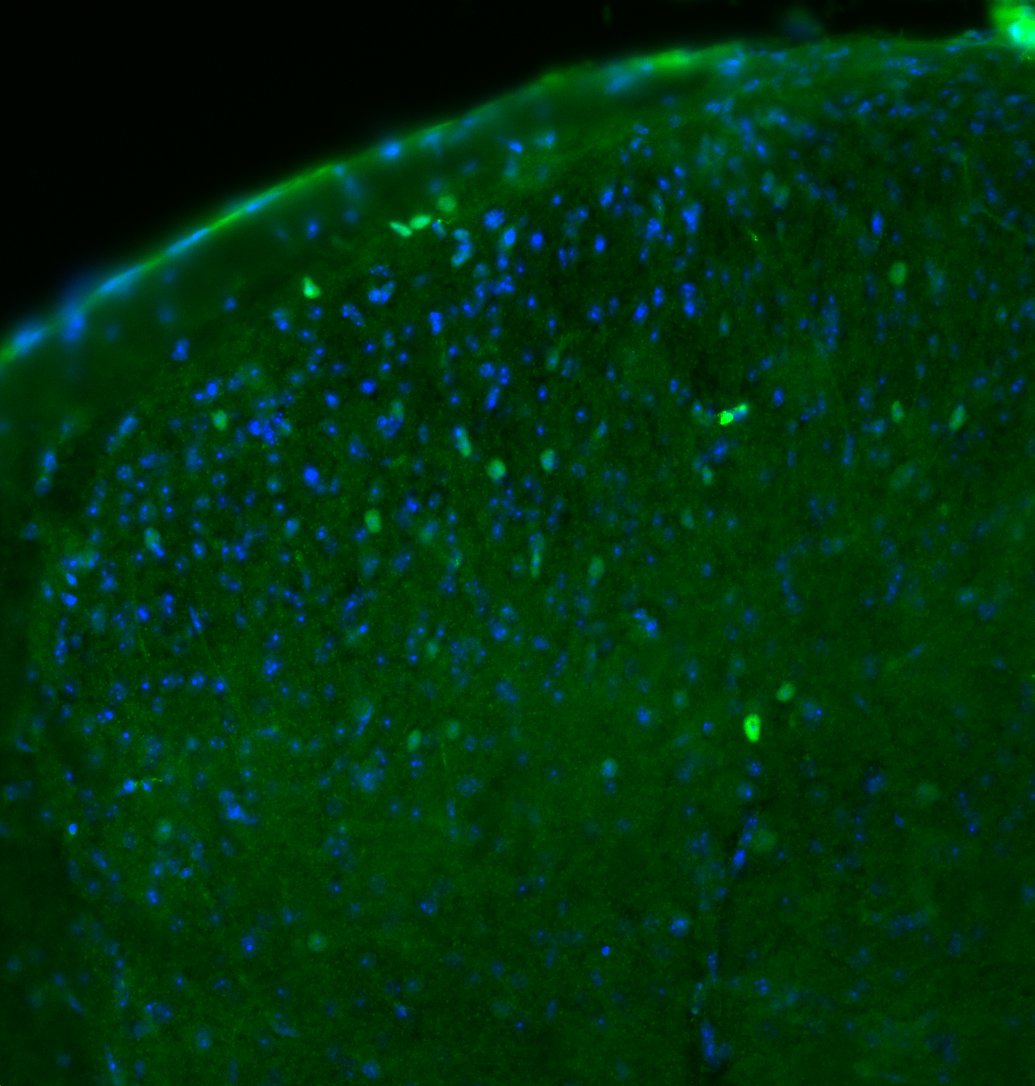
Immunohistochemistry: c-Fos Antibody (2H2) [NBP2-50037]
Immunohistochemistry: c-Fos Antibody (2H2) [NBP2-50037] - pAb 1:1000 (green), DAPI counterstain (blue) on 30 micron cryosection of mouse spinal cord. This image was submitted via customer Review.![Immunocytochemistry/ Immunofluorescence: c-Fos Antibody (2H2) [NBP2-50037]](https://resources.rndsystems.com/images/products/c-Fos-Antibody-2H2-Immunocytochemistry-Immunofluorescence-NBP2-50037-img0001.jpg "Immunocytochemistry/ Immunofluorescence: c-Fos Antibody (2H2) [NBP2-50037]")
Immunocytochemistry/ Immunofluorescence: c-Fos Antibody (2H2) [NBP2-50037]
Immunocytochemistry/Immunofluorescence: c-Fos Antibody (2H2) [NBP2-50037] - Left: NBP2-50037 staining (green) in HeLa cells, which were treated with serum-starvation for 36 hrs, followed by 2 hrs 20% FBS stimulation (bottom), or PBS treatment (top). Green c-Fos staining only localizes in the nuclei of stimulated cells, but not in un-stimulated cells. Cells are counter-stained with chicken pAb against Vimentin (NB300-223, red). Blue shows DAPI staining of nucleus. Middle: Mouse brain section (45 uM; fixed by transcardial perfusion with 4% PFA) labeled with NBP2-50037 using a standard HRP-DAB staining technique. Cells expressing c-Fos show dark color in nucleus. Right: Mouse cortical section labeled with NBP2-50037 (red) and rabbit polyclonal anti-NeuN (NBP1-92716, green) using IF confocal. Neurons positive for c-Fos and RBFOX3/NeuN appear to be yellow. Inset shows an enlarged image of NBP2-50037 staining. Nuclei are labeled with Dapi (blue).Applications for c-Fos Antibody (2H2) - BSA Free
Application
Recommended Usage
Immunocytochemistry/ Immunofluorescence
1:1000
Immunohistochemistry
1:1000
Western Blot
1:1000 - 1:2000
Reviewed Applications
Read 1 review rated 4 using NBP2-50037 in the following applications:
Formulation, Preparation, and Storage
Purification
Immunogen affinity purified
Formulation
50% PBS, 50% glycerol
Format
BSA Free
Preservative
5mM Sodium Azide
Concentration
1 mg/ml
Shipping
The product is shipped with polar packs. Upon receipt, store it immediately at the temperature recommended below.
Stability & Storage
Store at 4C short term. Aliquot and store at -20C long term. Avoid freeze-thaw cycles.
Background: c-Fos
In response to stimuli, c-Fos, which is encoded by protooncogenes, has a role in cell proliferation, differentiation, and transformation (3,6). A variety of stimuli can increase c-Fos expression such as growth factors, proinflammatory cytokines, UV radiation, neurotransmitters, hormones, injury, and stress (1,6). c-Fos has long been used as a marker for neuronal activity and is associated with neural and behavioral responses following stimuli (1-3, 6-7). Mouse studies have revealed that c-Fos is important for efficient neurogenesis and cortical development (3). Additionally, c-Fos signal can be used as a molecular marker for learning and memory, such as recognition and fear (2,7). Studies have found that repeated positive stimuli result in increased Fos expression while, conversely, repeated negative value stimuli are indicated by decreased signal (7). Intermediate early genes have also been implicated in neuropsychiatric disorders including showing altered c-Fos expression in a schizophrenia animal model (2). Furthermore, antipsychotics and antidepressants are both capable of impacting c-Fos expression (2).
References
1. Kovacs K. J. (1998). c-Fos as a transcription factor: a stressful (re)view from a functional map. Neurochemistry International. https://doi.org/10.1016/s0197-0186(98)00023-0
2. Gallo, F. T., Katche, C., Morici, J. F., Medina, J. H., & Weisstaub, N. V. (2018). Immediate Early Genes, Memory and Psychiatric Disorders: Focus on c-Fos, Egr1 and Arc. Frontiers in Behavioral Neuroscience. https://doi.org/10.3389/fnbeh.2018.00079
3. Velazquez, F. N., Caputto, B. L., & Boussin, F. D. (2015). c-Fos importance for brain development. Aging. https://doi.org/10.18632/aging.100862
4. Uniprot (P01100)
5. Wu, Z., Nicoll, M., & Ingham, R. J. (2021). AP-1 family transcription factors: a diverse family of proteins that regulate varied cellular activities in classical hodgkin lymphoma and ALK+ ALCL. Experimental Hematology & Oncology. https://doi.org/10.1186/s40164-020-00197-9
6. Shaulian, E., & Karin, M. (2001). AP-1 in cell proliferation and survival. Oncogene. https://doi.org/10.1038/sj.onc.1204383
7. Chung L. (2015). A Brief Introduction to the Transduction of Neural Activity into Fos Signal. Development & Reproduction. https://doi.org/10.12717/DR.2015.19.2.061
Long Name
FBJ/Finkel–Biskis–Jinkins Murine Osteosarcoma Viral Oncogene Homolog
Alternate Names
cFos, FOS, G0S7
Gene Symbol
FOS
UniProt
Additional c-Fos Products
Product Documents for c-Fos Antibody (2H2) - BSA Free
Certificate of Analysis
To download a Certificate of Analysis, please enter a lot or batch number in the search box below.
Product Specific Notices for c-Fos Antibody (2H2) - BSA Free
This product is for research use only and is not approved for use in humans or in clinical diagnosis. Primary Antibodies are guaranteed for 1 year from date of receipt.
Citations for c-Fos Antibody (2H2) - BSA Free
 Powered by Bioz
Customer Reviews for c-Fos Antibody (2H2) - BSA Free (1)
4 out of 5
1 Customer Rating
Have you used c-Fos Antibody (2H2) - BSA Free?
Submit a review and receive an Amazon gift card!
$25/€18/£15/$25CAN/¥2500 Yen for a review with an image
$10/€7/£6/$10CAN/¥1110 Yen for a review without an image
Submit a review
Customer Images
Showing
1
-
1 of
1 review
Showing All
Filter By:
-
Application: ImmunocytochemistrySample Tested: Adult spinal cordSpecies: MouseVerified Customer | Posted 06/20/2017NBP2-50037 used at 1:1000 (green), DAPI counterstain (blue) on 30 micron cryosection of mouse spinal cord.Immunofluorescence using NBP2-50037 at a dilution of 1:1000 on 4% PFA perfused 30 micron cryosections of mouse spinal cord. Tissue was blocked with 5% NGS/1% BSA/0.3% Triton X-100, then primary antibody was applied overnight at 4 C in the same diluent. Mouse on mouse avidin-biotin blocking kit with streptavidin-Alexa Fluor 488 conjugate was used for fluorescent detection.

There are no reviews that match your criteria.
Protocols
Find general support by application which include: protocols, troubleshooting, illustrated assays, videos and webinars.
- Antigen Retrieval Protocol (PIER)
- Antigen Retrieval for Frozen Sections Protocol
- Appropriate Fixation of IHC/ICC Samples
- Cellular Response to Hypoxia Protocols
- Chromogenic IHC Staining of Formalin-Fixed Paraffin-Embedded (FFPE) Tissue Protocol
- Chromogenic Immunohistochemistry Staining of Frozen Tissue
- ClariTSA™ Fluorophore Kits
- Detection & Visualization of Antibody Binding
- Fluorescent IHC Staining of Frozen Tissue Protocol
- Graphic Protocol for Heat-induced Epitope Retrieval
- Graphic Protocol for the Preparation and Fluorescent IHC Staining of Frozen Tissue Sections
- Graphic Protocol for the Preparation and Fluorescent IHC Staining of Paraffin-embedded Tissue Sections
- Graphic Protocol for the Preparation of Gelatin-coated Slides for Histological Tissue Sections
- ICC Cell Smear Protocol for Suspension Cells
- ICC Immunocytochemistry Protocol Videos
- ICC for Adherent Cells
- IHC Sample Preparation (Frozen sections vs Paraffin)
- Immunocytochemistry (ICC) Protocol
- Immunocytochemistry Troubleshooting
- Immunofluorescence of Organoids Embedded in Cultrex Basement Membrane Extract
- Immunofluorescent IHC Staining of Formalin-Fixed Paraffin-Embedded (FFPE) Tissue Protocol
- Immunohistochemistry (IHC) and Immunocytochemistry (ICC) Protocols
- Immunohistochemistry Frozen Troubleshooting
- Immunohistochemistry Paraffin Troubleshooting
- Preparing Samples for IHC/ICC Experiments
- Preventing Non-Specific Staining (Non-Specific Binding)
- Primary Antibody Selection & Optimization
- Protocol for Heat-Induced Epitope Retrieval (HIER)
- Protocol for Making a 4% Formaldehyde Solution in PBS
- Protocol for VisUCyte™ HRP Polymer Detection Reagent
- Protocol for the Fluorescent ICC Staining of Cell Smears - Graphic
- Protocol for the Fluorescent ICC Staining of Cultured Cells on Coverslips - Graphic
- Protocol for the Preparation & Fixation of Cells on Coverslips
- Protocol for the Preparation and Chromogenic IHC Staining of Frozen Tissue Sections
- Protocol for the Preparation and Chromogenic IHC Staining of Frozen Tissue Sections - Graphic
- Protocol for the Preparation and Chromogenic IHC Staining of Paraffin-embedded Tissue Sections
- Protocol for the Preparation and Chromogenic IHC Staining of Paraffin-embedded Tissue Sections - Graphic
- Protocol for the Preparation and Fluorescent ICC Staining of Cells on Coverslips
- Protocol for the Preparation and Fluorescent ICC Staining of Non-adherent Cells
- Protocol for the Preparation and Fluorescent ICC Staining of Stem Cells on Coverslips
- Protocol for the Preparation and Fluorescent IHC Staining of Frozen Tissue Sections
- Protocol for the Preparation and Fluorescent IHC Staining of Paraffin-embedded Tissue Sections
- Protocol for the Preparation of Gelatin-coated Slides for Histological Tissue Sections
- Protocol for the Preparation of a Cell Smear for Non-adherent Cell ICC - Graphic
- R&D Systems Quality Control Western Blot Protocol
- TUNEL and Active Caspase-3 Detection by IHC/ICC Protocol
- The Importance of IHC/ICC Controls
- Troubleshooting Guide: Immunohistochemistry
- Troubleshooting Guide: Western Blot Figures
- Western Blot Conditions
- Western Blot Protocol
- Western Blot Protocol for Cell Lysates
- Western Blot Troubleshooting
- Western Blot Troubleshooting Guide
- View all Protocols, Troubleshooting, Illustrated assays and Webinars
FAQs for c-Fos Antibody (2H2) - BSA Free
Showing
1
-
1 of
1 FAQ
Showing All
-
Q: I am looking for some c-fos antibodies that could work in IHC.
A:
Here are a list of products which might be of interest to you: Product List. We have additional filters on the left which may help you narrow down based on host species, target species, clonality, etc. We will guarantee these to work for all stated species and applications listed on the datasheet.
Loading...
Associated Pathways


NOD-like Receptor Signaling Pathways
 Pathogen or Damage-activated C-Type Lectin Receptor Signaling Pathways
 TGF-beta Signaling Pathways
 Th1 Differentiation Pathway
 Toll-Like Receptor Signaling Pathways
 VEGF - VEGF R2 Signaling Pathways
