c-jun [p Ser63] Antibody (SY0297)
Novus Biologicals | Catalog # NBP2-67471
Recombinant Monoclonal Antibody
Loading...
Key Product Details
Validated by
Biological Validation
Species Reactivity
Validated:
Human, Mouse, Rat
Cited:
Mouse
Applications
Validated:
Immunohistochemistry, Immunohistochemistry-Paraffin, Western Blot, Immunocytochemistry/ Immunofluorescence
Cited:
IF/IHC
Label
Unconjugated
Antibody Source
Recombinant Monoclonal Rabbit IgG Clone # SY0297 expressed in HEK293
Loading...
Product Specifications
Immunogen
Synthetic phospho-peptide corresponding to residues surrounding c-jun aa 31-80 / 331. (SwissProt: P05412 Human; SwissProt: P05627 Mouse; SwissProt: P17325 Rat)
Modification
p Ser63
Localization
Nucleus.
Clonality
Monoclonal
Host
Rabbit
Isotype
IgG
Scientific Data Images for c-jun [p Ser63] Antibody (SY0297)
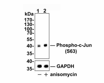
Western Blot: c-jun [p Ser63] Antibody (SY0297) [NBP2-67471] - Analysis of c-Jun [p Ser63] (whole cell lysate, 10 ug /lane. Lane 2: NIH/3T3 cells were treated with 250 ng/ml anisomycin for 30 minutes, whole cell lysates, 10 ug/lane. Proteins were transferred to a PVDF membrane and blocked with 5% BSA in PBS for 1 hour at room temperature. The primary antibody Anti-c-Jun [p Ser63] (1/500), Anti-c-Jun antibody (1/2000) and Anti-GAPDH antibody (1/10,000)was used in 5% BSA at room temperature for 2 hours. Goat Anti-Rabbit IgG H&L (HRP) Secondary Antibody at 1:200,000 dilution was used for 1 hour at room temperature. Predicted band size: 36 kDa Observed band size: 40 kDa Exposure time: 3 minutes 43 seconds
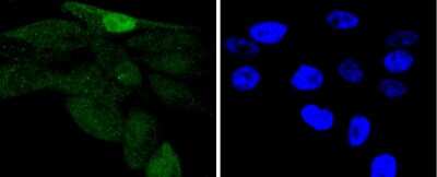
Immunocytochemistry/Immunofluorescence: c-jun [p Ser63] Antibody (SY0297) [NBP2-67471] - Staining Phospho-c-Jun(S63) in PC-3M cells (green). The nuclear counter stain is DAPI (blue). Cells were fixed in paraformaldehyde, permeabilised with 0.25% Triton X100/PBS.
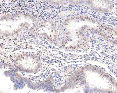
Immunohistochemistry-Paraffin: c-jun [p Ser63] Antibody (SY0297) [NBP2-67471] - Analysis of paraffin-embedded human endometrial tissue with Rabbit anti-c-jun [p Ser63] antibody washed with ddH2O and PBS, and then probed with the primary antibody at 1/500 dilution for 1 hour at room temperature. The detection was performed using an HRP conjugated compact polymer system. DAB was used as the chromogen. Tissues were counterstained with hematoxylin and mounted with DPX.
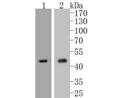
Western Blot: c-jun [p Ser63] Antibody (SY0297) [NBP2-67471] - Analysis of Phospho-c-Jun(S63) on different lysates using anti-Phospho-c-Jun(S63) antibody at 1/1,000 dilution. Positive control: Lane 1: NIH/3T3 Lane 2: 293T
Western Blot: c-jun [p Ser63] Antibody (SY0297) [NBP2-67471] - Western blot analysis of c-jun on different lysates. Proteins were transferred to a PVDF membrane and blocked with 5% BSA in PBS for 1 hour at room temperature. The primary antibody (1/500) was used in 5% BSA at room temperature for 2 hours. Goat Anti-Rab
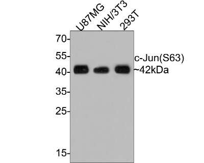
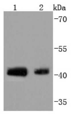
Western Blot: c-jun [p Ser63] Antibody (SY0297) [NBP2-67471] - Analysis of c-Jun (p Ser63) on different lysates with Rabbit anti-c-Jun (p Ser63) antibody at 1/500 dilution. Lane 1: U87MG cell lysate Lane 2: NIH/3T3 cell lysate Lane 3: 293T cell lysate Lysates/proteins at 10 ug/Lane. Predicted band size: 36 kDa Observed band size: 42 kDa Exposure time: 2 minutes; 12% SDS-PAGE gel. Proteins were transferred to a PVDF membrane and blocked with 5% NFDM/TBST for 1 hour at room temperature. The primary antibody at 1/500 dilution was used in 5% NFDM/TBST at room temperature for 2 hours. Goat Anti-Rabbit IgG - HRP Secondary Antibody at 1:300,000 dilution was used for 1 hour at room temperature.
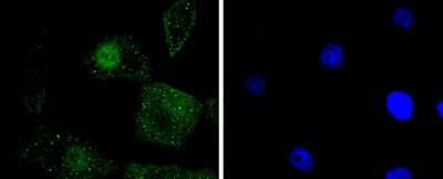
Immunocytochemistry/Immunofluorescence: c-jun [p Ser63] Antibody (SY0297) [NBP2-67471] - Staining Phospho-c-Jun(S63) in A549 cells (green). The nuclear counter stain is DAPI (blue). Cells were fixed in paraformaldehyde, permeabilised with 0.25% Triton X100/PBS.
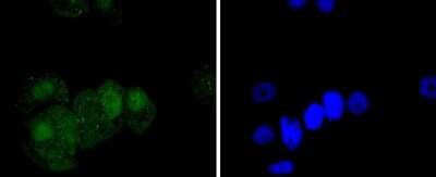
Immunocytochemistry/Immunofluorescence: c-jun [p Ser63] Antibody (SY0297) [NBP2-67471] - Staining Phospho-c-Jun(S63) in MCF-7 cells (green). The nuclear counter stain is DAPI (blue). Cells were fixed in paraformaldehyde, permeabilised with 0.25% Triton X100/PBS.
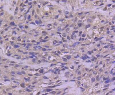
Immunohistochemistry-Paraffin: c-jun [p Ser63] Antibody (SY0297) [NBP2-67471] - Analysis of paraffin-embedded human breast carcinoma tissue using anti-Phospho-c-Jun(S63) antibody. Counter stained with hematoxylin.
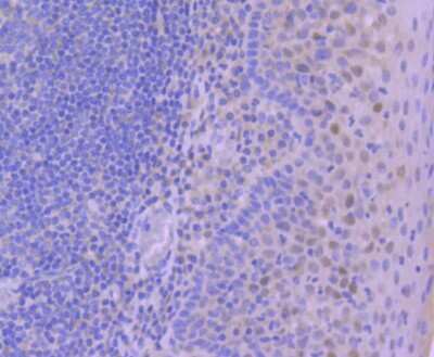
Immunohistochemistry-Paraffin: c-jun [p Ser63] Antibody (SY0297) [NBP2-67471] - Analysis of paraffin-embedded human tonsil tissue using anti-Phospho-c-Jun(S63) antibody. Counter stained with hematoxylin.
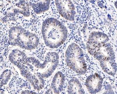
Immunohistochemistry-Paraffin: c-jun [p Ser63] Antibody (SY0297) [NBP2-67471] - Analysis of paraffin-embedded human colon carcinoma tissue with Rabbit anti-c-jun [Ser63] antibody washed with ddH2O and PBS, and then probed with the primary antibody at 1/500 dilution for 1 hour at room temperature. The detection was performed using an HRP conjugated compact polymer system. DAB was used as the chromogen. Tissues were counterstained with hematoxylin and mounted with DPX.
Applications for c-jun [p Ser63] Antibody (SY0297)
Application
Recommended Usage
Immunocytochemistry/ Immunofluorescence
1:50-1:200
Immunohistochemistry-Paraffin
1:50-1:500
Western Blot
1:500-1:2000
Reviewed Applications
Read 1 review rated 5 using NBP2-67471 in the following applications:
Formulation, Preparation, and Storage
Purification
Protein A purified
Formulation
TBS (pH7.4), 0.05% BSA, 40% Glycerol
Preservative
0.05% Sodium Azide
Concentration
1 mg/ml
Shipping
The product is shipped with polar packs. Upon receipt, store it immediately at the temperature recommended below.
Stability & Storage
Store at 4C short term. Aliquot and store at -20C long term. Avoid freeze-thaw cycles.
Background: c-Jun
Long Name
Cellular Repressor of E1A-stimulated Genes/Transcription Factor AP-1
Alternate Names
cJun, JUN
Gene Symbol
JUN
Additional c-Jun Products
Product Documents for c-jun [p Ser63] Antibody (SY0297)
Certificate of Analysis
To download a Certificate of Analysis, please enter a lot or batch number in the search box below.
Product Specific Notices for c-jun [p Ser63] Antibody (SY0297)
This product is for research use only and is not approved for use in humans or in clinical diagnosis. Primary Antibodies are guaranteed for 1 year from date of receipt.
Citations for c-jun [p Ser63] Antibody (SY0297)
 Powered by Bioz
Customer Reviews for c-jun [p Ser63] Antibody (SY0297) (1)
5 out of 5
1 Customer Rating
Have you used c-jun [p Ser63] Antibody (SY0297)?
Submit a review and receive an Amazon gift card!
$25/€18/£15/$25CAN/¥2500 Yen for a review with an image
$10/€7/£6/$10CAN/¥1110 Yen for a review without an image
Submit a review
Customer Images
![c-jun [p Ser63] Antibody (SY0297) NBP2-67471](https://resources.rndsystems.com/images/reviews/review_nbp2-67471_57406_0_0.png)
Showing
1
-
1 of
1 review
Showing All
Filter By:
-
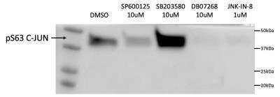
Application: Western BlotSample Tested: U251 glioma cell line; whole cell lysateSpecies: HumanVerified Customer | Posted 10/17/2021Western blot with lysates from U251 cells. Cells have been treated with JNK and p38 inhibitors.
![c-jun [p Ser63] Antibody (SY0297) NBP2-67471](data:image/png;base64,R0lGODlhAQABAAD/ACwAAAAAAQABAAACADs=)
There are no reviews that match your criteria.
Protocols
Find general support by application which include: protocols, troubleshooting, illustrated assays, videos and webinars.
- Antigen Retrieval Protocol (PIER)
- Antigen Retrieval for Frozen Sections Protocol
- Appropriate Fixation of IHC/ICC Samples
- Cellular Response to Hypoxia Protocols
- Chromogenic IHC Staining of Formalin-Fixed Paraffin-Embedded (FFPE) Tissue Protocol
- Chromogenic Immunohistochemistry Staining of Frozen Tissue
- ClariTSA™ Fluorophore Kits
- Detection & Visualization of Antibody Binding
- Fluorescent IHC Staining of Frozen Tissue Protocol
- Graphic Protocol for Heat-induced Epitope Retrieval
- Graphic Protocol for the Preparation and Fluorescent IHC Staining of Frozen Tissue Sections
- Graphic Protocol for the Preparation and Fluorescent IHC Staining of Paraffin-embedded Tissue Sections
- Graphic Protocol for the Preparation of Gelatin-coated Slides for Histological Tissue Sections
- ICC Cell Smear Protocol for Suspension Cells
- ICC Immunocytochemistry Protocol Videos
- ICC for Adherent Cells
- IHC Sample Preparation (Frozen sections vs Paraffin)
- Immunocytochemistry (ICC) Protocol
- Immunocytochemistry Troubleshooting
- Immunofluorescence of Organoids Embedded in Cultrex Basement Membrane Extract
- Immunofluorescent IHC Staining of Formalin-Fixed Paraffin-Embedded (FFPE) Tissue Protocol
- Immunohistochemistry (IHC) and Immunocytochemistry (ICC) Protocols
- Immunohistochemistry Frozen Troubleshooting
- Immunohistochemistry Paraffin Troubleshooting
- Preparing Samples for IHC/ICC Experiments
- Preventing Non-Specific Staining (Non-Specific Binding)
- Primary Antibody Selection & Optimization
- Protocol for Heat-Induced Epitope Retrieval (HIER)
- Protocol for Making a 4% Formaldehyde Solution in PBS
- Protocol for VisUCyte™ HRP Polymer Detection Reagent
- Protocol for the Fluorescent ICC Staining of Cell Smears - Graphic
- Protocol for the Fluorescent ICC Staining of Cultured Cells on Coverslips - Graphic
- Protocol for the Preparation & Fixation of Cells on Coverslips
- Protocol for the Preparation and Chromogenic IHC Staining of Frozen Tissue Sections
- Protocol for the Preparation and Chromogenic IHC Staining of Frozen Tissue Sections - Graphic
- Protocol for the Preparation and Chromogenic IHC Staining of Paraffin-embedded Tissue Sections
- Protocol for the Preparation and Chromogenic IHC Staining of Paraffin-embedded Tissue Sections - Graphic
- Protocol for the Preparation and Fluorescent ICC Staining of Cells on Coverslips
- Protocol for the Preparation and Fluorescent ICC Staining of Non-adherent Cells
- Protocol for the Preparation and Fluorescent ICC Staining of Stem Cells on Coverslips
- Protocol for the Preparation and Fluorescent IHC Staining of Frozen Tissue Sections
- Protocol for the Preparation and Fluorescent IHC Staining of Paraffin-embedded Tissue Sections
- Protocol for the Preparation of Gelatin-coated Slides for Histological Tissue Sections
- Protocol for the Preparation of a Cell Smear for Non-adherent Cell ICC - Graphic
- R&D Systems Quality Control Western Blot Protocol
- TUNEL and Active Caspase-3 Detection by IHC/ICC Protocol
- The Importance of IHC/ICC Controls
- Troubleshooting Guide: Immunohistochemistry
- Troubleshooting Guide: Western Blot Figures
- Western Blot Conditions
- Western Blot Protocol
- Western Blot Protocol for Cell Lysates
- Western Blot Troubleshooting
- Western Blot Troubleshooting Guide
- View all Protocols, Troubleshooting, Illustrated assays and Webinars
Loading...
Associated Pathways


NOD-like Receptor Signaling Pathways
 Pathogen or Damage-activated C-Type Lectin Receptor Signaling Pathways
 TGF-beta Signaling Pathways
 Th1 Differentiation Pathway
 Toll-Like Receptor Signaling Pathways
 VEGF - VEGF R2 Signaling Pathways
