Caspase-8 Antibody (90A992) - BSA Free
Novus Biologicals | Catalog # NB100-56527
Key Product Details
Validated by
Species Reactivity
Validated:
Cited:
Applications
Validated:
Cited:
Label
Antibody Source
Format
Product Specifications
Immunogen
Reactivity Notes
Specificity
Clonality
Host
Isotype
Scientific Data Images for Caspase-8 Antibody (90A992) - BSA Free
![Western Blot: Caspase-8 Antibody (90A992)BSA Free [NB100-56527]](https://resources.rndsystems.com/images/products/Caspase-8-Antibody-90A992-Western-Blot-NB100-56527-img0018.jpg "Western Blot: Caspase-8 Antibody (90A992)BSA Free [NB100-56527]")
Western Blot: Caspase-8 Antibody (90A992)BSA Free [NB100-56527]
Western Blot: Caspase-8 Antibody (90A992) [NB100-56527] - Western blot analysis of Caspase-8 in Jurkat cells using Caspase-8 antibody at 1 ug/ml. Cells were treated with 2 uM staurosporine for different time periods. Caspase-8 activation is detected in western blots by the presence of Caspase-8 cleavage fragments. The antibody detected both pro (full length) and active (cleaved) Caspase-8, depending on the treatment time points. A basal level of endogenously cleaved Caspase-8 can be see in untreated Jurkat cells. Goat anti-mouse lg HRP secondary antibody and PicoTect ECL substrate solution were used for this test.![Western Blot: Caspase-8 Antibody (90A992)BSA Free [NB100-56527]](https://resources.rndsystems.com/images/products/Caspase-8-Antibody-90A992-Western-Blot-NB100-56527-img0022.jpg "Western Blot: Caspase-8 Antibody (90A992)BSA Free [NB100-56527]")
Western Blot: Caspase-8 Antibody (90A992)BSA Free [NB100-56527]
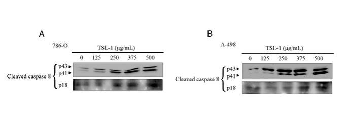
Western Blot: Caspase-8 Antibody (90A992) [NB100-56527] - Caspase-8 expression in 786-O and A-498 cellsClose![Immunohistochemistry-Paraffin: Caspase-8 Antibody (90A992) - BSA Free [NB100-56527]](https://resources.rndsystems.com/images/products/Caspase-8-Antibody-90A992-Immunohistochemistry-Paraffin-NB100-56527-img0014.jpg "Immunohistochemistry-Paraffin: Caspase-8 Antibody (90A992) - BSA Free [NB100-56527]")
Immunohistochemistry-Paraffin: Caspase-8 Antibody (90A992) - BSA Free [NB100-56527]
Immunohistochemistry-Paraffin: Caspase-8 Antibody (90A992) [NB100-56527] - Formalin-fixed, paraffin-embedded human spleen (top) and breast (bottom) stained with Caspase-8 antibody at 4 ug/ml. Localization can be cytoplasmic and nuclear. Cancer/normal adjacent tissue array was used for this test. Staining of formalin-fixed tissues is enhanced by boiling tissue sections in 10 mM sodium citrate buffer, pH 6.0 for 10-20 min followed by cooling at RT for 20 min.![Flow Cytometry: Caspase-8 Antibody (90A992) - BSA Free [NB100-56527]](https://resources.rndsystems.com/images/products/Caspase-8-Antibody-90A992-Flow-Cytometry-NB100-56527-img0008.jpg "Flow Cytometry: Caspase-8 Antibody (90A992) - BSA Free [NB100-56527]")
Flow Cytometry: Caspase-8 Antibody (90A992) - BSA Free [NB100-56527]
Flow Cytometry: Caspase-8 Antibody (90A992) [NB100-56527] - Flow cytometric analysis of Caspase-8 in HeLa cells using 0.1 ug of Caspase-8 antibody. Shaded histogram represents cells without antibody; green represents isotype control; red represents Caspase-8 antibody. Goat anti-mouse IgG-FITC secondary antibody was used for this test. IC-Flow (Intracellular Staining Flow Cytometry Kit) was used to fix and prepare the cells for staining.![Western Blot: Caspase-8 Antibody (90A992)BSA Free [NB100-56527]](https://resources.rndsystems.com/images/products/Caspase-8-Antibody-90A992-Western-Blot-NB100-56527-img0021.jpg "Western Blot: Caspase-8 Antibody (90A992)BSA Free [NB100-56527]")
Western Blot: Caspase-8 Antibody (90A992)BSA Free [NB100-56527]
Western Blot: Caspase-8 Antibody (90A992) [NB100-56527] - Analysis using the Biotin conjugate of NB100-56527. Detection of human Caspase-8 using Jurkat lysates with NB100-55786 at 2 ug/ml (lane 1) and 0.5 ug/ml (lane 2) dilution. NB100-55786 only detects 55 kDa Caspase-8 in Jurkat cells.![Simple Western: Caspase-8 Antibody (90A992)BSA Free [NB100-56527]](https://resources.rndsystems.com/images/products/Caspase-8-Antibody-90A992-Simple-Western-NB100-56527-img0019.jpg "Simple Western: Caspase-8 Antibody (90A992)BSA Free [NB100-56527]")
Simple Western: Caspase-8 Antibody (90A992)BSA Free [NB100-56527]
Simple Western: Caspase-8 Antibody (90A992) [NB100-56527] - Simple Western lane view shows a specific band for Caspase 8 in 0.5 mg/ml of Hek293 lysate. This experiment was performed under reducing conditions using the 12-230 kDa separation system. [NB100-56527] -")
Simple Western: Mouse Monoclonal Caspase-8 Antibody (90A992) [NB100-56527] -
Simple Western: Mouse Monoclonal Caspase-8 Antibody (90A992) [NB100-56527] - S2/IAPinh induces activation of the extrinsic apoptosis pathway via degradation of cIAP-1. (A) Schematic diagram of the intrinsic and extrinsic apoptosis pathways: arrows represent stimulation. TNFR, tumor necrosis factor receptor; DR4-5, death receptor 4–5; cIAP-1/2, cellular inhibitor of apoptosis protein-1/2; RIPK1, Receptor-interacting serine/threonine-protein kinase 1. (B) Protein expression of cIAP-1, cIAP-2, and XIAP in HPAC cells treated with vehicle, SW43 (10 µM), IAPinh (10 µM), or S2/IAPinh (10 µM) for 6 h. The precursor and cleaved forms of caspases 3, 8, and 9 were also analyzed for these cells using Wes automated capillary blotting system (Protein Simple). (C) Quantification of protein expression. Relative densitometry of each band normalized to the total protein. Data shown as means ± SEM. **P < 0.01, **** P < 0.0001. (D) Ratio of Caspase 3/7 counts to NucRed counts in HPAC cells treated with vehicle, SW43 (10 µM), IAPinh (10 µM), combination of SW43 (10 µM) and IAPinh (10 µM), or S2/IAPinh (10 µM) measured by the IncuCyte system (Sartorius). Bar graph shows the ratio of Caspase 3/7 at 48 h for each treatment. Data shown as means ± SEM. ****P < 0.001. (E) Activity of cell death in AsPC-1 cells was measured using YOYO-1 iodide on the IncuCyte (Sartorius). Representative images of AsPC-1 cells treated with or without Z-VAD-FMK and S2/IAPinh (10 uM) at baseline and 36 h after treatment. Scale bars are equal to 20 µm. (F) The AUC of lethal fraction at 36 h. Data shown as means ± SEM. ****P < 0.0001. Image collected and cropped by CiteAb under a CC-BY license from the following publication: The novel drug candidate S2/IAPinh improves survival in models of pancreatic and ovarian cancer. Sci Rep (2024). Not internally tested by Novus Biologicals. [NB100-56527] -")
Simple Western: Mouse Monoclonal Caspase-8 Antibody (90A992) [NB100-56527] -
Simple Western: Mouse Monoclonal Caspase-8 Antibody (90A992) [NB100-56527] - S2/IAPinh induces tumor cell death via activation of the extrinsic apoptosis pathway in vivo. KP2 tumor samples collected from animals at 48 h after the treatment with either vehicle, SW43, IAPinh, or S2/IAPinh, were used for analyses. (A) Representative images of TUNEL labeled apoptosis cells in KP2 tumor samples of each group. Nuclei were stained in blue with Hoechst, TUNEL positive cells are in red. Scale bars are equal to 20 µm. (B) Quantification of TUNEL positive cells per area in each group. Data are shown as means ± SEM; ****P < 0.0001. (C) Protein expression of cIAP-1, cIAP-2, and XIAP in KP2 tumor samples of each group. The precursor and cleaved forms of caspases 3, 8, and 9 were also analyzed for these cells using Wes automated capillary blotting system (Protein Simple). (D) Quantification of protein expression. Relative densitometry of each band normalized to the total protein. Data shown as means ± SEM. *P < 0.05, **P < 0.01, **** P < 0.0001. Image collected and cropped by CiteAb under a CC-BY license from the following publication: The novel drug candidate S2/IAPinh improves survival in models of pancreatic and ovarian cancer. Sci Rep (2024). Not internally tested by Novus Biologicals.Applications for Caspase-8 Antibody (90A992) - BSA Free
Flow Cytometry
Immunohistochemistry
Immunohistochemistry-Paraffin
Simple Western
Western Blot
See Simple Western Antibody Database for Simple Western validation: Tested in Hek293 lysate 0.5 mg/mL, separated by Size, antibody dilution of 1:100, apparent MW was 16 kDa.This antibody is CyTOF ready.
Reviewed Applications
Read 1 review rated 4 using NB100-56527 in the following applications:
Flow Cytometry Panel Builder
Bio-Techne Knows Flow Cytometry
Save time and reduce costly mistakes by quickly finding compatible reagents using the Panel Builder Tool.
Advanced Features
- Spectra Viewer - Custom analysis of spectra from multiple fluorochromes
- Spillover Popups - Visualize the spectra of individual fluorochromes
- Antigen Density Selector - Match fluorochrome brightness with antigen density

Formulation, Preparation, and Storage
Purification
Formulation
Format
Preservative
Concentration
Shipping
Stability & Storage
Background: Caspase-8
Alternate Names
Gene Symbol
Additional Caspase-8 Products
Product Documents for Caspase-8 Antibody (90A992) - BSA Free
Certificate of Analysis
To download a Certificate of Analysis, please enter a lot or batch number in the search box below.
Product Specific Notices for Caspase-8 Antibody (90A992) - BSA Free
This product is for research use only and is not approved for use in humans or in clinical diagnosis. Primary Antibodies are guaranteed for 1 year from date of receipt.
Related Research Areas
Citations for Caspase-8 Antibody (90A992) - BSA Free
 Powered by Bioz
Customer Reviews for Caspase-8 Antibody (90A992) - BSA Free (1)
Have you used Caspase-8 Antibody (90A992) - BSA Free?
Submit a review and receive an Amazon gift card!
$25/€18/£15/$25CAN/¥2500 Yen for a review with an image
$10/€7/£6/$10CAN/¥1110 Yen for a review without an image
Submit a review
Customer Images
-
Application: Western BlotSample Tested: 786-O and A-498 whole cell lysatesSpecies: HumanVerified Customer | Posted 04/29/2015Caspase-8 expression in 786-O and A-498 cells

There are no reviews that match your criteria.
Protocols
Find general support by application which include: protocols, troubleshooting, illustrated assays, videos and webinars.
- 7-Amino Actinomycin D (7-AAD) Cell Viability Flow Cytometry Protocol
- Antigen Retrieval Protocol (PIER)
- Antigen Retrieval for Frozen Sections Protocol
- Appropriate Fixation of IHC/ICC Samples
- Cellular Response to Hypoxia Protocols
- Chromogenic IHC Staining of Formalin-Fixed Paraffin-Embedded (FFPE) Tissue Protocol
- Chromogenic Immunohistochemistry Staining of Frozen Tissue
- ClariTSA™ Fluorophore Kits
- Detection & Visualization of Antibody Binding
- Extracellular Membrane Flow Cytometry Protocol
- Flow Cytometry Protocol for Cell Surface Markers
- Flow Cytometry Protocol for Staining Membrane Associated Proteins
- Flow Cytometry Staining Protocols
- Flow Cytometry Troubleshooting Guide
- Fluorescent IHC Staining of Frozen Tissue Protocol
- Graphic Protocol for Heat-induced Epitope Retrieval
- Graphic Protocol for the Preparation and Fluorescent IHC Staining of Frozen Tissue Sections
- Graphic Protocol for the Preparation and Fluorescent IHC Staining of Paraffin-embedded Tissue Sections
- Graphic Protocol for the Preparation of Gelatin-coated Slides for Histological Tissue Sections
- IHC Sample Preparation (Frozen sections vs Paraffin)
- Immunofluorescent IHC Staining of Formalin-Fixed Paraffin-Embedded (FFPE) Tissue Protocol
- Immunohistochemistry (IHC) and Immunocytochemistry (ICC) Protocols
- Immunohistochemistry Frozen Troubleshooting
- Immunohistochemistry Paraffin Troubleshooting
- Intracellular Flow Cytometry Protocol Using Alcohol (Methanol)
- Intracellular Flow Cytometry Protocol Using Detergents
- Intracellular Nuclear Staining Flow Cytometry Protocol Using Detergents
- Intracellular Staining Flow Cytometry Protocol Using Alcohol Permeabilization
- Intracellular Staining Flow Cytometry Protocol Using Detergents to Permeabilize Cells
- Preparing Samples for IHC/ICC Experiments
- Preventing Non-Specific Staining (Non-Specific Binding)
- Primary Antibody Selection & Optimization
- Propidium Iodide Cell Viability Flow Cytometry Protocol
- Protocol for Heat-Induced Epitope Retrieval (HIER)
- Protocol for Liperfluo
- Protocol for Making a 4% Formaldehyde Solution in PBS
- Protocol for VisUCyte™ HRP Polymer Detection Reagent
- Protocol for the Characterization of Human Th22 Cells
- Protocol for the Characterization of Human Th9 Cells
- Protocol for the Preparation & Fixation of Cells on Coverslips
- Protocol for the Preparation and Chromogenic IHC Staining of Frozen Tissue Sections
- Protocol for the Preparation and Chromogenic IHC Staining of Frozen Tissue Sections - Graphic
- Protocol for the Preparation and Chromogenic IHC Staining of Paraffin-embedded Tissue Sections
- Protocol for the Preparation and Chromogenic IHC Staining of Paraffin-embedded Tissue Sections - Graphic
- Protocol for the Preparation and Fluorescent IHC Staining of Frozen Tissue Sections
- Protocol for the Preparation and Fluorescent IHC Staining of Paraffin-embedded Tissue Sections
- Protocol for the Preparation of Gelatin-coated Slides for Histological Tissue Sections
- Protocol: Annexin V and PI Staining by Flow Cytometry
- Protocol: Annexin V and PI Staining for Apoptosis by Flow Cytometry
- R&D Systems Quality Control Western Blot Protocol
- TUNEL and Active Caspase-3 Detection by IHC/ICC Protocol
- The Importance of IHC/ICC Controls
- Troubleshooting Guide: Fluorokine Flow Cytometry Kits
- Troubleshooting Guide: Immunohistochemistry
- Troubleshooting Guide: Western Blot Figures
- Western Blot Conditions
- Western Blot Protocol
- Western Blot Protocol for Cell Lysates
- Western Blot Troubleshooting
- Western Blot Troubleshooting Guide
- View all Protocols, Troubleshooting, Illustrated assays and Webinars
FAQs for Caspase-8 Antibody (90A992) - BSA Free
-
Q: May we ask if NB600-1343, NB100-56527 and NB500-208 can recognize both active form and profor
A: Product NB100-56527 recognises both pro and active forms of Caspase 8. NB500-208 recognises only the active form, and NB600-1343 has not been tested for specificity at this time.
-
Q: May we ask the MW difference between the active form and the proform of Caspase 8 while performing WB?
A: In treated cells induced to undergo apoptosis, caspase-8 migrates as a 55/53 kDa (pro-form), 41/42 kDa (a cleaved/active or intermediate form), and 18 kDa (active form). The proform (55/53 kDa) is still seen in treated cells because not all cells undergo apoptosis at once.
-
Q: May we ask if NB600-1343, NB100-56527 and NB500-208 can recognize both active form and profor
A: Product NB100-56527 recognises both pro and active forms of Caspase 8. NB500-208 recognises only the active form, and NB600-1343 has not been tested for specificity at this time.
-
Q: May we ask the MW difference between the active form and the proform of Caspase 8 while performing WB?
A: In treated cells induced to undergo apoptosis, caspase-8 migrates as a 55/53 kDa (pro-form), 41/42 kDa (a cleaved/active or intermediate form), and 18 kDa (active form). The proform (55/53 kDa) is still seen in treated cells because not all cells undergo apoptosis at once.
Associated Pathways
