CD11b Antibody - BSA Free
Novus Biologicals | Catalog # NB110-89474
![Immunocytochemistry/ Immunofluorescence: CD11b Antibody - BSA Free [NB110-89474]](https://resources.rndsystems.com/images/products/CD11b-Antibody---BSA-Free-Immunocytochemistry-Immunofluorescence-NB110-89474-img0031.jpg "Immunocytochemistry/ Immunofluorescence: CD11b Antibody - BSA Free [NB110-89474]")
Key Product Details
Validated by
Biological Validation
Species Reactivity
Validated:
Human, Mouse, Rat, Bovine, C. elegans, Chinese Hamster, Monkey, Rhesus Macaque
Cited:
Human, Mouse, Rat, Bovine, Hamster - Cricetulus (Chinese Hamster), Nematode - Caenorhabditis elegans, Primate - Macaca mulatta (Rhesus Macaque)
Applications
Validated:
Immunohistochemistry, Immunohistochemistry-Paraffin, Immunohistochemistry-Frozen, Western Blot, Flow Cytometry, Flow (Cell Surface), Dual RNAscope ISH-IHC, Immunocytochemistry/ Immunofluorescence, Simple Western, In-situ Hybridization, Single Cell Western
Cited:
Immunohistochemistry, Immunohistochemistry-Paraffin, Immunohistochemistry-Frozen, Western Blot, Flow Cytometry, Dual RNAscope ISH-IHC, Immunofluorescence, Immunocytochemistry/ Immunofluorescence, Simple Western, IF/IHC
Label
Unconjugated
Antibody Source
Polyclonal Rabbit IgG
Format
BSA Free
Loading...
Product Specifications
Immunogen
Rabbit Polyclonal CD11b Antibody was made to a synthetic peptide within residues 250-350 of the mouse CD11b protein. [Swiss-Prot# P05555]
Reactivity Notes
Use in Mouse reported in scientific literature (PMID:35398596).Monkey reactivity reported in scientific literature (PMID: 26443820). Rhesus Macaque reactivity reported in scientific literature (PMID: 29760177). Use in C. elegans reported in scientific literature (PMID:32058942).
Localization
Monocytes and granulocytes.
Marker
Microglia Marker, Myeloid Marker
Clonality
Polyclonal
Host
Rabbit
Isotype
IgG
Theoretical MW
127.2 kDa.
Disclaimer note: The observed molecular weight of the protein may vary from the listed predicted molecular weight due to post translational modifications, post translation cleavages, relative charges, and other experimental factors.
Disclaimer note: The observed molecular weight of the protein may vary from the listed predicted molecular weight due to post translational modifications, post translation cleavages, relative charges, and other experimental factors.
Scientific Data Images for CD11b Antibody - BSA Free
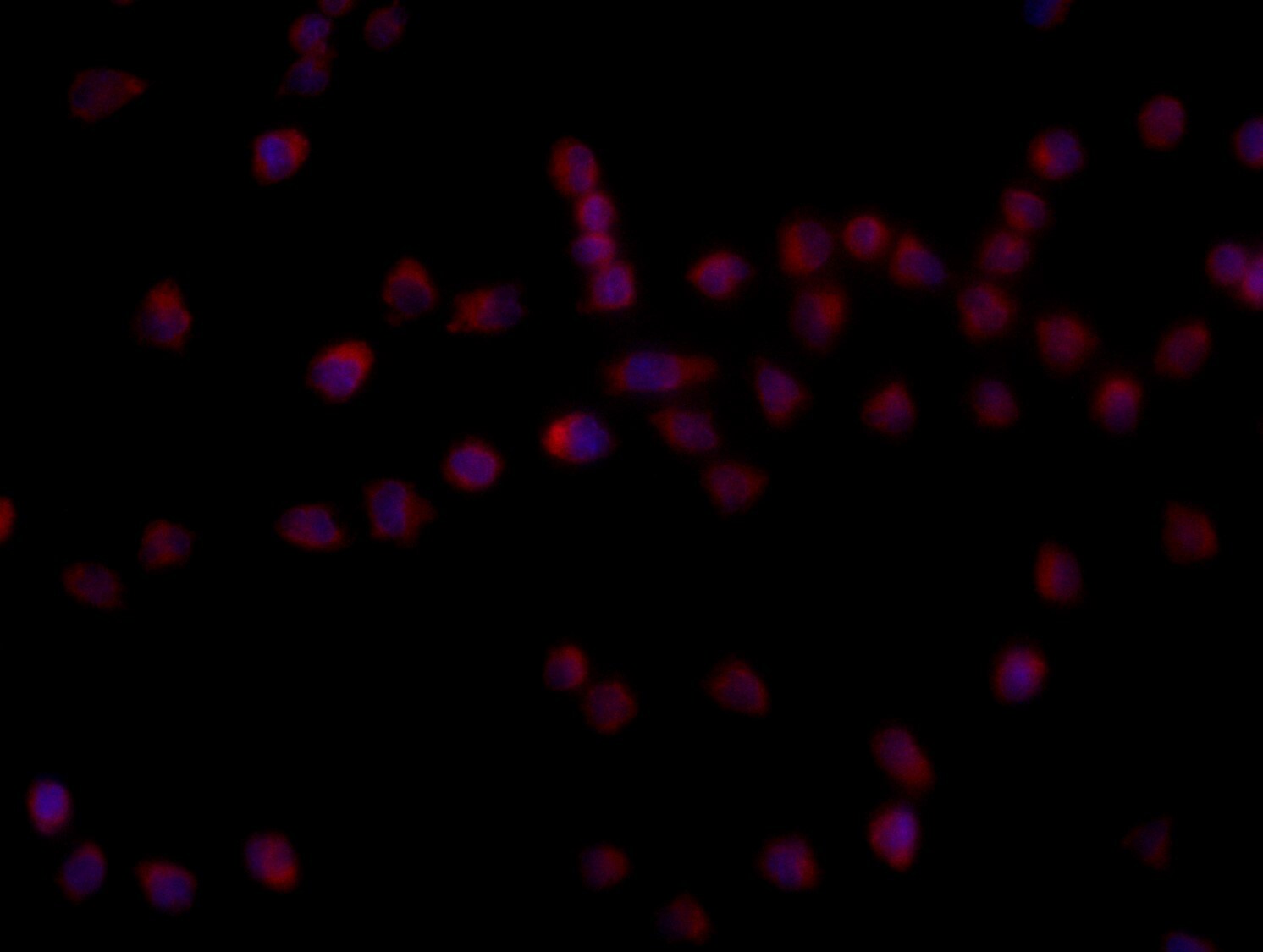
Immunocytochemistry/ Immunofluorescence: CD11b Antibody - BSA Free [NB110-89474]
Immunocytochemistry/Immunofluorescence: CD11b Antibody - BSA Free [NB110-89474] - Raw264.7 cells were fixed in 4% paraformaldehyde for 10 minutes and permeabilized in 0.05% Triton X-100 in PBS for 5 minutes. The cells were incubated with CD11b Antibody (NB110-89474) at 1ug/ml overnight at 4C and detected with an anti-rabbit DyLight 488 (Green) at a 1:1000 dilution for 60 minutes. Nuclei were counterstained with DAPI (Blue). Cells were imaged using a 100X objective and digitally deconvolved.![Immunocytochemistry/ Immunofluorescence: CD11b Antibody - BSA Free [NB110-89474]](https://resources.rndsystems.com/images/products/CD11b-Antibody---BSA-Free-Immunocytochemistry-Immunofluorescence-NB110-89474-img0028.jpg "Immunocytochemistry/ Immunofluorescence: CD11b Antibody - BSA Free [NB110-89474]")
Immunocytochemistry/ Immunofluorescence: CD11b Antibody - BSA Free [NB110-89474]
Immunocytochemistry/Immunofluorescence: CD11b Antibody - BSA Free [NB110-89474] - Raw264.7 cells were fixed in 4% paraformaldehyde for 10 minutes and permeabilized in 0.05% Triton X-100 in PBS for 5 minutes. The cells were incubated with CD11b Antibody conjugated to Alexa Fluor 488 (NB110-89474AF488) at 5 ug/ml for 1 hour at room temperature. Nuclei were counterstained with DAPI (Blue). Cells were imaged using a 100X objective and digitally deconvolved.![Western Blot: CD11b AntibodyBSA Free [NB110-89474]](https://resources.rndsystems.com/images/products/CD11b-Antibody---BSA-Free-Western-Blot-NB110-89474-img0013.jpg "Western Blot: CD11b AntibodyBSA Free [NB110-89474]")
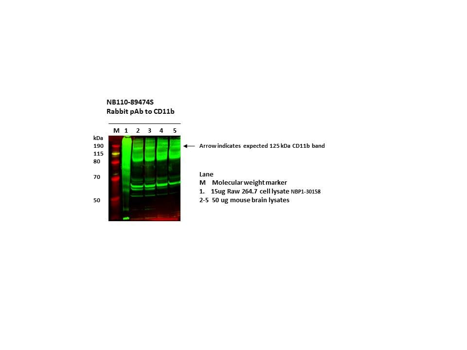
Western Blot: CD11b AntibodyBSA Free [NB110-89474]
Western Blot: CD11b Antibody - BSA Free [NB110-89474] - Western Blot detection of CD11b in RAW 264.7 whole cell lysates using [NB110-89474].![Immunocytochemistry/ Immunofluorescence: CD11b Antibody - BSA Free [NB110-89474]](https://resources.rndsystems.com/images/products/CD11b-Antibody---BSA-Free-Immunocytochemistry-Immunofluorescence-NB110-89474-img0010.jpg "Immunocytochemistry/ Immunofluorescence: CD11b Antibody - BSA Free [NB110-89474]")
Immunocytochemistry/ Immunofluorescence: CD11b Antibody - BSA Free [NB110-89474]
Immunocytochemistry/Immunofluorescence: CD11b Antibody - BSA Free [NB110-89474] - Staining of mouse pancreas using [NB110-89474] at 1:400 dilution. Nuclei counterstained with 4',6-diamidino-2-phenylindole (DAPI) (blue). ICC/IF image submitted by a verified customer review.![Immunohistochemistry: CD11b Antibody - BSA Free [NB110-89474]](https://resources.rndsystems.com/images/products/CD11b-Antibody---BSA-Free-Immunohistochemistry-NB110-89474-img0024.jpg "Immunohistochemistry: CD11b Antibody - BSA Free [NB110-89474]")
![Flow (Cell Surface): CD11b Antibody - BSA Free [NB110-89474]](https://resources.rndsystems.com/images/products/CD11b-Antibody-BSA-Free-Flow-Cell-Surface-NB110-89474-img0033.jpg "Flow (Cell Surface): CD11b Antibody - BSA Free [NB110-89474]")
Flow (Cell Surface): CD11b Antibody - BSA Free [NB110-89474]
Flow (Cell Surface): CD11b Antibody - BSA Free [NB110-89474] - Surface staining of CD11b in CT26 colorectal carcinoma tumor model. Using Alexa Fluor 405 conjugated version of the antibody (NB110-89474AF405). Image from verified customer review.![Immunocytochemistry/ Immunofluorescence: CD11b Antibody - BSA Free [NB110-89474]](https://resources.rndsystems.com/images/products/CD11b-Antibody---BSA-Free-Immunocytochemistry-Immunofluorescence-NB110-89474-img0029.jpg "Immunocytochemistry/ Immunofluorescence: CD11b Antibody - BSA Free [NB110-89474]")
Immunocytochemistry/ Immunofluorescence: CD11b Antibody - BSA Free [NB110-89474]
Immunocytochemistry/Immunofluorescence: CD11b Antibody - BSA Free [NB110-89474] - Raw264.7 cells were fixed in 4% paraformaldehyde for 10 minutes and permeabilized in 0.05% Triton X-100 in PBS for 5 minutes. The cells were incubated with CD11b Antibody conjugated to Alexa Fluor 647 (NB110-89474AF647) at 5 ug/ml for 1 hour at room temperature. Nuclei were counterstained with DAPI (Blue). Cells were imaged using a 100X objective and digitally deconvolved.![Immunocytochemistry/ Immunofluorescence: CD11b Antibody - BSA Free [NB110-89474]](https://resources.rndsystems.com/images/products/CD11b-Antibody---BSA-Free-Immunocytochemistry-Immunofluorescence-NB110-89474-img0030.jpg "Immunocytochemistry/ Immunofluorescence: CD11b Antibody - BSA Free [NB110-89474]")
Immunocytochemistry/ Immunofluorescence: CD11b Antibody - BSA Free [NB110-89474]
Immunocytochemistry/Immunofluorescence: CD11b Antibody - BSA Free [NB110-89474] - Raw264.7 cells were fixed in 4% paraformaldehyde for 10 minutes and permeabilized in 0.05% Triton X-100 in PBS for 5 minutes. The cells were incubated with CD11b Antibody conjugated to DyLight 650 (NB110-8947C) at 5 ug/ml for 1 hour at room temperature. Nuclei were counterstained with DAPI (Blue). Cells were imaged using a 100X objective and digitally deconvolved.![Immunocytochemistry/ Immunofluorescence: CD11b Antibody - BSA Free [NB110-89474]](https://resources.rndsystems.com/images/products/CD11b-Antibody---BSA-Free-Immunocytochemistry-Immunofluorescence-NB110-89474-img0009.jpg "Immunocytochemistry/ Immunofluorescence: CD11b Antibody - BSA Free [NB110-89474]")
Immunocytochemistry/ Immunofluorescence: CD11b Antibody - BSA Free [NB110-89474]
Immunocytochemistry/Immunofluorescence: CD11b Antibody - BSA Free [NB110-89474] - CD11b Antibody [NB110-89474] - CD11b antibody [NB110-89474] was tested in Raw264.7 cells with DyLight 488 (green). Nuclei and alpha-tubulin were counterstained with 4',6-diamidino-2-phenylindole (DAPI) (blue) and DyLight 550 (red).![Immunohistochemistry: CD11b Antibody - BSA Free [NB110-89474]](https://resources.rndsystems.com/images/products/CD11b%20Antibody%20-%20BSA%20Free-Immunohistochemistry-NB110-89474-img0034.jpg "Immunohistochemistry: CD11b Antibody - BSA Free [NB110-89474]")
Immunohistochemistry: CD11b Antibody - BSA Free [NB110-89474]
CD11b Antibody - BSA Free-Immunohistochemistry-NB110-89474-img0034.jpg![Immunohistochemistry: CD11b Antibody - BSA Free [NB110-89474]](https://resources.rndsystems.com/images/products/CD11b-Antibody---BSA-Free-Immunohistochemistry-NB110-89474-img0011.jpg "Immunohistochemistry: CD11b Antibody - BSA Free [NB110-89474]")
Immunohistochemistry: CD11b Antibody - BSA Free [NB110-89474]
Immunohistochemistry: CD11b Antibody - BSA Free [NB110-89474] - Immunohistochemical analysis of CD11b in human renal cancer with [NB110-89474] using 3,3'-Diaminobenzidine (DAB) with hematoxylin counterstain.![Immunohistochemistry-Frozen: CD11b Antibody - BSA Free [NB110-89474]](https://resources.rndsystems.com/images/products/CD11b-Antibody---BSA-Free-Immunohistochemistry-Frozen-NB110-89474-img0021.jpg "Immunohistochemistry-Frozen: CD11b Antibody - BSA Free [NB110-89474]")
Immunohistochemistry-Frozen: CD11b Antibody - BSA Free [NB110-89474]
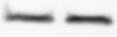
Immunohistochemistry-Frozen: CD11b Antibody - BSA Free [NB110-89474] - Immunohistochemical analysis of frozen sections of mouse spleen using [NB110-89474]. IHC-Fr image submitted by a verified customer review.![Immunohistochemistry: CD11b Antibody - BSA Free [NB110-89474]](https://resources.rndsystems.com/images/products/CD11b-Antibody---BSA-Free-Immunohistochemistry-NB110-89474-img0023.jpg "Immunohistochemistry: CD11b Antibody - BSA Free [NB110-89474]")
Immunohistochemistry: CD11b Antibody - BSA Free [NB110-89474]
CD11b-Antibody---BSA-Free-Immunohistochemistry-NB110-89474-img0023.jpg![Flow (Cell Surface): CD11b Antibody - BSA Free [NB110-89474]](https://resources.rndsystems.com/images/products/CD11b-Antibody-BSA-Free-Flow-Cell-Surface-NB110-89474-img0022.jpg "Flow (Cell Surface): CD11b Antibody - BSA Free [NB110-89474]")
Flow (Cell Surface): CD11b Antibody - BSA Free [NB110-89474]
Flow (Cell Surface): CD11b Antibody - BSA Free [NB110-89474] - CD11b Antibody [NB110-89474] - A surface stain was performed on Raw264.7 cells with DyLight 550-conjugated [NB110-89474R] (blue) and a matched isotype control (orange). Cells were incubated in an antibody dilution of 10 ug/mL for 20 minutes at room temperature. Both antibodies were conjugated to DyLight 550.![Flow (Cell Surface): CD11b Antibody - BSA Free [NB110-89474]](https://resources.rndsystems.com/images/products/CD11b-Antibody-BSA-Free-Flow-Cell-Surface-NB110-89474-img0032.jpg "Flow (Cell Surface): CD11b Antibody - BSA Free [NB110-89474]")
Flow (Cell Surface): CD11b Antibody - BSA Free [NB110-89474]
Flow (Cell Surface): CD11b Antibody - BSA Free [NB110-89474] - An intracellular stain was performed on THP-1 cells with CD11b NB110-89474 (blue) and a matched isotype control NBP2-24891 (orange). Cells were incubated in an antibody dilution of 2.5 ug/mL for 30 minutes at room temperature, followed by Rabbit IgG (H+L) Cross-Adsorbed Secondary Antibody, Dylight 550 (SA5-10033, Thermo Fisher).![Simple Western: CD11b AntibodyBSA Free [NB110-89474]](https://resources.rndsystems.com/images/products/CD11b-Antibody---BSA-Free-Simple-Western-NB110-89474-img0012.jpg "Simple Western: CD11b AntibodyBSA Free [NB110-89474]")
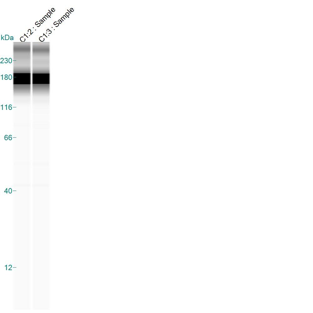
Simple Western: CD11b AntibodyBSA Free [NB110-89474]
Simple Western: CD11b Antibody - BSA Free [NB110-89474] - Specific band for Cd11b in 1.0 mg/mL of Dentate Gyrus from Rat Brain. [NB110-89474] was used at a dilution of 1:50. This experiment was performed under reducing conditions using the 12-230 kDa separation system. Simple Western image submitted by a verified customer review.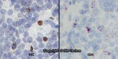
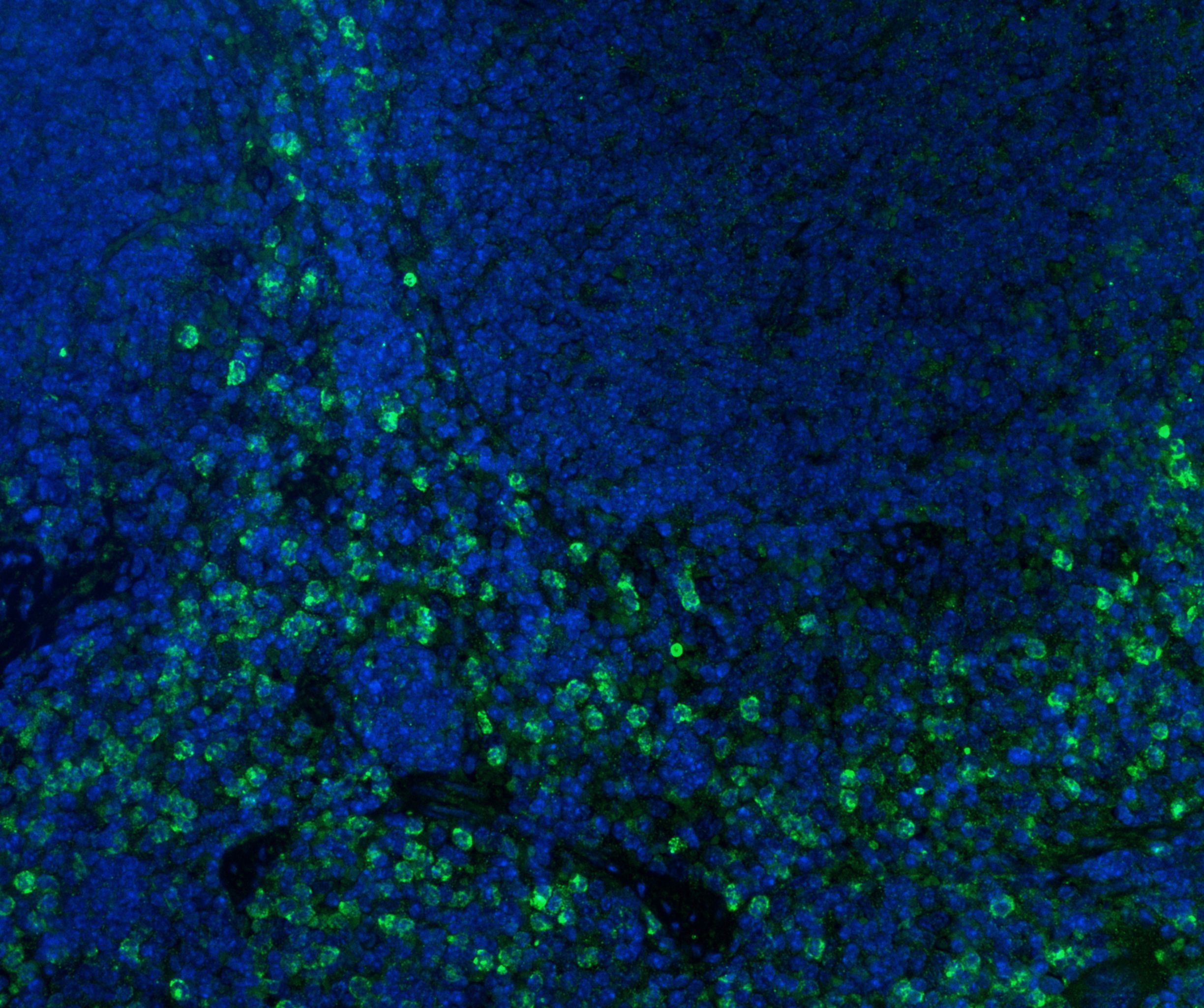
Dual RNAscope ISH-IHC: CD11b Antibody - BSA Free [NB110-89474] - Formalin-fixed paraffin-embedded tissue sections of human lymph node were probed for CD11b mRNA (ACD RNAScope Probe, catalog #555098; Fast Red chromogen, ACD catalog # 322750). Adjacent tissue section was processed for immunohistochemistry using Rabbit Polyclonal (Novus Biologicals catalog #NB110-89474) at 1:50 dilution with 1 hour incubation at room temperature followed by incubation with anti-rabbit IgG VisUCyte HRP Polymer Antibody (Catalog # VC003) and DAB chromogen (yellow-brown). Tissue was counterstained with hematoxylin (blue). Specific staining was localized to lymphocytes.
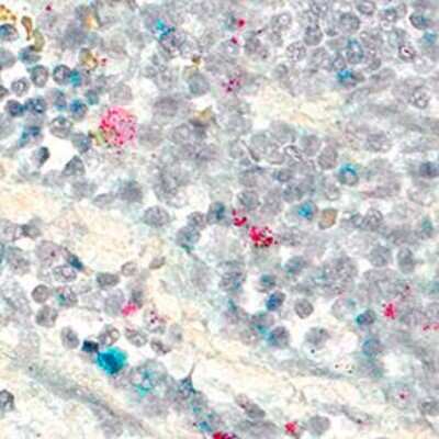
Dual RNAscope ISH-IHC: CD11b Antibody - BSA Free [NB110-89474] - CD14 mRNA (red) and CD11b protein (green) were detected in formalin-fixed paraffin-embedded tissue sections of human malignant lymph node. ACD's Integrated Co-Detection Workflow was performed using ACD RNAScope Probe Hs-CD14 and CD11b antibody at 1:200 dilution. Tissue was stained on Leica Bond RX using RNAscope (TM) 2.5 LS Reagent Kit-RED, BOND Polymer Refine Detection (DAB) and Hematoxylin, BOND Polymer Refine Red Detection and Hematoxylin and RNA scope (TM) 2.5 LS Green Accessory Pack. Tissue was counterstained with 50% hematoxylin (blue).

Immunocytochemistry/Immunofluorescence: CD11b Antibody - BSA Free [NB110-89474] -
CD11b staining in IMhu. Cells were stained with antibodies against the CD11b surface antigen. (A) Panel A shows positive staining for CD11b and (B) panel B shows nuclear DAPI staining. Magnitude 40×.
Western Blot: CD11b Antibody - BSA Free [NB110-89474] -
Western Blot: CD11b Antibody - BSA Free [NB110-89474] - Immunodensities of (a) CD11b, (b) GFAP & (c) NF-kappa B (p65) proteins with representative immunoblots in striatum from Munc18-OE (n = 5) & wild-type (n = 5) mice. Bar graphs are ratios of optical densities of our proteins of interest to beta -actin (42 kDa band), expressed as immunoreactivity in percentage of mean value of the WT group (100%). No differences were detected between groups for any of the analyzed proteins. Right panels are representative immunoblots for target proteins & beta -actin which included Munc18-OE (OE) & wild-type (WT) mice samples. The molecular masses were estimated from referenced standards. Image collected & cropped by CiteAb from the following publication (https://pubmed.ncbi.nlm.nih.gov/25069615), licensed under a CC-BY license. Not internally tested by Novus Biologicals.
Immunohistochemistry: CD11b Antibody - BSA Free [NB110-89474] -
Immunohistochemistry: CD11b Antibody - BSA Free [NB110-89474] - Comparison of Ki-67 or CD11b immunoreactivity of Aldara-treated dorsal skin samples of C57BL/6 mice using OP or MP. Nuclear proliferation marker Ki-67 immunostaining on OP (a) & MP (b) dorsal skin tissue samples at 400x magnification. Dermal dendritic cells were labelled with anti-CD11b antibody on OP (c) & MP samples (d) at 400x magnification. Image collected & cropped by CiteAb from the following publication (https://pubmed.ncbi.nlm.nih.gov/30842501), licensed under a CC-BY license. Not internally tested by Novus Biologicals.
Western Blot: CD11b Antibody - BSA Free [NB110-89474] -
Western Blot: CD11b Antibody - BSA Free [NB110-89474] - Immunodensities of (a) CD11b, (b) GFAP & (c) NF-kappa B (p65) proteins with representative immunoblots in cerebral cortex from Munc18-OE (n = 5) & wild-type (n = 5) mice. Bar graphs are ratios of optical densities of our proteins of interest to beta -actin (42 kDa band), expressed as immunoreactivity in percentage of the mean value of the WT group (100%). CD11b was significantly decreased in Munc18-OE mice compared to the WT (t = 3.01; P <0.05). No differences between groups were observed in the levels of GFAP or NF-kappa B. Right panels are representative immunoblots for target proteins & beta -actin which included Munc18-OE (OE) & wild-type (WT) mice samples. The molecular masses were estimated from referenced standards. Image collected & cropped by CiteAb from the following publication (https://pubmed.ncbi.nlm.nih.gov/25069615), licensed under a CC-BY license. Not internally tested by Novus Biologicals.
Immunocytochemistry/ Immunofluorescence: CD11b Antibody - BSA Free [NB110-89474] -
Immunocytochemistry/ Immunofluorescence: CD11b Antibody - BSA Free [NB110-89474] - CD11b staining in IMhu. Cells were stained with antibodies against the CD11b surface antigen. (A) Panel A shows positive staining for CD11b & (B) panel B shows nuclear DAPI staining. Magnitude 40×. Image collected & cropped by CiteAb from the following publication (https://pubmed.ncbi.nlm.nih.gov/31096716), licensed under a CC-BY license. Not internally tested by Novus Biologicals.
Immunohistochemistry-Paraffin: CD11b Antibody - BSA Free [NB110-89474] -
Population of CD11b + myeloid progenitor cells differentiate into SMA + stromal cells within tumors and in vitro. (A) Representative image of red fluorescent protein (RFP) + stromal cells in tumor from CCR2-RFP heterozygous SCID mouse. (B) RFP + SMA + double positive cells within tumor stroma. (C) CD11b + SMA + double positive cells within tumor stroma. (D) CD45 + CD11b + CD34+ myeloid progenitor cells in the mammary gland at 1.5 (n = 5 empty vector (EV), 7 CCL2) and 2.5 weeks (n = 5 EV, 7 CCL2) post-transplantation were quantified by flow cytometry. (E) CD45 + CD11b + CD34 + myeloid progenitor cells in the bone marrow at 1.5 weeks (n = 6 EV, 7 CCL2) and 2.5 weeks (n = 3 mice/group) post-transplantation were quantified by flow cytometry. (F) Representative brightfield image of colony formed by CD45 + CD11b + CD34 + myeloid progenitor cells isolated using fluorescence-activated cell sorting (FACS). (G) Colonies in culture co-stained with SMA and collagen I. Statistical differences determined by Mann–Whitney U test. Magnification bars = 50 um. Image collected and cropped by CiteAb from the following open publication (https://pubmed.ncbi.nlm.nih.gov/32731354), licensed under a CC-BY license. Not internally tested by Novus Biologicals.Applications for CD11b Antibody - BSA Free
Application
Recommended Usage
Flow (Cell Surface)
1:10 - 1:1000
Flow Cytometry
reported in scientific literature (PMID 21422470)
Immunocytochemistry/ Immunofluorescence
1:200. Use reported in multiple pieces of scientific literature (PMID 23980916)
Immunohistochemistry
1:400
Immunohistochemistry-Frozen
reported in scientific literature (PMID 23980916)
Immunohistochemistry-Paraffin
1:400. Use reported in scientific literature (PMID 31022918)
In-situ Hybridization
reported in scientific literature (PMID 27133471)
Simple Western
1:50
Single Cell Western
1:10
Western Blot
2 ug/mL. Use reported in scientific literature (PMID 31082627)
Application Notes
In WB a specific band is observed ~160 kDa and an apparant non-specific band is observed ~56 kDa. Prior to immunostaining paraffin tissues, antigen retrieval with sodium citrate buffer (pH 6.0) is recommended. In ICC/IF, membrane staining was observed in Raw 264.7 cells. This antibody does not appear to work in human samples with WB. The observed molecular weight of the protein may vary from the listed predicted molecular weight due to post translational modifications, post translation cleavages, relative charges, and other experimental factors.
Reviewed Applications
Read 12 reviews rated 4.5 using NB110-89474 in the following applications:
Flow Cytometry Panel Builder
Bio-Techne Knows Flow Cytometry
Save time and reduce costly mistakes by quickly finding compatible reagents using the Panel Builder Tool.
Advanced Features
- Spectra Viewer - Custom analysis of spectra from multiple fluorochromes
- Spillover Popups - Visualize the spectra of individual fluorochromes
- Antigen Density Selector - Match fluorochrome brightness with antigen density

Formulation, Preparation, and Storage
Purification
Immunogen affinity purified
Formulation
PBS
Format
BSA Free
Preservative
0.02% Sodium Azide
Concentration
1 mg/ml
Shipping
The product is shipped with polar packs. Upon receipt, store it immediately at the temperature recommended below.
Stability & Storage
Aliquot and store at -20C or -80C. Avoid freeze-thaw cycles.
Background: CD11b/Integrin alpha M
References
1. Rosetti, F., & Mayadas, T. N. (2016). The many faces of Mac-1 in autoimmune disease. Immunol Rev, 269(1), 175-193. doi:10.1111/imr.12373
2. Kim, D., Kim, T. H., Wu, G., Park, B. K., Ha, J. H., Kim, Y. S.,... Kwon, H. J. (2016). Extracellular Release of CD11b by TLR9 Stimulation in Macrophages. PLoS One, 11(3), e0150677. doi:10.1371/journal.pone.0150677
3. Christensen, J. E., Andreasen, S. O., Christensen, J. P., & Thomsen, A. R. (2001). CD11b expression as a marker to distinguish between recently activated effector CD8(+) T cells and memory cells. Int Immunol, 13(4), 593-600. doi:10.1093/intimm/13.4.593
4. Nath, S. K., Han, S., Kim-Howard, X., Kelly, J. A., Viswanathan, P., Gilkeson, G. S.,... Harley, J. B. (2008). A nonsynonymous functional variant in integrin-alpha(M) (encoded by ITGAM) is associated with systemic lupus erythematosus. Nat Genet, 40(2), 152-154. doi:10.1038/ng.71
Alternate Names
CD11b, Integrin alpha M, ITGAM, MAC 1, MAC1, Mac1, alpha subunit, Mac-1a, Mo1, alpha subunit
Gene Symbol
ITGAM
UniProt
Additional CD11b/Integrin alpha M Products
Product Documents for CD11b Antibody - BSA Free
Certificate of Analysis
To download a Certificate of Analysis, please enter a lot or batch number in the search box below.
Product Specific Notices for CD11b Antibody - BSA Free
This product is for research use only and is not approved for use in humans or in clinical diagnosis. Primary Antibodies are guaranteed for 1 year from date of receipt.
Related Research Areas
Citations for CD11b Antibody - BSA Free
 Powered by Bioz
Customer Reviews for CD11b Antibody - BSA Free (12)
4.5 out of 5
12 Customer Ratings
Have you used CD11b Antibody - BSA Free?
Submit a review and receive an Amazon gift card!
$25/€18/£15/$25CAN/¥2500 Yen for a review with an image
$10/€7/£6/$10CAN/¥1110 Yen for a review without an image
Submit a review
Customer Images


Showing
1
-
5 of
12 reviews
Showing All
Filter By:
-
Application: ImmunocytochemistrySample Tested: Human microgliaSpecies: HumanVerified Customer | Posted 05/20/2021hMAN MICROGLIA CELL LINE STAINED FOR cd11B WIH DAPI

-
Application: Western BlotSample Tested: B cell enriched peripheral blood mononuclear cells (PBMC)Species: MouseVerified Customer | Posted 05/03/2020
-
Application: Western BlotSample Tested: Hepa 1-6 mouse hepatoma cell lineSpecies: MouseVerified Customer | Posted 12/27/2019
-
Application: Western BlotSample Tested: Adult mouse brain tissueSpecies: MouseVerified Customer | Posted 11/30/2018Western blot of mouse brain RIPA lysate with NB110-89474SS rabbit pAb to CD11b. Image detection using Licor Odyssey IR scanner.
-
Application: Western BlotSample Tested: Jurkat and THP1 cellsSpecies: HumanVerified Customer | Posted 10/28/2018
-
Application: Immunohistochemistry-FrozenSample Tested: Frozen mouse spleenSpecies: MouseVerified Customer | Posted 05/12/2018
-
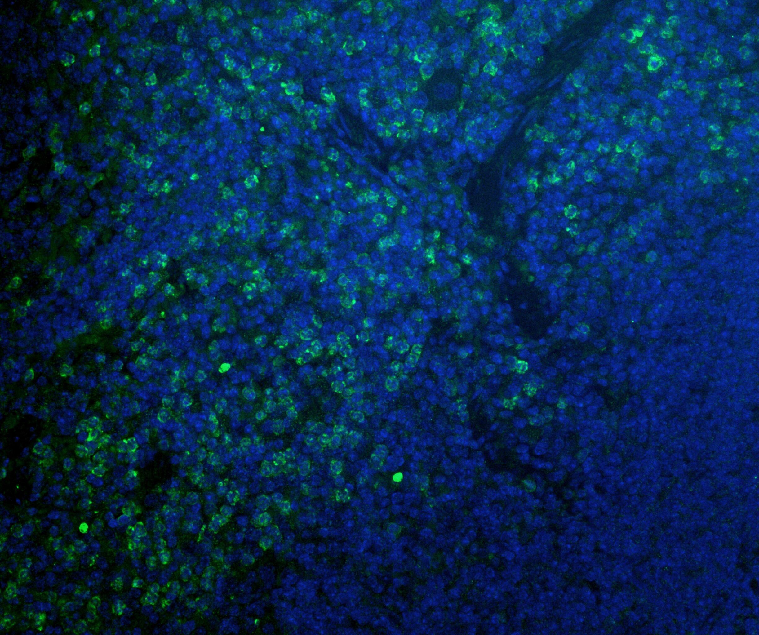
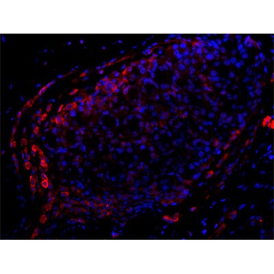
Application: Immunohistochemistry-FrozenSample Tested: Human lung graftSpecies: HumanVerified Customer | Posted 02/22/2018Blue: DAPI+ nuclei Red: CD11bHuman lung tissue was stained with CD11b. Primary antibody (1:100) was diluted in a blocking buffer (PBS with 0.5%). A secondary Alexa647-conjugated Goat anti-Rabbit antibody (1:400) was used to detect CD11b.
-
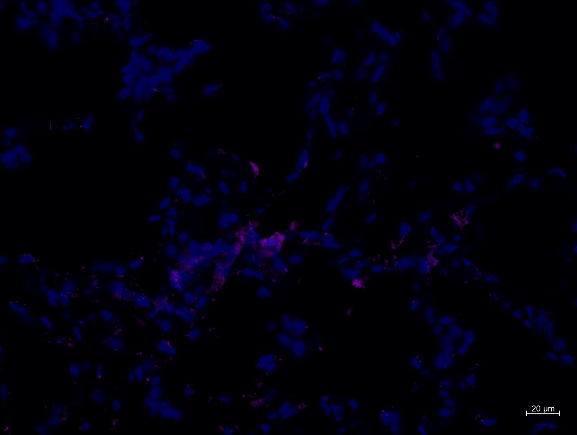
Application: Immunohistochemistry-FrozenSample Tested: Lung graft and IL1b-KO monocyte treated lungSpecies: MouseVerified Customer | Posted 02/22/2018Blue: DAPU+ nuclei Red: CD11bLung collected from an IL1b-KO monocytes-treated recipient was stained with CD11b. Primary antibody (1:100) was diluted in a blocking buffer (PBS with 0.5%). A secondary Alexa647-conjugated Goat anti-Rabbit antibody (1:400) was used to detect CD11b.
-
Application: Immunohistochemistry-ParaffinSample Tested: Mouse spleen cellsSpecies: MouseVerified Customer | Posted 09/15/2017Permeabillized with 3% Triton-X100 Antigen Retrieval with Citrate buffer, 20 min, 90C 1st ab dilution: 1:200
-
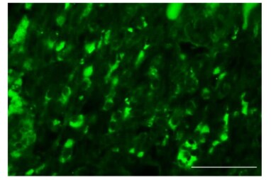
Application: ImmunofluorescenceSample Tested: cardiac tissueSpecies: MouseVerified Customer | Posted 11/29/2016Cd11b staining in the infarct of a mouse at day 7 post-MIIn order to visualize staining the secondary method was used. Blocking was performed in goat serum and secondary antibodies used were Alexa Fluor 488 conjugate.
-
Application: Western BlotVerified Customer | Posted 01/21/2015Simple Western showing CD11b in 1 mg/ml of denate gyrus from rat brain lysate
-
Application: ImmunofluorescenceSample Tested: Mouse PancreasSpecies: MouseVerified Customer | Posted 03/05/2013
There are no reviews that match your criteria.
Protocols
View specific protocols for CD11b Antibody - BSA Free (NB110-89474):
Immunocytochemistry Protocol
Culture cells to appropriate density in 35 mm culture dishes or 6-well plates.
1. Remove culture medium and add 10% formalin to the dish. Fix at room temperature for 30 minutes.
2. Remove the formalin and add ice cold methanol. Incubate for 5-10 minutes.
3. Remove methanol and add washing solution (i.e. PBS). Be sure to not let the specimen dry out. Wash three times for 10 minutes.
4. To block nonspecific antibody binding incubate in 10% normal goat serum from 1 hour to overnight at room temperature.
5. Add primary antibody at appropriate dilution and incubate at room temperature from 2 hours to overnight at room temperature.
6. Remove primary antibody and replace with washing solution. Wash three times for 10 minutes.
7. Add secondary antibody at appropriate dilution. Incubate for 1 hour at room temperature.
8. Remove antibody and replace with wash solution, then wash for 10 minutes. Add Hoechst 33258 to wash solution at 1:25,0000 and incubate for 10 minutes. Wash a third time for 10 minutes.
9. Cells can be viewed directly after washing. The plates can also be stored in PBS containing Azide covered in Parafilm (TM). Cells can also be cover-slipped using Fluoromount, with appropriate sealing.
*The above information is only intended as a guide. The researcher should determine what protocol best meets their needs. Please follow safe laboratory procedures.
Immunohistochemistry-Paraffin Embedded Sections
Antigen Unmasking:
Bring slides to a boil in 10 mM sodium citrate buffer (pH 6.0) then maintain at a sub-boiling temperature for 10 minutes. Cool slides on bench-top for 30 minutes.
Staining:
1. Wash sections in deionized water three times for 5 minutes each.
2. Wash sections in wash buffer for 5 minutes.
3. Block each section with 100-400 ul blocking solution for 1 hour at room temperature.
4. Remove blocking solution and add 100-400 ul diluted primary antibody. Incubate overnight at 4C.
5. Remove antibody solution and wash sections in wash buffer three times for 5 minutes each.
6. Add 100-400 ul biotinylated diluted secondary antibody. Incubate 30 minutes at room temperature.
7. Remove secondary antibody solution and wash sections three times with wash buffer for 5 minutes each.
8. Add 100-400 ul Streptavidin-HRP reagent to each section and incubate for 30 minutes at room temperature.
9. Wash sections three times in wash buffer for 5 minutes each.
10. Add 100-400 ul DAB substrate to each section and monitor staining closely.
11. As soon as the sections develop, immerse slides in deionized water.
12. Counterstain sections in hematoxylin.
13. Wash sections in deionized water two times for 5 minutes each.
14. Dehydrate sections.
15. Mount coverslips.
Western Blot Protocol
1. Perform SDS-PAGE on samples to be analyzed, loading 40 ug of total protein per lane.
2. Transfer proteins to membrane according to the instructions provided by the manufacturer of the membrane and transfer apparatus.
3. Stain according to standard Ponceau S procedure (or similar product) to assess transfer success, and mark molecular weight standards where appropriate.
4. Rinse the blot.
5. Block the membrane using standard blocking buffer for at least 1 hour.
6. Wash the membrane in wash buffer three times for 10 minutes each.
7. Dilute primary antibody in blocking buffer and incubate 1 hour at room temperature.
8. Wash the membrane in wash buffer three times for 10 minutes each.
9. Apply the diluted HRP conjugated secondary antibody in blocking buffer (as per manufacturers instructions) and incubate 1 hour at room temperature.
10. Wash the blot in wash buffer three times for 10 minutes each (this step can be repeated as required to reduce background).
11. Apply the detection reagent of choice in accordance with the manufacturers instructions.
**Note: Tween-20 can be added to the blocking or antibody dilution buffer at a final concentration of 0.05-0.2%.
Find general support by application which include: protocols, troubleshooting, illustrated assays, videos and webinars.
- 7-Amino Actinomycin D (7-AAD) Cell Viability Flow Cytometry Protocol
- Antigen Retrieval Protocol (PIER)
- Antigen Retrieval for Frozen Sections Protocol
- Appropriate Fixation of IHC/ICC Samples
- Cellular Response to Hypoxia Protocols
- Chromogenic IHC Staining of Formalin-Fixed Paraffin-Embedded (FFPE) Tissue Protocol
- Chromogenic Immunohistochemistry Staining of Frozen Tissue
- ClariTSA™ Fluorophore Kits
- Detection & Visualization of Antibody Binding
- Extracellular Membrane Flow Cytometry Protocol
- Flow Cytometry Protocol for Cell Surface Markers
- Flow Cytometry Protocol for Staining Membrane Associated Proteins
- Flow Cytometry Staining Protocols
- Flow Cytometry Troubleshooting Guide
- Fluorescent IHC Staining of Frozen Tissue Protocol
- Graphic Protocol for Heat-induced Epitope Retrieval
- Graphic Protocol for the Preparation and Fluorescent IHC Staining of Frozen Tissue Sections
- Graphic Protocol for the Preparation and Fluorescent IHC Staining of Paraffin-embedded Tissue Sections
- Graphic Protocol for the Preparation of Gelatin-coated Slides for Histological Tissue Sections
- ICC Cell Smear Protocol for Suspension Cells
- ICC Immunocytochemistry Protocol Videos
- ICC for Adherent Cells
- IHC Sample Preparation (Frozen sections vs Paraffin)
- ISH-IHC Protocol for Chromogenic Detection on Formalin Fixed Paraffin Embedded (FFPE) Tissue
- Immunocytochemistry (ICC) Protocol
- Immunocytochemistry Troubleshooting
- Immunofluorescence of Organoids Embedded in Cultrex Basement Membrane Extract
- Immunofluorescent IHC Staining of Formalin-Fixed Paraffin-Embedded (FFPE) Tissue Protocol
- Immunohistochemistry (IHC) and Immunocytochemistry (ICC) Protocols
- Immunohistochemistry Frozen Troubleshooting
- Immunohistochemistry Paraffin Troubleshooting
- Intracellular Flow Cytometry Protocol Using Alcohol (Methanol)
- Intracellular Flow Cytometry Protocol Using Detergents
- Intracellular Nuclear Staining Flow Cytometry Protocol Using Detergents
- Intracellular Staining Flow Cytometry Protocol Using Alcohol Permeabilization
- Intracellular Staining Flow Cytometry Protocol Using Detergents to Permeabilize Cells
- Preparing Samples for IHC/ICC Experiments
- Preventing Non-Specific Staining (Non-Specific Binding)
- Primary Antibody Selection & Optimization
- Propidium Iodide Cell Viability Flow Cytometry Protocol
- Protocol for Heat-Induced Epitope Retrieval (HIER)
- Protocol for Liperfluo
- Protocol for Making a 4% Formaldehyde Solution in PBS
- Protocol for VisUCyte™ HRP Polymer Detection Reagent
- Protocol for the Characterization of Human Th22 Cells
- Protocol for the Characterization of Human Th9 Cells
- Protocol for the Fluorescent ICC Staining of Cell Smears - Graphic
- Protocol for the Fluorescent ICC Staining of Cultured Cells on Coverslips - Graphic
- Protocol for the Preparation & Fixation of Cells on Coverslips
- Protocol for the Preparation and Chromogenic IHC Staining of Frozen Tissue Sections
- Protocol for the Preparation and Chromogenic IHC Staining of Frozen Tissue Sections - Graphic
- Protocol for the Preparation and Chromogenic IHC Staining of Paraffin-embedded Tissue Sections
- Protocol for the Preparation and Chromogenic IHC Staining of Paraffin-embedded Tissue Sections - Graphic
- Protocol for the Preparation and Fluorescent ICC Staining of Cells on Coverslips
- Protocol for the Preparation and Fluorescent ICC Staining of Non-adherent Cells
- Protocol for the Preparation and Fluorescent ICC Staining of Stem Cells on Coverslips
- Protocol for the Preparation and Fluorescent IHC Staining of Frozen Tissue Sections
- Protocol for the Preparation and Fluorescent IHC Staining of Paraffin-embedded Tissue Sections
- Protocol for the Preparation of Gelatin-coated Slides for Histological Tissue Sections
- Protocol for the Preparation of a Cell Smear for Non-adherent Cell ICC - Graphic
- Protocol: Annexin V and PI Staining by Flow Cytometry
- Protocol: Annexin V and PI Staining for Apoptosis by Flow Cytometry
- R&D Systems Quality Control Western Blot Protocol
- TUNEL and Active Caspase-3 Detection by IHC/ICC Protocol
- The Importance of IHC/ICC Controls
- Troubleshooting Guide: Fluorokine Flow Cytometry Kits
- Troubleshooting Guide: Immunohistochemistry
- Troubleshooting Guide: Western Blot Figures
- Western Blot Conditions
- Western Blot Protocol
- Western Blot Protocol for Cell Lysates
- Western Blot Troubleshooting
- Western Blot Troubleshooting Guide
- View all Protocols, Troubleshooting, Illustrated assays and Webinars
FAQs for CD11b Antibody - BSA Free
Showing
1
-
5 of
8 FAQs
Showing All
-
Q: Do you know if this CD11b antibody can be used in CD11b positive cell depletion?
A: This particular CD11b antibody is not reccomended for depletion studies due to the preservative used; however, our CD11b antibody NBP1-06650 is offered in a preservative free format and has been validated for blocking.
-
Q: I am looking for a Mac-1 (microglia cell marker) antibody to do immunostaining with my mouse brain and I checked, you have a lot of antibodies for this biomarker. I don't know which one is the best to be used for immunohistology on mouse brain tissue. Could you please help with this?
A:
I would recommend NB110-89474. This antibody has been published in 11 scientific articles, including several times in IHC in mouse. As long as CD11b is expressed in your sample, it should work just fine and is backed by our 100% Novus Guarantee.
-
Q: I'm not sure if this will work for me, can I get a free sample of your CD11b antibody to test?
A: We don't offer free samples but we do offer this CD11b antibody in a trial size of 0.025 ml at a reduced cost, all antibodies are backed by our 100% guarantee to work in all validated species and applications on the product's web page.
-
Q: Is CD11b antibody also staining neutrophils in the brain
A: CD11b is typically used as a marker for granulocytes and macrophages. You may need a combination of markers to specifically identify neutrophils
-
Q: Looking for a bone marrow macrophage marker can you recommend a CD11b antibody for ICC?
A: I would recommend NB110-89474 which is our most popular CD11b antibody and has been validated in ICC and cited in 18 publications.
-
Q: The protocol says to do enzymatic antigen retrieval before incubating with the CD11b antibody, which enzyme is used?
A: The protocol is referencting the PIER method using Proteinase K as the enzyme.
-
Q: We need a CD11b antibody for IHC-P on mouse tissue conjugated to FITC, do you have one that will work?
A: We have 3 CD11b antibodies for IHC-P and just one CD11 b antibody we offer conjugated to FITC, NB110-89474F.
-
Q: What types of cells can this CD11b antibody be used as a marker for?
A: A study used this CD11b antibody to stain granulocytes in mouse uterine tissue in IHC. CD11b is also expressed on other leukocytes such as monocytes, macrophages, and natural killer cells.
-
Q: Do you know if this CD11b antibody can be used in CD11b positive cell depletion?
A: This particular CD11b antibody is not reccomended for depletion studies due to the preservative used; however, our CD11b antibody NBP1-06650 is offered in a preservative free format and has been validated for blocking.
-
Q: I am looking for a Mac-1 (microglia cell marker) antibody to do immunostaining with my mouse brain and I checked, you have a lot of antibodies for this biomarker. I don't know which one is the best to be used for immunohistology on mouse brain tissue. Could you please help with this?
A:
I would recommend NB110-89474. This antibody has been published in 11 scientific articles, including several times in IHC in mouse. As long as CD11b is expressed in your sample, it should work just fine and is backed by our 100% Novus Guarantee.
-
Q: I'm not sure if this will work for me, can I get a free sample of your CD11b antibody to test?
A: We don't offer free samples but we do offer this CD11b antibody in a trial size of 0.025 ml at a reduced cost, all antibodies are backed by our 100% guarantee to work in all validated species and applications on the product's web page.
-
Q: Is CD11b antibody also staining neutrophils in the brain
A: CD11b is typically used as a marker for granulocytes and macrophages. You may need a combination of markers to specifically identify neutrophils
-
Q: Looking for a bone marrow macrophage marker can you recommend a CD11b antibody for ICC?
A: I would recommend NB110-89474 which is our most popular CD11b antibody and has been validated in ICC and cited in 18 publications.
-
Q: The protocol says to do enzymatic antigen retrieval before incubating with the CD11b antibody, which enzyme is used?
A: The protocol is referencting the PIER method using Proteinase K as the enzyme.
-
Q: We need a CD11b antibody for IHC-P on mouse tissue conjugated to FITC, do you have one that will work?
A: We have 3 CD11b antibodies for IHC-P and just one CD11 b antibody we offer conjugated to FITC, NB110-89474F.
-
Q: What types of cells can this CD11b antibody be used as a marker for?
A: A study used this CD11b antibody to stain granulocytes in mouse uterine tissue in IHC. CD11b is also expressed on other leukocytes such as monocytes, macrophages, and natural killer cells.
Loading...
Associated Pathways


