CD34 Antibody (4H11[APG]) - BSA Free
Novus Biologicals | Catalog # NB500-608
Clone 4H11[APG] was used by HLDA to establish CD designation.
Key Product Details
Species Reactivity
Validated:
Human
Cited:
Human
Applications
Validated:
Immunohistochemistry, Immunohistochemistry-Paraffin, Western Blot, Flow Cytometry, Flow (Cell Surface), Immunocytochemistry/ Immunofluorescence
Cited:
Immunocytochemistry/ Immunofluorescence
Label
Unconjugated
Antibody Source
Monoclonal Mouse IgG1 Clone # 4H11[APG]
Format
BSA Free
Loading...
Product Specifications
Immunogen
Permanent human cell line derived from peripheral leucocytes of a patient suffering from chronic myeloid leukaemia.
Localization
Membrane; Single-pass type I membrane protein.
Marker
Hematopoietic Stem Cell Marker
Specificity
The antibody 4H11[APG] reacts with Class III epitope on CD34 (Mucosialin), a 110-115 kDa monomeric transmembrane phosphoglycoprotein expressed on hematopoietic progenitors cells and on the most pluripotential stem cells; it is gradually lost on progenitor cells. The antibody 4H11[APG] completely blocks binding of Class II antibody QBEnd10 and Class III antibodies BIRMA K3 and 8G12 on KG1a cell line. HLDA VI; WS Code M MA58
Clonality
Monoclonal
Host
Mouse
Isotype
IgG1
Theoretical MW
115 kDa.
Disclaimer note: The observed molecular weight of the protein may vary from the listed predicted molecular weight due to post translational modifications, post translation cleavages, relative charges, and other experimental factors.
Disclaimer note: The observed molecular weight of the protein may vary from the listed predicted molecular weight due to post translational modifications, post translation cleavages, relative charges, and other experimental factors.
Scientific Data Images for CD34 Antibody (4H11[APG]) - BSA Free
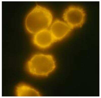
Immunocytochemistry/Immunofluorescence: CD34 Antibody (4H11[APG]) [NB500-608] - Human chronic myeloid leukemia cell line MOLM-7 with anti-human CD34 (4H11[APG]) PE.
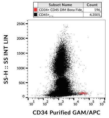
Flow (Cell Surface): CD34 Antibody (4H11[APG]) [NB500-608] - Surface staining of CD34+ cells in human peripheral blood
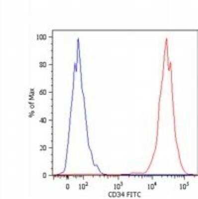
Flow (Cell Surface): CD34 Antibody (4H11[APG]) [NB500-608] - Analysis using the FITC conjugate of NB500-608. Staining of Kg-1a human acute myelogenous leukemia cell line with anti-human CD34 (4H11[APG]) FITC. Total viable cells were used for analysis.
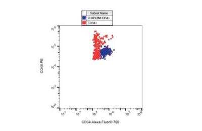
Flow Cytometry: CD34 Antibody (4H11[APG]) [NB500-608] - Staining of CD34+ cells in human peripheral blood with anti-human CD34 (4H11[APG]) Alexa Fluor® 700.
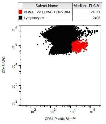
Flow (Cell Surface): CD34 Antibody (4H11[APG]) [NB500-608] - Surface staining of CD34+ cells in human peripheral blood with anti-human CD34 (4H11[APG]) Pacific BlueTM
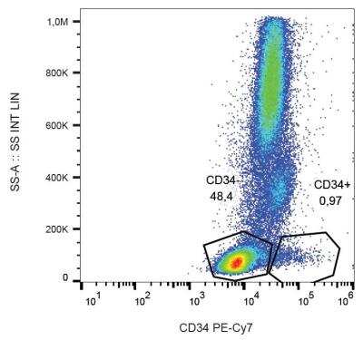
Flow (Cell Surface): CD34 Antibody (4H11[APG]) [NB500-608] - Surface staining of CD34+ cells in human peripheral blood with anti-human CD34 (4H11[APG]) PE-CyTM7.
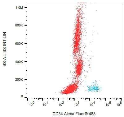
Flow (Cell Surface): CD34 Antibody (4H11[APG]) [NB500-608] - Surface staining of CD34+ cells in human peripheral blood with anti-human CD34 (4H11[APG]) Alexa Fluor® 488.
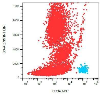
Flow (Cell Surface): CD34 Antibody (4H11[APG]) [NB500-608] - Surface staining of CD34+ cells in human peripheral blood with anti-human CD34 (4H11[APG]) APC.
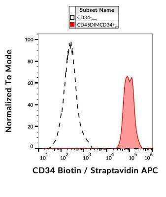
Flow (Cell Surface): CD34 Antibody (4H11[APG]) [NB500-608] - Surface staining of CD34+ cells in human peripheral blood with anti-CD34 (4H11[APG]) biotin / streptavidin-APC.
Applications for CD34 Antibody (4H11[APG]) - BSA Free
Application
Recommended Usage
Flow Cytometry
2 ug/ml
Immunohistochemistry
1:10-1:500
Immunohistochemistry-Paraffin
10 ug/ml
Western Blot
1-2 ug/ml
Application Notes
Western Blot - Sample preparation: Resuspend approx. 50 mil. cells in 1 ml cold Lysis buffer (1% laurylmaltoside in 20 mM Tris/Cl, 100 mM NaCl pH 8.2, 50 mM NaF including Protease inhibitor Cocktail). Incubate 60 min on ice. Centrifuge to remove cell debris. Mix lysate with non-reducing SDS-PAGE sample buffer. Application note: Non-reducing conditions.
Flow Cytometry Panel Builder
Bio-Techne Knows Flow Cytometry
Save time and reduce costly mistakes by quickly finding compatible reagents using the Panel Builder Tool.
Advanced Features
- Spectra Viewer - Custom analysis of spectra from multiple fluorochromes
- Spillover Popups - Visualize the spectra of individual fluorochromes
- Antigen Density Selector - Match fluorochrome brightness with antigen density

Formulation, Preparation, and Storage
Purification
Protein A purified
Formulation
Phosphate buffered saline (PBS), pH 7.4
Format
BSA Free
Preservative
15mM Sodium Azide
Concentration
1.0 mg/ml
Shipping
The product is shipped with polar packs. Upon receipt, store it immediately at the temperature recommended below.
Stability & Storage
Store at 4C. Do not freeze.
Background: CD34
CD34 has commonly been used as a marker for the diagnosis and classification of various diseases and pathologies including leukemia and solitary fibrous tumor (SFT) (2,5). In terms of immunohistochemistry and histopathology, CD34 has been the most common marker for SFT and is expressed in ~79% of cases (5). In addition to its use as a cell marker, CD34-postive (CD34+) hematopoietic stem cells have been used therapeutically in patients following radiation or chemotherapy due to their regenerative potential (6). There are several clinical trials showing promising results for CD34+ cell therapy for cardiovascular diseases including heart failure, ischemia, dilated cardiomyopathy, acute myocardial infarction, and angina (6). Besides hematopoietic lineages, CD34 is also expressed in non-hematopoietic cells including mesenchymal stem cells, endothelial cells and progenitors, fibrocytes, muscle satellite cells, and some cancer stem cells (1,3). While the clinical and cell therapy applications of CD34 as a cell marker is well documented, the function of CD34 is less understood but has been implicated in many cellular processes such as adhesion, proliferation, signal transduction, differentiation, and progenitor phenotype maintenance (1,3).
References
1. Sidney, L. E., Branch, M. J., Dunphy, S. E., Dua, H. S., & Hopkinson, A. (2014). Concise review: evidence for CD34 as a common marker for diverse progenitors. Stem cells (Dayton, Ohio), 32(6), 1380-1389. https://doi.org/10.1002/stem.1661
2. Krause, D. S., Fackler, M. J., Civin, C. I., & May, W. S. (1996). CD34: structure, biology, and clinical utility. Blood, 87(1), 1-13
3. Kapoor, S., Shenoy, S. P., & Bose, B. (2020). CD34 cells in somatic, regenerative and cancer stem cells: Developmental biology, cell therapy, and omics big data perspective. Journal of cellular biochemistry, 121(5-6), 3058-3069. https://doi.org/10.1002/jcb.29571
4. Uniprot (P28906)
5. Davanzo, B., Emerson, R. E., Lisy, M., Koniaris, L. G., & Kays, J. K. (2018). Solitary fibrous tumor. Translational gastroenterology and hepatology, 3, 94. https://doi.org/10.21037/tgh.2018.11.02
6. Prasad, M., Corban, M. T., Henry, T. D., Dietz, A. B., Lerman, L. O., & Lerman, A. (2020). Promise of autologous CD34+ stem/progenitor cell therapy for treatment of cardiovascular disease. Cardiovascular research, 116(8), 1424-1433. https://doi.org/10.1093/cvr/cvaa027
Alternate Names
CD34, HPCA1
Gene Symbol
CD34
Additional CD34 Products
Product Documents for CD34 Antibody (4H11[APG]) - BSA Free
Certificate of Analysis
To download a Certificate of Analysis, please enter a lot or batch number in the search box below.
Product Specific Notices for CD34 Antibody (4H11[APG]) - BSA Free
This product is for research use only and is not approved for use in humans or in clinical diagnosis. Primary Antibodies are guaranteed for 1 year from date of receipt.
Related Research Areas
Citations for CD34 Antibody (4H11[APG]) - BSA Free
 Powered by Bioz
Customer Reviews for CD34 Antibody (4H11[APG]) - BSA Free
There are currently no reviews for this product. Be the first to review CD34 Antibody (4H11[APG]) - BSA Free and earn rewards!
Have you used CD34 Antibody (4H11[APG]) - BSA Free?
Submit a review and receive an Amazon gift card!
$25/€18/£15/$25CAN/¥2500 Yen for a review with an image
$10/€7/£6/$10CAN/¥1110 Yen for a review without an image
Submit a review
Protocols
View specific protocols for CD34 Antibody (4H11[APG]) - BSA Free (NB500-608):
Western Blot Protocol for CD34 Antibody (NB500-608):
Use Kg-1a human leukemia cell lysate as a positive control and JURKAT human leukemia T-cell line as a negative control.
Sample preparation: Resuspend approx. 50 million cells in 1 ml cold Lysis buffer (1% laurylmaltoside in 20 mM Tris/Cl, 100 mM NaCl pH 8.2, 50 mM NaF including Protease inhibitor Cocktail). Incubate 60 min on ice. Centrifuge to remove cell debris. Mix lysate with non-reducing SDS-PAGE sample buffer.
Application note: non-reducing conditions.
Find general support by application which include: protocols, troubleshooting, illustrated assays, videos and webinars.
- 7-Amino Actinomycin D (7-AAD) Cell Viability Flow Cytometry Protocol
- Antigen Retrieval Protocol (PIER)
- Antigen Retrieval for Frozen Sections Protocol
- Appropriate Fixation of IHC/ICC Samples
- Cellular Response to Hypoxia Protocols
- Chromogenic IHC Staining of Formalin-Fixed Paraffin-Embedded (FFPE) Tissue Protocol
- Chromogenic Immunohistochemistry Staining of Frozen Tissue
- ClariTSA™ Fluorophore Kits
- Detection & Visualization of Antibody Binding
- Extracellular Membrane Flow Cytometry Protocol
- Flow Cytometry Protocol for Cell Surface Markers
- Flow Cytometry Protocol for Staining Membrane Associated Proteins
- Flow Cytometry Staining Protocols
- Flow Cytometry Troubleshooting Guide
- Fluorescent IHC Staining of Frozen Tissue Protocol
- Graphic Protocol for Heat-induced Epitope Retrieval
- Graphic Protocol for the Preparation and Fluorescent IHC Staining of Frozen Tissue Sections
- Graphic Protocol for the Preparation and Fluorescent IHC Staining of Paraffin-embedded Tissue Sections
- Graphic Protocol for the Preparation of Gelatin-coated Slides for Histological Tissue Sections
- ICC Cell Smear Protocol for Suspension Cells
- ICC Immunocytochemistry Protocol Videos
- ICC for Adherent Cells
- IHC Sample Preparation (Frozen sections vs Paraffin)
- Immunocytochemistry (ICC) Protocol
- Immunocytochemistry Troubleshooting
- Immunofluorescence of Organoids Embedded in Cultrex Basement Membrane Extract
- Immunofluorescent IHC Staining of Formalin-Fixed Paraffin-Embedded (FFPE) Tissue Protocol
- Immunohistochemistry (IHC) and Immunocytochemistry (ICC) Protocols
- Immunohistochemistry Frozen Troubleshooting
- Immunohistochemistry Paraffin Troubleshooting
- Intracellular Flow Cytometry Protocol Using Alcohol (Methanol)
- Intracellular Flow Cytometry Protocol Using Detergents
- Intracellular Nuclear Staining Flow Cytometry Protocol Using Detergents
- Intracellular Staining Flow Cytometry Protocol Using Alcohol Permeabilization
- Intracellular Staining Flow Cytometry Protocol Using Detergents to Permeabilize Cells
- Preparing Samples for IHC/ICC Experiments
- Preventing Non-Specific Staining (Non-Specific Binding)
- Primary Antibody Selection & Optimization
- Propidium Iodide Cell Viability Flow Cytometry Protocol
- Protocol for Heat-Induced Epitope Retrieval (HIER)
- Protocol for Liperfluo
- Protocol for Making a 4% Formaldehyde Solution in PBS
- Protocol for VisUCyte™ HRP Polymer Detection Reagent
- Protocol for the Characterization of Human Th22 Cells
- Protocol for the Characterization of Human Th9 Cells
- Protocol for the Fluorescent ICC Staining of Cell Smears - Graphic
- Protocol for the Fluorescent ICC Staining of Cultured Cells on Coverslips - Graphic
- Protocol for the Preparation & Fixation of Cells on Coverslips
- Protocol for the Preparation and Chromogenic IHC Staining of Frozen Tissue Sections
- Protocol for the Preparation and Chromogenic IHC Staining of Frozen Tissue Sections - Graphic
- Protocol for the Preparation and Chromogenic IHC Staining of Paraffin-embedded Tissue Sections
- Protocol for the Preparation and Chromogenic IHC Staining of Paraffin-embedded Tissue Sections - Graphic
- Protocol for the Preparation and Fluorescent ICC Staining of Cells on Coverslips
- Protocol for the Preparation and Fluorescent ICC Staining of Non-adherent Cells
- Protocol for the Preparation and Fluorescent ICC Staining of Stem Cells on Coverslips
- Protocol for the Preparation and Fluorescent IHC Staining of Frozen Tissue Sections
- Protocol for the Preparation and Fluorescent IHC Staining of Paraffin-embedded Tissue Sections
- Protocol for the Preparation of Gelatin-coated Slides for Histological Tissue Sections
- Protocol for the Preparation of a Cell Smear for Non-adherent Cell ICC - Graphic
- Protocol: Annexin V and PI Staining by Flow Cytometry
- Protocol: Annexin V and PI Staining for Apoptosis by Flow Cytometry
- R&D Systems Quality Control Western Blot Protocol
- TUNEL and Active Caspase-3 Detection by IHC/ICC Protocol
- The Importance of IHC/ICC Controls
- Troubleshooting Guide: Fluorokine Flow Cytometry Kits
- Troubleshooting Guide: Immunohistochemistry
- Troubleshooting Guide: Western Blot Figures
- Western Blot Conditions
- Western Blot Protocol
- Western Blot Protocol for Cell Lysates
- Western Blot Troubleshooting
- Western Blot Troubleshooting Guide
- View all Protocols, Troubleshooting, Illustrated assays and Webinars
FAQs for CD34 Antibody (4H11[APG]) - BSA Free
Showing
1
-
1 of
1 FAQ
Showing All
-
Q: I wonder if you have a CD105 or CD34 antibody suitable for IHC that is specific for human and do not bind mouse?
A: We do not have any anti-human CD34 or CD105 antibodies that are confirmed to NOT detect the mouse protein. When we have tested an antibody and confirmed that it will not react with mouse samples, we will add Mu(-) to the datasheet, and unfortunately all of our CD105 and CD34 antibodies will either detect the mouse protein, or they have not been used in mouse samples before.


