CXCR4 Antibody - BSA Free
Novus Biologicals | Catalog # NB100-56437
![Knockdown Validated: CXCR4 Antibody [NB100-56437]](https://resources.rndsystems.com/images/products/CXCR4-Antibody-Knockdown-Validated-NB100-56437-img0007.jpg "Western Blot: CXCR4 Antibody [NB100-56437]")
Key Product Details
Validated by
Species Reactivity
Validated:
Cited:
Applications
Validated:
Cited:
Label
Antibody Source
Format
Product Specifications
Immunogen
Clonality
Host
Isotype
Scientific Data Images for CXCR4 Antibody - BSA Free
Western Blot: CXCR4 Antibody [NB100-56437]
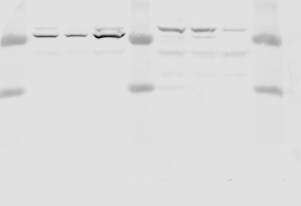
Western Blot: CXCR4 Antibody [NB100-56437] - Western blot shows lysates of Jurkat human T lymphocyte parental cell line and CXCR4 knockout (KO) Jurkat cell line. PVDF membrane was probed with 1 ug/ml of Rabbit Anti-Human CXCR4 Polyclonal Antibody (Catalog # NB100-56437) followed by HRP-conjugated Anti-Rabbit IgG Secondary Antibody (Catalog #HAF008). Specific band was detected for CXCR4 at approximately 38 kDa (as indicated) in the parental Jurkat cell line, but is not detectable in the knockout Jurkat cell line. This experiment was conducted under reducing conditions.![Western Blot: CXCR4 Antibody [NB100-56437]](https://resources.rndsystems.com/images/products/CXCR4-Antibody-Western-Blot-NB100-56437-img0005.jpg "Western Blot: CXCR4 Antibody [NB100-56437]")
Western Blot: CXCR4 Antibody [NB100-56437]
Western Blot: CXCR4 Antibody [NB100-56437] - Detection of 15 ug in HeLa cells with antibody concentration at 1 ug/mL.![Immunocytochemistry/ Immunofluorescence: CXCR4 Antibody [NB100-56437]](https://resources.rndsystems.com/images/products/CXCR4-Antibody-Immunocytochemistry-Immunofluorescence-NB100-56437-img0003.jpg "Immunocytochemistry/ Immunofluorescence: CXCR4 Antibody [NB100-56437]")
Immunocytochemistry/ Immunofluorescence: CXCR4 Antibody [NB100-56437]
Immunocytochemistry/Immunofluorescence: CXCR4 Antibody [NB100-56437] - Jurkat cells were fixed for 10 minutes using 10% formalin and then permeabilized for 5 minutes using 1X TBS + 0.5% Triton X-100. The cells were incubated with anti-CXCR4 (NB100-56437) at a 1:25 dilution for 1 hour at room temperature and detected with an anti-rabbit DyLight 488 (Green) at a 1:500 dilution. Alpha tubulin was used as a co-stain at a 1:1000 dilution and detected with and anti-mouse DyLight 550 (Red) at a 1:500 dilution. Nuclei were counterstained with DAPI (Blue). Cells were imaged using a 40X objective.![Immunohistochemistry-Paraffin: CXCR4 Antibody [NB100-56437]](https://resources.rndsystems.com/images/products/CXCR4-Antibody-Immunohistochemistry-Paraffin-NB100-56437-img0006.jpg "Immunohistochemistry-Paraffin: CXCR4 Antibody [NB100-56437]")
Immunohistochemistry-Paraffin: CXCR4 Antibody [NB100-56437]
Immunohistochemistry-Paraffin: CXCR4 Antibody [NB100-56437] - CXCR4 was detected in immersion sections of human tonsil using rabbit anti-human antibody (Catalog # NB100-56437) at 1:300 dilution overnight at 4C. Tissue was stained using the VisuCyte anti-rabbit HRP polymer detection reagent with DAB chromogen (brown) and counterstained with hematoxylin (blue).
Western Blot: CXCR4 Antibody - BSA Free [NB100-56437] -
CXCL12 secreted from DFs induces AR and CXCR4 in DPCs. The effects of rCXCL12 on the expression of AR and CXCR4 in DPCs were observed using qRT-PCR (A,B) and Western blot analysis (C). rCXCL12 increased the mRNA and protein expression of the AR and CXCR4 in DPCs. $ p < 0.05, $$ p < 0.01 vs. Control. (D) DFs were treated with 100 nM TP or DHT for 48 h and the culture medium (CM) was collected. CM from DFs treated with TP and DHT (DFCMTP and DFCMDHT) significantly reduced hair length in human hair organ culture. $ p < 0.05 vs. DFCM, n = 10. The dollar sign ($) indicates differences in a one-way ANOVA. Image collected and cropped by CiteAb from the following open publication (https://www.mdpi.com/1422-0067/25/3/1705), licensed under a CC-BY license. Not internally tested by Novus Biologicals.Applications for CXCR4 Antibody - BSA Free
Immunohistochemistry
Immunohistochemistry-Frozen
Immunohistochemistry-Paraffin
Western Blot
Reviewed Applications
Read 1 review rated 5 using NB100-56437 in the following applications:
Formulation, Preparation, and Storage
Purification
Formulation
Format
Preservative
Concentration
Shipping
Stability & Storage
Background: CXCR4
Long Name
Alternate Names
Gene Symbol
UniProt
Additional CXCR4 Products
Product Documents for CXCR4 Antibody - BSA Free
Certificate of Analysis
To download a Certificate of Analysis, please enter a lot or batch number in the search box below.
Product Specific Notices for CXCR4 Antibody - BSA Free
This product is for research use only and is not approved for use in humans or in clinical diagnosis. Primary Antibodies are guaranteed for 1 year from date of receipt.
Related Research Areas
Citations for CXCR4 Antibody - BSA Free
 Powered by Bioz
Customer Reviews for CXCR4 Antibody - BSA Free (1)
Have you used CXCR4 Antibody - BSA Free?
Submit a review and receive an Amazon gift card!
$25/€18/£15/$25CAN/¥2500 Yen for a review with an image
$10/€7/£6/$10CAN/¥1110 Yen for a review without an image
Submit a review
Customer Images
-
Application: Western BlotSample Tested: Human MDA-MB-231 cellsSpecies: HumanVerified Customer | Posted 03/07/2020

There are no reviews that match your criteria.
Protocols
Find general support by application which include: protocols, troubleshooting, illustrated assays, videos and webinars.
- Antigen Retrieval Protocol (PIER)
- Antigen Retrieval for Frozen Sections Protocol
- Appropriate Fixation of IHC/ICC Samples
- Cellular Response to Hypoxia Protocols
- Chromogenic IHC Staining of Formalin-Fixed Paraffin-Embedded (FFPE) Tissue Protocol
- Chromogenic Immunohistochemistry Staining of Frozen Tissue
- ClariTSA™ Fluorophore Kits
- Detection & Visualization of Antibody Binding
- Fluorescent IHC Staining of Frozen Tissue Protocol
- Graphic Protocol for Heat-induced Epitope Retrieval
- Graphic Protocol for the Preparation and Fluorescent IHC Staining of Frozen Tissue Sections
- Graphic Protocol for the Preparation and Fluorescent IHC Staining of Paraffin-embedded Tissue Sections
- Graphic Protocol for the Preparation of Gelatin-coated Slides for Histological Tissue Sections
- ICC Cell Smear Protocol for Suspension Cells
- ICC Immunocytochemistry Protocol Videos
- ICC for Adherent Cells
- IHC Sample Preparation (Frozen sections vs Paraffin)
- Immunocytochemistry (ICC) Protocol
- Immunocytochemistry Troubleshooting
- Immunofluorescence of Organoids Embedded in Cultrex Basement Membrane Extract
- Immunofluorescent IHC Staining of Formalin-Fixed Paraffin-Embedded (FFPE) Tissue Protocol
- Immunohistochemistry (IHC) and Immunocytochemistry (ICC) Protocols
- Immunohistochemistry Frozen Troubleshooting
- Immunohistochemistry Paraffin Troubleshooting
- Preparing Samples for IHC/ICC Experiments
- Preventing Non-Specific Staining (Non-Specific Binding)
- Primary Antibody Selection & Optimization
- Protocol for Heat-Induced Epitope Retrieval (HIER)
- Protocol for Making a 4% Formaldehyde Solution in PBS
- Protocol for VisUCyte™ HRP Polymer Detection Reagent
- Protocol for the Fluorescent ICC Staining of Cell Smears - Graphic
- Protocol for the Fluorescent ICC Staining of Cultured Cells on Coverslips - Graphic
- Protocol for the Preparation & Fixation of Cells on Coverslips
- Protocol for the Preparation and Chromogenic IHC Staining of Frozen Tissue Sections
- Protocol for the Preparation and Chromogenic IHC Staining of Frozen Tissue Sections - Graphic
- Protocol for the Preparation and Chromogenic IHC Staining of Paraffin-embedded Tissue Sections
- Protocol for the Preparation and Chromogenic IHC Staining of Paraffin-embedded Tissue Sections - Graphic
- Protocol for the Preparation and Fluorescent ICC Staining of Cells on Coverslips
- Protocol for the Preparation and Fluorescent ICC Staining of Non-adherent Cells
- Protocol for the Preparation and Fluorescent ICC Staining of Stem Cells on Coverslips
- Protocol for the Preparation and Fluorescent IHC Staining of Frozen Tissue Sections
- Protocol for the Preparation and Fluorescent IHC Staining of Paraffin-embedded Tissue Sections
- Protocol for the Preparation of Gelatin-coated Slides for Histological Tissue Sections
- Protocol for the Preparation of a Cell Smear for Non-adherent Cell ICC - Graphic
- R&D Systems Quality Control Western Blot Protocol
- TUNEL and Active Caspase-3 Detection by IHC/ICC Protocol
- The Importance of IHC/ICC Controls
- Troubleshooting Guide: Immunohistochemistry
- Troubleshooting Guide: Western Blot Figures
- Western Blot Conditions
- Western Blot Protocol
- Western Blot Protocol for Cell Lysates
- Western Blot Troubleshooting
- Western Blot Troubleshooting Guide
- View all Protocols, Troubleshooting, Illustrated assays and Webinars
FAQs for CXCR4 Antibody - BSA Free
-
Q: Which is your best CXCR4 for immunohistochemistry in paraffin tissues? I would like to detect it in paraffin embedded tissues from human breast cancer samples.
A:
CXCR4 antibody (NB100-74396) is our best selling product among all the CXCR4 antibodies and it has been validated for IHC-P in human cervical carcinoma tissue sections. NB100-74396 has been cited in at least 7 research publications. Additionally, here is a list of all of our CXCR4 antibodies that has been verified in IHC-P in human samples.


