E-Cadherin Antibody (7H12)
Novus Biologicals | Catalog # NBP2-19051

Loading...
Key Product Details
Validated by
Knockout/Knockdown, Biological Validation
Species Reactivity
Validated:
Human, Mouse, Monkey
Cited:
Human, Mouse
Applications
Validated:
Multiplex Immunofluorescence, Immunohistochemistry, Immunohistochemistry-Paraffin, Western Blot, ELISA, Flow Cytometry, Immunocytochemistry/ Immunofluorescence, COMET
Cited:
Immunohistochemistry-Paraffin, Immunohistochemistry-Frozen, Western Blot, Immunocytochemistry/ Immunofluorescence, IF/IHC
Label
Unconjugated
Antibody Source
Monoclonal Mouse IgG1 Clone # 7H12
Loading...
Product Specifications
Immunogen
Purified recombinant fragment of human E-Cadherin expressed in E. Coli.
Reactivity Notes
Please note that this antibody is reactive to Mouse and derived from the same host, Mouse. Mouse-On-Mouse blocking reagent may be needed for IHC and ICC experiments to reduce high background signal. You can find these reagents under catalog numbers PK-2200-NB and MP-2400-NB. Please contact Technical Support if you have any questions.
Marker
Epithelial Cell Marker, Adherens Junctions Marker
Clonality
Monoclonal
Host
Mouse
Isotype
IgG1
Theoretical MW
135 kDa.
Disclaimer note: The observed molecular weight of the protein may vary from the listed predicted molecular weight due to post translational modifications, post translation cleavages, relative charges, and other experimental factors.
Disclaimer note: The observed molecular weight of the protein may vary from the listed predicted molecular weight due to post translational modifications, post translation cleavages, relative charges, and other experimental factors.
Scientific Data Images for E-Cadherin Antibody (7H12)
Detection of E-Cadherin in Human Breast via Multiplex Immunofluorescence staining on COMET™
E-Cadherin was detected in immersion fixed paraffin-embedded sections of human breast using Mouse Anti-Human E-Cadherin Monoclonal Antibody (Novus Catalog # NBP2-19051) at 1:1000 at 37 ° Celsius for 4 minutes. Before incubation with the primary antibody, tissue underwent an all-in-one dewaxing and antigen retrieval preprocessing using PreTreatment Module (PT Module) and Dewax and HIER Buffer H (pH 9). Tissue was stained using the Alexa Fluor™ Plus 647 Goat anti-Mouse IgG Secondary Antibody at 1:200 at 37 ° Celsius for 2 minutes. (Yellow; Lunaphore Catalog # DR647MS) and counterstained with DAPI (blue; Lunaphore Catalog # DR100). Specific staining was localized to the membrane. Protocol available in COMET™ Panel Builder.
Detection of E-Cadherin in Human Liver via Multiplex Immunofluorescence staining on COMET™
E-Cadherin was detected in immersion fixed paraffin-embedded sections of human liver using Mouse Anti-Human E-Cadherin Monoclonal Antibody (Catalog # NBP2-19051) at 1:1000 at 37 ° Celsius for 4 minutes. Before incubation with the primary antibody, tissue underwent an all-in-one dewaxing and antigen retrieval preprocessing using PreTreatment Module (PT Module) and Dewax and HIER Buffer H (pH 9). Tissue was stained using the Alexa Fluor™ 647 Goat anti-Mouse IgG Secondary Antibody at 1:200 at 37 ° Celsius for 2 minutes. (Yellow; Lunaphore Catalog # DR647MS) and counterstained with DAPI (blue; Lunaphore Catalog # DR100). Specific staining was localized to the membrane. Protocol available in COMET™ Panel Builder.
Detection of E-Cadherin in Human Colon Cancer via Multiplex Immunofluorescence staining on COMET™
E-Cadherin was detected in immersion fixed paraffin-embedded sections of human colon cancer using Mouse Anti-Human E-Cadherin Monoclonal Antibody (Catalog # NBP2-19051) at 1:1000 at 37 ° Celsius for 4 minutes. Before incubation with the primary antibody, tissue underwent an all-in-one dewaxing and antigen retrieval preprocessing using PreTreatment Module (PT Module) and Dewax and HIER Buffer H (pH 9). Tissue was stained using the Alexa Fluor™ 647 Goat anti-Mouse IgG Secondary Antibody at 1:200 at 37 ° Celsius for 2 minutes. (Yellow; Lunaphore Catalog # DR647MS) and counterstained with DAPI (blue; Lunaphore Catalog # DR100). Specific staining was localized to the membrane. Protocol available in COMET™ Panel Builder.![Immunohistochemistry-Paraffin: E-Cadherin Antibody (7H12) - BSA Free [NBP2-19051]](https://resources.rndsystems.com/images/products/E-Cadherin-Antibody-7H12-Immunohistochemistry-Paraffin-NBP2-19051-img0011.jpg "Immunohistochemistry-Paraffin: E-Cadherin Antibody (7H12) - BSA Free [NBP2-19051]")
Immunohistochemistry-Paraffin: E-Cadherin Antibody (7H12) - BSA Free [NBP2-19051]
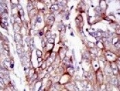
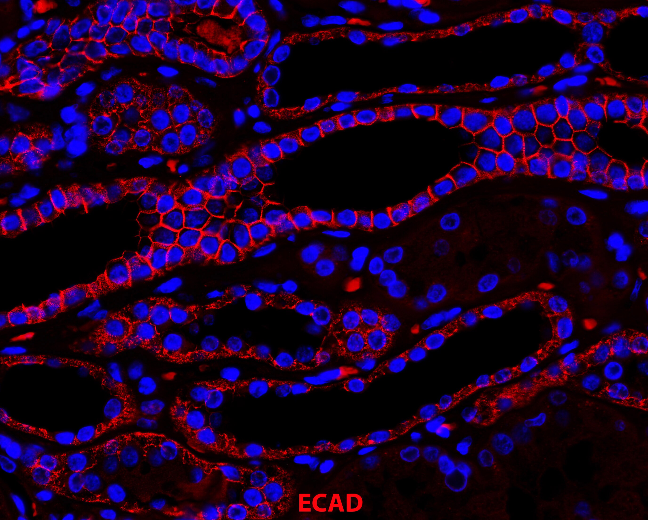
Immunohistochemistry-Paraffin: E-Cadherin Antibody (7H12) [NBP2-19051] - Human kidney distal tubules stained with E-CAD antibody. Secondary antibody: Donkey anti-Mouse Alexa Fluor 555. Imaging with a 40x objective from Zeiss Observer microscope with Apotome2. Image from verified customer review.![Western Blot: E-Cadherin Antibody (7H12)BSA Free [NBP2-19051]](https://resources.rndsystems.com/images/products/E-Cadherin-Antibody-7H12-Western-Blot-NBP2-19051-img0013.jpg "Western Blot: E-Cadherin Antibody (7H12)BSA Free [NBP2-19051]")
![Western Blot: E-Cadherin Antibody (7H12)BSA Free [NBP2-19051]](https://resources.rndsystems.com/images/products/E-Cadherin-Antibody-7H12-Western-Blot-NBP2-19051-img0002.jpg "Western Blot: E-Cadherin Antibody (7H12)BSA Free [NBP2-19051]")
Western Blot: E-Cadherin Antibody (7H12)BSA Free [NBP2-19051]
Western Blot: E-Cadherin Antibody (7H12) [NBP2-19051] - Analysis of E-Cadherin in LNCAP (1), A431 (2), DU145 (3), PC-3 (4), MCF-7 (5), PC-12 (6), NIH/3T3 (7), C6 (8) and COS7 (9) cell lysate.![Immunocytochemistry/ Immunofluorescence: E-Cadherin Antibody (7H12) - BSA Free [NBP2-19051]](https://resources.rndsystems.com/images/products/E-Cadherin-Antibody-7H12-Immunocytochemistry-Immunofluorescence-NBP2-19051-img0010.jpg "Immunocytochemistry/ Immunofluorescence: E-Cadherin Antibody (7H12) - BSA Free [NBP2-19051]")
Immunocytochemistry/ Immunofluorescence: E-Cadherin Antibody (7H12) - BSA Free [NBP2-19051]
Immunocytochemistry/Immunofluorescence: E-Cadherin Antibody (7H12) [NBP2-19051] - ICC staining of E-Cadherin in human colon cancer sphere. Image from verified customer review.![Western Blot: E-Cadherin Antibody (7H12)BSA Free [NBP2-19051]](https://resources.rndsystems.com/images/products/E-Cadherin-Antibody-7H12-Western-Blot-NBP2-19051-img0014.jpg "Western Blot: E-Cadherin Antibody (7H12)BSA Free [NBP2-19051]")
![Immunocytochemistry/ Immunofluorescence: E-Cadherin Antibody (7H12) - BSA Free [NBP2-19051]](https://resources.rndsystems.com/images/products/E-Cadherin-Antibody-7H12-Immunocytochemistry-Immunofluorescence-NBP2-19051-img0009.jpg "Immunocytochemistry/ Immunofluorescence: E-Cadherin Antibody (7H12) - BSA Free [NBP2-19051]")
Immunocytochemistry/ Immunofluorescence: E-Cadherin Antibody (7H12) - BSA Free [NBP2-19051]
Immunocytochemistry/Immunofluorescence: E-Cadherin Antibody (7H12) [NBP2-19051] - Human colonic tissue was stained with E-Cadherin antibody. Image from verified customer review.![Immunohistochemistry-Paraffin: E-Cadherin Antibody (7H12) - BSA Free [NBP2-19051]](https://resources.rndsystems.com/images/products/E-Cadherin-Antibody-7H12-Immunohistochemistry-Paraffin-NBP2-19051-img0012.jpg "Immunohistochemistry-Paraffin: E-Cadherin Antibody (7H12) - BSA Free [NBP2-19051]")
Immunohistochemistry-Paraffin: E-Cadherin Antibody (7H12) - BSA Free [NBP2-19051]
Immunohistochemistry-Paraffin: E-Cadherin Antibody (7H12) [NBP2-19051] - IHC staining of E-Cadherin antibody in mouse colon cancer tissue. Antibody dilution 1:500. Image from verified customer review.![Flow Cytometry: E-Cadherin Antibody (7H12) - BSA Free [NBP2-19051]](https://resources.rndsystems.com/images/products/E-Cadherin-Antibody-7H12-Flow-Cytometry-NBP2-19051-img0004.jpg "Flow Cytometry: E-Cadherin Antibody (7H12) - BSA Free [NBP2-19051]")
Flow Cytometry: E-Cadherin Antibody (7H12) - BSA Free [NBP2-19051]
Flow Cytometry: E-Cadherin Antibody (7H12) [NBP2-19051] - Analysis of Hela cells using E-Cadherin mouse mAb (green) and negative control (purple).![Western Blot: E-Cadherin Antibody (7H12)BSA Free [NBP2-19051]](https://resources.rndsystems.com/images/products/E-Cadherin-Antibody-7H12-Western-Blot-NBP2-19051-img0015.jpg "Western Blot: E-Cadherin Antibody (7H12)BSA Free [NBP2-19051]")
![Immunohistochemistry-Paraffin: E-Cadherin Antibody (7H12) - BSA Free [NBP2-19051]](https://resources.rndsystems.com/images/products/E-Cadherin-Antibody-7H12-Immunohistochemistry-Paraffin-NBP2-19051-img0003.jpg "Immunohistochemistry-Paraffin: E-Cadherin Antibody (7H12) - BSA Free [NBP2-19051]")
Immunohistochemistry-Paraffin: E-Cadherin Antibody (7H12) - BSA Free [NBP2-19051]
Immunohistochemistry-Paraffin: E-Cadherin Antibody (7H12) [NBP2-19051] - IHC staining of E-Cadherin in gastric cancer tissues (left) and lung cancer tissues (right) with DAB staining. - BSA Free [NBP2-19051] -")
Western Blot: E-Cadherin Antibody (7H12) - BSA Free [NBP2-19051] -
Mitochondrial fission regulates migration & invasion in BRAFV600E CRC cells through glucose metabolic reprograming. (A) Relative cell invasion post vemurafenib treatment in BRAFV600E cells (n=3, mean ± SEM); statistical analysis was done by unpaired t-test followed by Welch’s correction assuming unequal variance in means, corresponding p values shown. (B) Western blot showing a reduction in EMT markers upon vemurafenib treatment in BRAFV600E cells. Relative cellular invasion post (C) DRP1 silencing, (D) PDK1 silencing, & (E) 2-deoxyglucose treatment in Colo205 & HT29 cells (n=3, mean± SEM); statistical analysis was done by unpaired t-test followed by Welch’s correction assuming unequal variance in means, corresponding p values shown. (F) Western blots showing a reduction in epithelial mesenchymal transition (EMT) markers upon DRP1 silencing, PDK1 knockdown, & 2-DG treatment in Colo205 & HT29 cells. Image collected & cropped by CiteAb from the following publication (https://pubmed.ncbi.nlm.nih.gov/33738242), licensed under a CC-BY license. Not internally tested by Novus Biologicals.Applications for E-Cadherin Antibody (7H12)
Application
Recommended Usage
ELISA
1:10000
Flow Cytometry
1:200-1:400
Immunocytochemistry/ Immunofluorescence
1:10-1:500
Immunohistochemistry
1:200-1:1000
Immunohistochemistry-Paraffin
1:200-1:1000
Multiplex Immunofluorescence
1:1000
Western Blot
1:500-1:2000
Application Notes
Use in ICC/IF reported in scientific literature (PMID: 20605541).
Reviewed Applications
Read 7 reviews rated 4.4 using NBP2-19051 in the following applications:
Flow Cytometry Panel Builder
Bio-Techne Knows Flow Cytometry
Save time and reduce costly mistakes by quickly finding compatible reagents using the Panel Builder Tool.
Advanced Features
- Spectra Viewer - Custom analysis of spectra from multiple fluorochromes
- Spillover Popups - Visualize the spectra of individual fluorochromes
- Antigen Density Selector - Match fluorochrome brightness with antigen density

Formulation, Preparation, and Storage
Purification
Ascites
Formulation
Ascites
Preservative
0.03% Sodium Azide
Concentration
This product is unpurified. The exact concentration of antibody is not quantifiable.
Shipping
The product is shipped with polar packs. Upon receipt, store it immediately at the temperature recommended below.
Stability & Storage
Store at 4C short term. Aliquot and store at -20C long term. Avoid freeze-thaw cycles.
Background: E-Cadherin
Alternate Names
Arc-1, CAD1, Cadherin-1, CD324, CDH1, Cell-CAM120/80, ECAD, ECadherin, L-CAM, Uvomorulin
Entrez Gene IDs
999 (Human)
Gene Symbol
CDH1
UniProt
Additional E-Cadherin Products
Product Documents for E-Cadherin Antibody (7H12)
Certificate of Analysis
To download a Certificate of Analysis, please enter a lot or batch number in the search box below.
Product Specific Notices for E-Cadherin Antibody (7H12)
This product is for research use only and is not approved for use in humans or in clinical diagnosis. Primary Antibodies are guaranteed for 1 year from date of receipt.
Citations for E-Cadherin Antibody (7H12)
 Powered by Bioz
Customer Reviews for E-Cadherin Antibody (7H12) (7)
4.4 out of 5
7 Customer Ratings
Have you used E-Cadherin Antibody (7H12)?
Submit a review and receive an Amazon gift card!
$25/€18/£15/$25CAN/¥2500 Yen for a review with an image
$10/€7/£6/$10CAN/¥1110 Yen for a review without an image
Submit a review
Customer Images
-(01-ml)_NBP2-19051_11681.jpg)
-(01-ml)_NBP2-19051_11671.jpg)
-(01-ml)_NBP2-19051_11036.jpg)
-(01-ml)_NBP2-19051_8291.jpg)
Showing
1
-
5 of
7 reviews
Showing All
Filter By:
-
Application: Immunohistochemistry-ParaffinSample Tested: Colon cancer tissueSpecies: MouseVerified Customer | Posted 02/01/2022Mouse colon cancer tissueAntibody dilution 1:500

-
Application: Immunohistochemistry-ParaffinSample Tested: Human kidneySpecies: HumanVerified Customer | Posted 08/08/2018Human Kidney Distal Tubules Stained with ECAD AntibodyTaken with a 40x Objective from Zeiss Observer microscope with Apotome2 Secondary antibody: Donkey Anti Mouse 555
-
Application: Western BlotSample Tested: Human breast cancer cell linesSpecies: HumanVerified Customer | Posted 09/02/2015
-
Application: ImmunohistochemistrySample Tested: Human colonic tissueSpecies: HumanVerified Customer | Posted 11/04/2014e-Cadherin IHC
-
Application: Flow CytometrySample Tested: Human colon cancer cell line HT29Species: HumanVerified Customer | Posted 11/04/2014E-Cadherin Flow
-
Application: ImmunocytochemistrySample Tested: Human colon cancer sphereSpecies: HumanVerified Customer | Posted 10/08/2014E-Cadherin
-
Application: Western BlotSample Tested:Species: HumanVerified Customer | Posted 06/13/2014Western blot analysis of extracts from MCF7 cells using E-Cadherin antibody (NBP2-19051, 1:100).
There are no reviews that match your criteria.
Protocols
Find general support by application which include: protocols, troubleshooting, illustrated assays, videos and webinars.
- 7-Amino Actinomycin D (7-AAD) Cell Viability Flow Cytometry Protocol
- Antigen Retrieval Protocol (PIER)
- Antigen Retrieval for Frozen Sections Protocol
- Appropriate Fixation of IHC/ICC Samples
- Cellular Response to Hypoxia Protocols
- Chromogenic IHC Staining of Formalin-Fixed Paraffin-Embedded (FFPE) Tissue Protocol
- Chromogenic Immunohistochemistry Staining of Frozen Tissue
- ClariTSA™ Fluorophore Kits
- Detection & Visualization of Antibody Binding
- ELISA Sample Preparation & Collection Guide
- ELISA Troubleshooting Guide
- Extracellular Membrane Flow Cytometry Protocol
- Flow Cytometry Protocol for Cell Surface Markers
- Flow Cytometry Protocol for Staining Membrane Associated Proteins
- Flow Cytometry Staining Protocols
- Flow Cytometry Troubleshooting Guide
- Fluorescent IHC Staining of Frozen Tissue Protocol
- Graphic Protocol for Heat-induced Epitope Retrieval
- Graphic Protocol for the Preparation and Fluorescent IHC Staining of Frozen Tissue Sections
- Graphic Protocol for the Preparation and Fluorescent IHC Staining of Paraffin-embedded Tissue Sections
- Graphic Protocol for the Preparation of Gelatin-coated Slides for Histological Tissue Sections
- How to Run an R&D Systems DuoSet ELISA
- How to Run an R&D Systems Quantikine ELISA
- How to Run an R&D Systems Quantikine™ QuicKit™ ELISA
- ICC Cell Smear Protocol for Suspension Cells
- ICC Immunocytochemistry Protocol Videos
- ICC for Adherent Cells
- IHC Sample Preparation (Frozen sections vs Paraffin)
- Immunocytochemistry (ICC) Protocol
- Immunocytochemistry Troubleshooting
- Immunofluorescence of Organoids Embedded in Cultrex Basement Membrane Extract
- Immunofluorescent IHC Staining of Formalin-Fixed Paraffin-Embedded (FFPE) Tissue Protocol
- Immunohistochemistry (IHC) and Immunocytochemistry (ICC) Protocols
- Immunohistochemistry Frozen Troubleshooting
- Immunohistochemistry Paraffin Troubleshooting
- Intracellular Flow Cytometry Protocol Using Alcohol (Methanol)
- Intracellular Flow Cytometry Protocol Using Detergents
- Intracellular Nuclear Staining Flow Cytometry Protocol Using Detergents
- Intracellular Staining Flow Cytometry Protocol Using Alcohol Permeabilization
- Intracellular Staining Flow Cytometry Protocol Using Detergents to Permeabilize Cells
- Preparing Samples for IHC/ICC Experiments
- Preventing Non-Specific Staining (Non-Specific Binding)
- Primary Antibody Selection & Optimization
- Propidium Iodide Cell Viability Flow Cytometry Protocol
- Protocol for Heat-Induced Epitope Retrieval (HIER)
- Protocol for Liperfluo
- Protocol for Making a 4% Formaldehyde Solution in PBS
- Protocol for VisUCyte™ HRP Polymer Detection Reagent
- Protocol for the Characterization of Human Th22 Cells
- Protocol for the Characterization of Human Th9 Cells
- Protocol for the Fluorescent ICC Staining of Cell Smears - Graphic
- Protocol for the Fluorescent ICC Staining of Cultured Cells on Coverslips - Graphic
- Protocol for the Preparation & Fixation of Cells on Coverslips
- Protocol for the Preparation and Chromogenic IHC Staining of Frozen Tissue Sections
- Protocol for the Preparation and Chromogenic IHC Staining of Frozen Tissue Sections - Graphic
- Protocol for the Preparation and Chromogenic IHC Staining of Paraffin-embedded Tissue Sections
- Protocol for the Preparation and Chromogenic IHC Staining of Paraffin-embedded Tissue Sections - Graphic
- Protocol for the Preparation and Fluorescent ICC Staining of Cells on Coverslips
- Protocol for the Preparation and Fluorescent ICC Staining of Non-adherent Cells
- Protocol for the Preparation and Fluorescent ICC Staining of Stem Cells on Coverslips
- Protocol for the Preparation and Fluorescent IHC Staining of Frozen Tissue Sections
- Protocol for the Preparation and Fluorescent IHC Staining of Paraffin-embedded Tissue Sections
- Protocol for the Preparation of Gelatin-coated Slides for Histological Tissue Sections
- Protocol for the Preparation of a Cell Smear for Non-adherent Cell ICC - Graphic
- Protocol: Annexin V and PI Staining by Flow Cytometry
- Protocol: Annexin V and PI Staining for Apoptosis by Flow Cytometry
- Quantikine HS ELISA Kit Assay Principle, Alkaline Phosphatase
- Quantikine HS ELISA Kit Principle, Streptavidin-HRP Polymer
- R&D Systems Quality Control Western Blot Protocol
- Sandwich ELISA (Colorimetric) – Biotin/Streptavidin Detection Protocol
- Sandwich ELISA (Colorimetric) – Direct Detection Protocol
- TUNEL and Active Caspase-3 Detection by IHC/ICC Protocol
- The Importance of IHC/ICC Controls
- Troubleshooting Guide: ELISA
- Troubleshooting Guide: Fluorokine Flow Cytometry Kits
- Troubleshooting Guide: Immunohistochemistry
- Troubleshooting Guide: Western Blot Figures
- Western Blot Conditions
- Western Blot Protocol
- Western Blot Protocol for Cell Lysates
- Western Blot Troubleshooting
- Western Blot Troubleshooting Guide
- View all Protocols, Troubleshooting, Illustrated assays and Webinars
Loading...
Associated Pathways
