FoxO1/FKHR Antibody (83N7F8) - BSA Free
Novus Biologicals | Catalog # NBP2-31376
Key Product Details
Species Reactivity
Validated:
Human, Mouse, Rat
Cited:
Human, Mouse, Rat
Predicted:
Bovine (91%), Porcine (90%), Squirrel (91%). Backed by our 100% Guarantee.
Applications
Validated:
Immunohistochemistry, Immunohistochemistry-Paraffin, Western Blot, Flow Cytometry, Immunocytochemistry/ Immunofluorescence, Simple Western
Cited:
Western Blot, Flow Cytometry
Label
Unconjugated
Antibody Source
Monoclonal Mouse IgG2b Kappa Clone # 83N7F8
Format
BSA Free
Loading...
Product Specifications
Immunogen
FOXO1A
Reactivity Notes
Use in Mouse reported in scientific literature (PMID:33662610). Rat reactivity reported in scientific literature (PMID: 31553623).
Localization
Cytoplasm. Nucleus. Note: Shuttles between the cytoplasm and nucleus.
Clonality
Monoclonal
Host
Mouse
Isotype
IgG2b Kappa
Scientific Data Images for FoxO1/FKHR Antibody (83N7F8) - BSA Free
![Western Blot: FoxO1/FKHR Antibody (83N7F8)BSA Free [NBP2-31376]](https://resources.rndsystems.com/images/products/FoxO1-FKHR-Antibody-83N7F8-Western-Blot-NBP2-31376-img0007.jpg "Western Blot: FoxO1/FKHR Antibody (83N7F8)BSA Free [NBP2-31376]")
Western Blot: FoxO1/FKHR Antibody (83N7F8)BSA Free [NBP2-31376]
Western Blot: FoxO1/FKHR Antibody (83N7F8) [NBP2-31376] - Analysis of (A) Ntera2 cell lysate, (B) HEK293 cell lysate, and (C) human placenta tissue using FOXO1A antibody at a concentration of 2 ug/ml.![Immunocytochemistry/ Immunofluorescence: FoxO1/FKHR Antibody (83N7F8) - BSA Free [NBP2-31376]](https://resources.rndsystems.com/images/products/FoxO1-FKHR-Antibody-83N7F8-Immunocytochemistry-Immunofluorescence-NBP2-31376-img0009.jpg "Immunocytochemistry/ Immunofluorescence: FoxO1/FKHR Antibody (83N7F8) - BSA Free [NBP2-31376]")
Immunocytochemistry/ Immunofluorescence: FoxO1/FKHR Antibody (83N7F8) - BSA Free [NBP2-31376]
Immunocytochemistry/Immunofluorescence: FoxO1/FKHR Antibody (83N7F8) [NBP2-31376] - FOXO1A antibody was tested in Ntera2 cells with DyLight 488 (green). Nuclei and alpha-tubulin were counterstained with DAPI (blue) and DyLight 550 (red). An antibody concentration of 0.1 ug/ml was used. Image objective 40x.![Immunohistochemistry-Paraffin: FoxO1/FKHR Antibody (83N7F8) - BSA Free [NBP2-31376]](https://resources.rndsystems.com/images/products/FoxO1-FKHR-Antibody-83N7F8-Immunohistochemistry-Paraffin-NBP2-31376-img0011.jpg "Immunohistochemistry-Paraffin: FoxO1/FKHR Antibody (83N7F8) - BSA Free [NBP2-31376]")
Immunohistochemistry-Paraffin: FoxO1/FKHR Antibody (83N7F8) - BSA Free [NBP2-31376]
Immunohistochemistry-Paraffin: FoxO1/FKHR Antibody (83N7F8) [NBP2-31376] - Analysis of FOXO1A protein in a section of normal human uterus by using FOXO1A antibody at a concentration of 5 ug/ml. The columnar cells of straight non-convoluted glands in uterous showed specific staining for FOXO1A in the cellular cytoplasm as well as nuclei, whereas the stromal cells showed mild cytoplasmic staining only.![Flow Cytometry: FoxO1/FKHR Antibody (83N7F8) - BSA Free [NBP2-31376]](https://resources.rndsystems.com/images/products/FoxO1-FKHR-Antibody-83N7F8-Flow-Cytometry-NBP2-31376-img0016.jpg "Flow Cytometry: FoxO1/FKHR Antibody (83N7F8) - BSA Free [NBP2-31376]")
Flow Cytometry: FoxO1/FKHR Antibody (83N7F8) - BSA Free [NBP2-31376]
Flow Cytometry: FoxO1/FKHR Antibody (83N7F8) [NBP2-31376] - An intracellular stain was performed on RH-30 cells with FoxO1/FKHR Antibody [83N7F8] NBP2-31376AF488 (blue) and a matched isotype control (orange). Cells were fixed with 4% PFA and then permeabilized with 0.1% saponin. Cells were incubated in an antibody dilution of 5 ug/mL for 30 minutes at room temperature. Both antibodies were conjugated to Alexa Fluor 488.![Western Blot: FoxO1/FKHR Antibody (83N7F8)BSA Free [NBP2-31376]](https://resources.rndsystems.com/images/products/FoxO1-FKHR-Antibody-83N7F8-Western-Blot-NBP2-31376-img0004.jpg "Western Blot: FoxO1/FKHR Antibody (83N7F8)BSA Free [NBP2-31376]")
Western Blot: FoxO1/FKHR Antibody (83N7F8)BSA Free [NBP2-31376]
Western Blot: FoxO1/FKHR Antibody (83N7F8) [NBP2-31376] - Detection of FOXO1A / FOXO1 protein in (A) Hela cells lysate, (B) Jurkat cell lysate, and (C) partial recombinant protein by using FOXO1A antibody at a concentration of 0.5 ug/ml for lysate and 0.05 ug/ml for the recombinant protein.![Immunohistochemistry-Paraffin: FoxO1/FKHR Antibody (83N7F8) - BSA Free [NBP2-31376]](https://resources.rndsystems.com/images/products/FoxO1-FKHR-Antibody-83N7F8-Immunohistochemistry-Paraffin-NBP2-31376-img0010.jpg "Immunohistochemistry-Paraffin: FoxO1/FKHR Antibody (83N7F8) - BSA Free [NBP2-31376]")
Immunohistochemistry-Paraffin: FoxO1/FKHR Antibody (83N7F8) - BSA Free [NBP2-31376]
Immunohistochemistry-Paraffin: FoxO1/FKHR Antibody (83N7F8) [NBP2-31376] - Analysis of FOXO1A protein in a section of normal human kidney by using FOXO1A antibody at a concentration of 5 ug/ml. Cytoplasmic and nuclear positivity was observed in the cells of various tubules/ducts of renal medulla.![Flow Cytometry: FoxO1/FKHR Antibody (83N7F8) - BSA Free [NBP2-31376]](https://resources.rndsystems.com/images/products/FoxO1-FKHR-Antibody-83N7F8-Flow-Cytometry-NBP2-31376-img0013.jpg "Flow Cytometry: FoxO1/FKHR Antibody (83N7F8) - BSA Free [NBP2-31376]")
Flow Cytometry: FoxO1/FKHR Antibody (83N7F8) - BSA Free [NBP2-31376]
Flow Cytometry: FoxO1/FKHR Antibody (83N7F8) [NBP2-31376] - Analysis using the Alexa Fluor 488 conjugated FoxO1 antibody, NBP2-31376AF488 (blue) and fluorescence minus one control (red). Staining of FoxO1 in primary CD4+ T-cells isolated from human peripheral blood using 1uL of antibody per 1X10e5 cells. Image from verified customer review.![Flow Cytometry: FoxO1/FKHR Antibody (83N7F8) - BSA Free [NBP2-31376]](https://resources.rndsystems.com/images/products/FoxO1-FKHR-Antibody-83N7F8-Flow-Cytometry-NBP2-31376-img0014.jpg "Flow Cytometry: FoxO1/FKHR Antibody (83N7F8) - BSA Free [NBP2-31376]")
Flow Cytometry: FoxO1/FKHR Antibody (83N7F8) - BSA Free [NBP2-31376]
Flow Cytometry: FoxO1/FKHR Antibody (83N7F8) [NBP2-31376] - Using the Alexa Fluor 488 direct conjugate An intracellular stain was performed on Daudi cells with FoxO1/FKHR (83N7F8) antibody NBP2-31376AF488 (blue) and a matched isotype control NBP2-27231AF488 (orange). Cells were fixed with 4% PFA and then permeablized with 0.1% saponin. Cells were incubated in an antibody dilution of 5 ug/mL for 30 minutes at room temperature. Both antibodies were conjugated to Alexa Fluor 488.![Flow Cytometry: FoxO1/FKHR Antibody (83N7F8) - BSA Free [NBP2-31376]](https://resources.rndsystems.com/images/products/FoxO1-FKHR-Antibody-83N7F8-Flow-Cytometry-NBP2-31376-img0015.jpg "Flow Cytometry: FoxO1/FKHR Antibody (83N7F8) - BSA Free [NBP2-31376]")
Flow Cytometry: FoxO1/FKHR Antibody (83N7F8) - BSA Free [NBP2-31376]
Flow Cytometry: FoxO1/FKHR Antibody (83N7F8) [NBP2-31376] - An intracellular stain was performed on U2-OS cells with FoxO1/FKHR [83N7F8] Antibody NBP2-31376AF488 (blue) and a matched isotype control (orange). Cells were fixed with 4% PFA and then permeabilized with 0.1% saponin. Cells were incubated in an antibody dilution of 10 ug/mL for 30 minutes at room temperature. Both antibodies were conjugated to Alexa Fluor 488.![Simple Western: FoxO1/FKHR Antibody (83N7F8)BSA Free [NBP2-31376]](https://resources.rndsystems.com/images/products/FoxO1-FKHR-Antibody-83N7F8-Simple-Western-NBP2-31376-img0012.jpg "Simple Western: FoxO1/FKHR Antibody (83N7F8)BSA Free [NBP2-31376]")
Simple Western: FoxO1/FKHR Antibody (83N7F8)BSA Free [NBP2-31376]
Simple Western: FoxO1/FKHR Antibody (83N7F8) [NBP2-31376] - Lane view shows a specific band for FoxO1/FKHR in 0.5 mg/ml of Hek293 lysate. This experiment was performed under reducing conditions using the 12-230 kDa separation system.Applications for FoxO1/FKHR Antibody (83N7F8) - BSA Free
Application
Recommended Usage
Immunocytochemistry/ Immunofluorescence
0.1-2 ug/ml
Immunohistochemistry
5 ug/ml
Immunohistochemistry-Paraffin
5 ug/ml
Simple Western
1:50
Western Blot
0.5-2 ug/ml
Application Notes
In Simple Western only 10 - 15 uL of the recommended dilution is used per data point.
See Simple Western Antibody Database for Simple Western validation: Tested in Hek293 lysate 0.5 mg/mL, separated by Size, antibody dilution of 1:50, apparent MW was 107 kDa.
See Simple Western Antibody Database for Simple Western validation: Tested in Hek293 lysate 0.5 mg/mL, separated by Size, antibody dilution of 1:50, apparent MW was 107 kDa.
Reviewed Applications
Read 1 review rated 4 using NBP2-31376 in the following applications:
Flow Cytometry Panel Builder
Bio-Techne Knows Flow Cytometry
Save time and reduce costly mistakes by quickly finding compatible reagents using the Panel Builder Tool.
Advanced Features
- Spectra Viewer - Custom analysis of spectra from multiple fluorochromes
- Spillover Popups - Visualize the spectra of individual fluorochromes
- Antigen Density Selector - Match fluorochrome brightness with antigen density

Formulation, Preparation, and Storage
Purification
Protein G purified
Formulation
PBS
Format
BSA Free
Preservative
0.05% Sodium Azide
Concentration
1.0 mg/ml
Shipping
The product is shipped with polar packs. Upon receipt, store it immediately at the temperature recommended below.
Stability & Storage
Store at 4C short term. Aliquot and store at -20C long term. Avoid freeze-thaw cycles.
Background: FoxO1/FKHR
Long Name
Forkhead Box O1
Alternate Names
FKH1, FKHR
Entrez Gene IDs
2308 (Human)
Gene Symbol
FOXO1
UniProt
Additional FoxO1/FKHR Products
Product Documents for FoxO1/FKHR Antibody (83N7F8) - BSA Free
Certificate of Analysis
To download a Certificate of Analysis, please enter a lot or batch number in the search box below.
Product Specific Notices for FoxO1/FKHR Antibody (83N7F8) - BSA Free
This product is for research use only and is not approved for use in humans or in clinical diagnosis. Primary Antibodies are guaranteed for 1 year from date of receipt.
Related Research Areas
Citations for FoxO1/FKHR Antibody (83N7F8) - BSA Free
 Powered by Bioz
Customer Reviews for FoxO1/FKHR Antibody (83N7F8) - BSA Free (1)
4 out of 5
1 Customer Rating
Have you used FoxO1/FKHR Antibody (83N7F8) - BSA Free?
Submit a review and receive an Amazon gift card!
$25/€18/£15/$25CAN/¥2500 Yen for a review with an image
$10/€7/£6/$10CAN/¥1110 Yen for a review without an image
Submit a review
Customer Images
Showing
1
-
1 of
1 review
Showing All
Filter By:
-
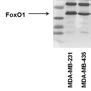
Application: Western BlotSample Tested: Human cancer cell whole cell lysateSpecies: HumanVerified Customer | Posted 08/23/2015FoxO1 expression in human breast cancer cell lines. NBP2-31376

There are no reviews that match your criteria.
Protocols
Find general support by application which include: protocols, troubleshooting, illustrated assays, videos and webinars.
- 7-Amino Actinomycin D (7-AAD) Cell Viability Flow Cytometry Protocol
- Antigen Retrieval Protocol (PIER)
- Antigen Retrieval for Frozen Sections Protocol
- Appropriate Fixation of IHC/ICC Samples
- Cellular Response to Hypoxia Protocols
- Chromogenic IHC Staining of Formalin-Fixed Paraffin-Embedded (FFPE) Tissue Protocol
- Chromogenic Immunohistochemistry Staining of Frozen Tissue
- ClariTSA™ Fluorophore Kits
- Detection & Visualization of Antibody Binding
- Extracellular Membrane Flow Cytometry Protocol
- Flow Cytometry Protocol for Cell Surface Markers
- Flow Cytometry Protocol for Staining Membrane Associated Proteins
- Flow Cytometry Staining Protocols
- Flow Cytometry Troubleshooting Guide
- Fluorescent IHC Staining of Frozen Tissue Protocol
- Graphic Protocol for Heat-induced Epitope Retrieval
- Graphic Protocol for the Preparation and Fluorescent IHC Staining of Frozen Tissue Sections
- Graphic Protocol for the Preparation and Fluorescent IHC Staining of Paraffin-embedded Tissue Sections
- Graphic Protocol for the Preparation of Gelatin-coated Slides for Histological Tissue Sections
- ICC Cell Smear Protocol for Suspension Cells
- ICC Immunocytochemistry Protocol Videos
- ICC for Adherent Cells
- IHC Sample Preparation (Frozen sections vs Paraffin)
- Immunocytochemistry (ICC) Protocol
- Immunocytochemistry Troubleshooting
- Immunofluorescence of Organoids Embedded in Cultrex Basement Membrane Extract
- Immunofluorescent IHC Staining of Formalin-Fixed Paraffin-Embedded (FFPE) Tissue Protocol
- Immunohistochemistry (IHC) and Immunocytochemistry (ICC) Protocols
- Immunohistochemistry Frozen Troubleshooting
- Immunohistochemistry Paraffin Troubleshooting
- Intracellular Flow Cytometry Protocol Using Alcohol (Methanol)
- Intracellular Flow Cytometry Protocol Using Detergents
- Intracellular Nuclear Staining Flow Cytometry Protocol Using Detergents
- Intracellular Staining Flow Cytometry Protocol Using Alcohol Permeabilization
- Intracellular Staining Flow Cytometry Protocol Using Detergents to Permeabilize Cells
- Preparing Samples for IHC/ICC Experiments
- Preventing Non-Specific Staining (Non-Specific Binding)
- Primary Antibody Selection & Optimization
- Propidium Iodide Cell Viability Flow Cytometry Protocol
- Protocol for Heat-Induced Epitope Retrieval (HIER)
- Protocol for Liperfluo
- Protocol for Making a 4% Formaldehyde Solution in PBS
- Protocol for VisUCyte™ HRP Polymer Detection Reagent
- Protocol for the Characterization of Human Th22 Cells
- Protocol for the Characterization of Human Th9 Cells
- Protocol for the Fluorescent ICC Staining of Cell Smears - Graphic
- Protocol for the Fluorescent ICC Staining of Cultured Cells on Coverslips - Graphic
- Protocol for the Preparation & Fixation of Cells on Coverslips
- Protocol for the Preparation and Chromogenic IHC Staining of Frozen Tissue Sections
- Protocol for the Preparation and Chromogenic IHC Staining of Frozen Tissue Sections - Graphic
- Protocol for the Preparation and Chromogenic IHC Staining of Paraffin-embedded Tissue Sections
- Protocol for the Preparation and Chromogenic IHC Staining of Paraffin-embedded Tissue Sections - Graphic
- Protocol for the Preparation and Fluorescent ICC Staining of Cells on Coverslips
- Protocol for the Preparation and Fluorescent ICC Staining of Non-adherent Cells
- Protocol for the Preparation and Fluorescent ICC Staining of Stem Cells on Coverslips
- Protocol for the Preparation and Fluorescent IHC Staining of Frozen Tissue Sections
- Protocol for the Preparation and Fluorescent IHC Staining of Paraffin-embedded Tissue Sections
- Protocol for the Preparation of Gelatin-coated Slides for Histological Tissue Sections
- Protocol for the Preparation of a Cell Smear for Non-adherent Cell ICC - Graphic
- Protocol: Annexin V and PI Staining by Flow Cytometry
- Protocol: Annexin V and PI Staining for Apoptosis by Flow Cytometry
- R&D Systems Quality Control Western Blot Protocol
- TUNEL and Active Caspase-3 Detection by IHC/ICC Protocol
- The Importance of IHC/ICC Controls
- Troubleshooting Guide: Fluorokine Flow Cytometry Kits
- Troubleshooting Guide: Immunohistochemistry
- Troubleshooting Guide: Western Blot Figures
- Western Blot Conditions
- Western Blot Protocol
- Western Blot Protocol for Cell Lysates
- Western Blot Troubleshooting
- Western Blot Troubleshooting Guide
- View all Protocols, Troubleshooting, Illustrated assays and Webinars
Loading...
Associated Pathways


IL-4 Signaling Pathways
 IL-7 Signaling Pathways
 IL-9 Signaling Pathways
 IL-15 Signaling Pathways
 IL-21 Signaling Pathways
 MAPK Signaling: Oxidative Stress Pathway
 TGF-beta Signaling Pathways
 VEGF - VEGF R2 Signaling Pathways
