CCR5 is a G protein-linked seven transmembrane domain chemokine receptor. CCR5 serves as a receptor for several chemokines including MIP-1 alpha, MIP-1 beta, MCP-2, and RANTES. It also functions as a coreceptor for Macrophage Tropic HIV-1 infection.

Key Product Details
Species Reactivity
Validated:
Human
Cited:
Human
Applications
Validated:
Western Blot, Flow Cytometry, CyTOF-ready
Cited:
Western Blot, Flow Cytometry, Immunocytochemistry, Competitive Binding Assay, ELISA Development, Functional Assay
Label
Unconjugated
Antibody Source
Monoclonal Mouse IgG1 Clone # CTC5
Loading...
Product Specifications
Immunogen
CHO Chinese hamster ovary cell line transfected with human CCR5
Specificity
Detects human CCR5 in Western blots. For additional information regarding epitope specificity for this antibody and other R&D Systems anti‑human CCR5 antibodies, see Reference 1.
Clonality
Monoclonal
Host
Mouse
Isotype
IgG1
Scientific Data Images for Human CCR5 Antibody (CTC5)
Detection of CCR5 in Human Peripheral Blood Lymphocytes by Flow Cytometry.
Human peripheral blood lymphocytes were stained with (A) Mouse Anti-Human CCR5 Monoclonal Antibody (Catalog # MAB1802) or (B) control antibody (Catalog # MAB002), followed by Phycoerythrin-conjugated Anti-Mouse IgG Secondary Antibody (Catalog # F0102B) and Mouse anti-Human CD3 APC-conjugated monoclonal antibody (Catalog # FAB100A). View our protocol for Staining Membrane-associated Proteins.
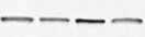
Detection of Human CCR5 by Western Blot
TGF-beta signaling regulated the expression of CCR5(A) 3×10⁵ MDA-MB-231 and MCF-7 cells were stimulated with 1-5ng/ml TGF-beta 1 for 24 h, and total RNA was isolated and tested for CCR5 mRNA by quantitative PCR. (B) Western blot for CCR5 protein in breast cancer cells (106) under TGF-beta 1 stimulation for 48 h. Data presented were representatives of at least three independent experiments. (C) MDA-MB-231 and MCF-7 cells (3×10⁵) were co-transfected with pGL3-CCR5 and pRL-TK and exposed to different concentrations of TGF-beta 1 for 24 h, and luciferase activities were determined. (D) MDA-MB-231 and MCF-7 cells were pre-treated with 5μM SIS3 for 2 h, and cells were subjected to luciferase assay. (E) 106 MCF-7 cells were transfected with TGF beta RI/ALK5 siRNA, and were then co-cultured with lactate-activated THP-1 macrophages (ratio 1:1) for 24 h. The protein levels of CCR5 were assayed by western blot. (F) The expression of TGF-beta 1, CCL5 and CCR5 in clinical samples obtained from breast cancer patients. The mRNA levels were measured by quantitative PCR, and the correlation between TGF-beta 1 and CCL5-CCR5 axis was shown. (G) Representative IHC staining for TGF-beta 1, CCL5 and CCR5 in breast cancer samples. The sample used was derived from 28 breast cancer cases. Scale bars represent 50 μm. *, P<0.05; **, P<0.01. Image collected and cropped by CiteAb from the following publication (https://www.oncotarget.com/lookup/doi/10.18632/oncotarget.22786), licensed under a CC-BY license. Not internally tested by R&D Systems.
Detection of Human CCR5 by Immunohistochemistry
CCL5-CCR5 axis induced aerobic glycolysis by regulation of AMPK signaling(A) Western blot for AMPK, c-Myc, HIF-1 alpha and Akt in breast cancer cells co-cultured with 15mM lactic acid-activated THP-1 macrophages (ratio 1:1) for 72 h. Results presented were representatives of at least three independent experiments. (B) The expression of AMPK downstream signaling target ACC in breast cancer cells co-cultured as in (A). (C) MDA-MB-231 and MCF-7 cells were transfected with 50 nM AMPK alpha 1 siRNA, or pretreated with 10μM compound C for 4 h, and then incubated with 15mM lactic acid-activated THP-1 macrophages (ratio 1:1) for 48 h. The glucose uptake, lactic acid production and ATP levels were detected. (D) The inhibition of AMPK abrogated macrophage-induced EMT in MCF-7 cells. Cells were treated as described in (C). After co-culture, the expression of EMT markers, E-cadherin and vimentin, was measured by western blot. (E) Recombinant human CCL5 induced the phosphorylation of AMPK in MDA-MB-231 and MCF-7/CCR5 cells. 106 cells were treated with 50ng/ml CCL5 for defferent time points as indicated, and phosphorylated AMPK and total AMPK were investigated by western blot. (F) Inhibition of CCR5 in MDA-MB-231 cells significantly attenuated macrophage-induced AMPK phosphorylation. MDA-MB-231 cells were transfected with shRNAs designed against CCR5, or pre-treated with 5μM Maraviroc for 2 h, then co-cultured with 15 mM lactate-activated macrophages as described in (A). After co-culture, the phosphorylation of AMPK was detected by western blot. (G) Expressions of CCL5, CCR5 and p-AMPK in samples obtained from breast cancer patients (n =28). Scale bars represent 50 μm. *, P<0.05; **, P<0.01. Image collected and cropped by CiteAb from the following publication (https://www.oncotarget.com/lookup/doi/10.18632/oncotarget.22786), licensed under a CC-BY license. Not internally tested by R&D Systems.
Detection of Human CCR5 by Western Blot
Lactate-activated macrophages induced EMT in breast cancer cells through CCL5-CCR5 axis(A) 106 THP-1 macrophages were treated with 15 mM lactate for 72 h, and then cells were washed twice and fresh media were added. Macrophages were cultured for another 24 h and the conditional media (lactate CM) was collected. The effect of CM on breast cancer cell migration was measured by double chamber transwell assay. 5μg/ml anti-CCL5 neutralizing antibody significantly decreased lactate CM-induced cell migration. (B) 106 MCF-7 cells were co-cultured with 15 mM lactate-activated macrophages in the presence of 5μg/ml anti-CCL5 antibody or not, and protein levels of EMT markers were tested by western blot. (C) 106 breast cancer cells were co-cultured with 106 lactate-activated THP-1 macrophages (or 106 lactate-activated primary macrophages) for different time points, and the expression of CCR5 was monitored by western blot. (D) MDA-MB-231 and MCF-7 cells were transfected with shCCR5 plasmids, or pre-treated with 5μM Maraviroc for 2 h, then cell migration induced by lactate CM was detected by double chamber transwell assay. Lactate CM was described in (A). (E) MCF-7 cells (106) were transfected with pcDNA3.1-CCR5, and then cultured with 10ng/ml CCL5 for 24 h. The expression of E-cadherin, N-cadherin and vimentin was investigated by western blot. (F) 106 Human primary macrophages (No. 4 and No. 9) were treated with 15 mM lactate for 72 h and CM was collected as described in (A). The migration of MDA-MB-231 cells was measured in the presence of primary macrophage CM. 5μg/ml anti-CCL5 neutralizing antibody, shRNAs designed against CCR5, or 5μM Maraviroc, significantly reduced primary macrophage CM-induced cell migration. *, P<0.05; **, P<0.01. Image collected and cropped by CiteAb from the following publication (https://www.oncotarget.com/lookup/doi/10.18632/oncotarget.22786), licensed under a CC-BY license. Not internally tested by R&D Systems.
Detection of Human CCR5 by Immunohistochemistry
Macrophages promoted breast cancer metastasis through CCL5(A) MDA-MB-231 cells were co-cultured with 15 mM lactate-activated THP-1 macrophages for 7 days, in the presence of 5μg/ml anti-CCL5 neutralizing antibody or not. MDA-MB-231 cells were then collected and injected into the tail vein of nude mice. After two weeks, animals were sacrificed and metastatic nodules on lung surfaces were counted. (B) CCR5, HK2 and p-AMPK were immunostained in MDA-MB-231 metastases. Scale bars represent 50 μm. *, P<0.05; **, P<0.01. Image collected and cropped by CiteAb from the following publication (https://www.oncotarget.com/lookup/doi/10.18632/oncotarget.22786), licensed under a CC-BY license. Not internally tested by R&D Systems.Applications for Human CCR5 Antibody (CTC5)
Application
Recommended Usage
CyTOF-ready
Ready to be labeled using established conjugation methods. No BSA or other carrier proteins that could interfere with conjugation.
Flow Cytometry
0.25 µg/106 cells
Sample: Human peripheral blood lymphocytes
Sample: Human peripheral blood lymphocytes
Western Blot
Lee, B. et al. (1999) J. Biol. Chem. 274:9617. This application was not tested by R&D Systems.
Reviewed Applications
Read 3 reviews rated 4.7 using MAB1802 in the following applications:
Flow Cytometry Panel Builder
Bio-Techne Knows Flow Cytometry
Save time and reduce costly mistakes by quickly finding compatible reagents using the Panel Builder Tool.
Advanced Features
- Spectra Viewer - Custom analysis of spectra from multiple fluorochromes
- Spillover Popups - Visualize the spectra of individual fluorochromes
- Antigen Density Selector - Match fluorochrome brightness with antigen density

Formulation, Preparation, and Storage
Purification
Protein A or G purified from hybridoma culture supernatant
Reconstitution
Reconstitute at 0.5 mg/mL in sterile PBS. For liquid material, refer to CoA for concentration.
Loading...
Formulation
Lyophilized from a 0.2 μm filtered solution in PBS with Trehalose. *Small pack size (SP) is supplied either lyophilized or as a 0.2 µm filtered solution in PBS.
Shipping
Lyophilized product is shipped at ambient temperature. Liquid small pack size (-SP) is shipped with polar packs. Upon receipt, store immediately at the temperature recommended below.
Stability & Storage
Use a manual defrost freezer and avoid repeated freeze-thaw cycles.
- 12 months from date of receipt, -20 to -70 °C as supplied.
- 1 month, 2 to 8 °C under sterile conditions after reconstitution.
- 6 months, -20 to -70 °C under sterile conditions after reconstitution.
Calculators
Background: CCR5
References
- Lee, B. et al. (1999) J. Biol. Chem. 274:9617.
Alternate Names
CCR5, CD195
Gene Symbol
CCR5
Additional CCR5 Products
Product Documents for Human CCR5 Antibody (CTC5)
Certificate of Analysis
To download a Certificate of Analysis, please enter a lot or batch number in the search box below.
Note: Certificate of Analysis not available for kit components.
Product Specific Notices for Human CCR5 Antibody (CTC5)
For research use only
Citations for Human CCR5 Antibody (CTC5)
 Powered by Bioz
Customer Reviews for Human CCR5 Antibody (CTC5) (3)
4.7 out of 5
3 Customer Ratings
Have you used Human CCR5 Antibody (CTC5)?
Submit a review and receive an Amazon gift card!
$25/€18/£15/$25CAN/¥2500 Yen for a review with an image
$10/€7/£6/$10CAN/¥1110 Yen for a review without an image
Submit a review
Customer Images
Showing
1
-
3 of
3 reviews
Showing All
Filter By:
-
Application: Western BlotSample Tested: Dendritic cellsSpecies: HumanVerified Customer | Posted 11/29/2021

-
Application: Western BlotSample Tested: U937 human histiocytic lymphoma cell lineSpecies: HumanVerified Customer | Posted 06/28/2017
-
Application: Flow CytometrySample Tested: See PMID 22811524Species: HumanVerified Customer | Posted 02/10/2015
There are no reviews that match your criteria.
Protocols
Find general support by application which include: protocols, troubleshooting, illustrated assays, videos and webinars.
- 7-Amino Actinomycin D (7-AAD) Cell Viability Flow Cytometry Protocol
- Cellular Response to Hypoxia Protocols
- Extracellular Membrane Flow Cytometry Protocol
- Flow Cytometry Protocol for Cell Surface Markers
- Flow Cytometry Protocol for Staining Membrane Associated Proteins
- Flow Cytometry Staining Protocols
- Flow Cytometry Troubleshooting Guide
- Intracellular Flow Cytometry Protocol Using Alcohol (Methanol)
- Intracellular Flow Cytometry Protocol Using Detergents
- Intracellular Nuclear Staining Flow Cytometry Protocol Using Detergents
- Intracellular Staining Flow Cytometry Protocol Using Alcohol Permeabilization
- Intracellular Staining Flow Cytometry Protocol Using Detergents to Permeabilize Cells
- Propidium Iodide Cell Viability Flow Cytometry Protocol
- Protocol for Liperfluo
- Protocol for the Characterization of Human Th22 Cells
- Protocol for the Characterization of Human Th9 Cells
- Protocol: Annexin V and PI Staining by Flow Cytometry
- Protocol: Annexin V and PI Staining for Apoptosis by Flow Cytometry
- R&D Systems Quality Control Western Blot Protocol
- Troubleshooting Guide: Fluorokine Flow Cytometry Kits
- Troubleshooting Guide: Western Blot Figures
- Western Blot Conditions
- Western Blot Protocol
- Western Blot Protocol for Cell Lysates
- Western Blot Troubleshooting
- Western Blot Troubleshooting Guide
- View all Protocols, Troubleshooting, Illustrated assays and Webinars
Loading...
Associated Pathways
