CD47 (also integrin-associated protein/IAP and OA3) is a variably glycosylated, 40‑60 kDa atypical member of the Ig-Superfamily. It is expressed on almost all cell types, including erythrocytes. CD47 binds to TSP-1 and SIRP alpha, and forms a membrane complex with CD36 and alpha v beta 3. Mature human CD47 is a 305 amino acid (aa), five-transmembrane glycoprotein. It contains a 123 aa extracellular region (aa 19‑141) that is characterized by the presence of a V-type Ig-like domain (aa 19‑127), and a 34 aa C-terminal cytoplasmic tail that interacts with Gi alpha subunits. Three splice variants occur over aa 293‑323. Over aa 19‑139, human CD47 shares 61%, 71% and 66% aa identity with mouse, porcine and canine CD47, respectively.

Key Product Details
Validated by
Knockout/Knockdown, Biological Validation
Species Reactivity
Validated:
Human
Cited:
Human, Mouse
Applications
Validated:
Knockout Validated, Immunohistochemistry, Western Blot, Neutralization, Flow Cytometry, CyTOF-ready
Cited:
Immunohistochemistry, Immunohistochemistry-Paraffin, Western Blot, Neutralization, Immunocytochemistry, Dot Blot
Label
Unconjugated
Antibody Source
Polyclonal Sheep IgG
Loading...
Product Specifications
Immunogen
Mouse myeloma cell line NS0-derived recombinant human CD47
Gln19-Pro139
Accession # Q08722
Gln19-Pro139
Accession # Q08722
Specificity
Detects human CD47 in direct ELISAs and Western blots. In direct ELISAs, less than 5% cross-reactivity with recombinant mouse CD47 is observed.
Clonality
Polyclonal
Host
Sheep
Isotype
IgG
Endotoxin Level
<0.10 EU per 1 μg of the antibody by the LAL method.
Scientific Data Images for Human CD47 Antibody
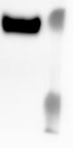
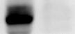
Detection of Human CD47 by Western Blot.
fWestern blot shows lysates of U937 human histiocytic lymphoma cell line and human placenta tissue, not heated to minimize aggregation. PVDF membrane was probed with 1 µg/mL of Sheep Anti-Human CD47 Antigen Affinity-purified Polyclonal Antibody (Catalog # AF4670) followed by HRP-conjugated Anti-Sheep IgG Secondary Antibody (Catalog # HAF016). Specific bands were detected for CD47 at approximately 45-70 kDa (as indicated). This experiment was conducted under reducing conditions and using Immunoblot Buffer Group 1.
Detection of CD47 in Human Lymphocytes by Flow Cytometry.
Human whole blood lymphocytes were stained with Sheep Anti-Human CD47 Antigen Affinity-purified Polyclonal Antibody (Catalog # AF4670, filled histogram) or control antibody (Catalog # 5-001-A, open histogram), followed by Northern-Lights™ 557-conjugated Anti-Sheep IgG Secondary Antibody (Catalog # NL010).
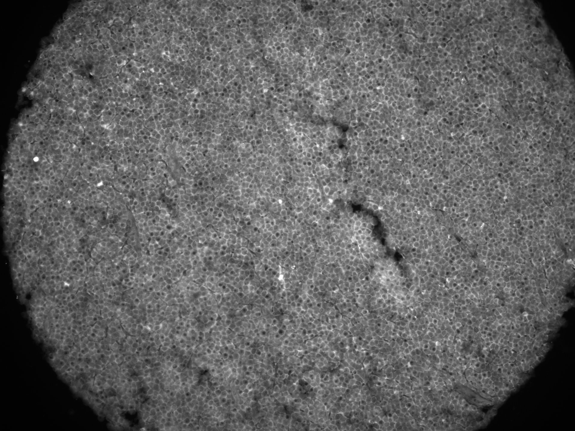
CD47 in Human Placenta.
CD47 was detected in immersion fixed paraffin-embedded sections of human placenta using 5 µg/mL Sheep Anti-Human CD47 Antigen Affinity-purified Polyclonal Antibody (Catalog # AF4670) overnight at 4 °C. Tissue was stained with the Anti-Sheep HRP-DAB Cell & Tissue Staining Kit (brown; Catalog # CTS019) and counterstained with hematoxylin (blue). View our protocol for Chromogenic IHC Staining of Paraffin-embedded Tissue Sections.
Cell Adhesion Mediated by SIRP alpha /CD172a and Neutral-ization by Human CD47 Antibody.
Recombinant Human SIRPa/CD172a (Catalog # 4546-SA), immobilized onto a microplate, supports the adhesion of the human erythrocytes in a dose-dependent manner (orange line). Adhesion elicited by Recombinant Human SIRP-a (2 µg/mL) is neutralized (green line) by increasing concentrations of Sheep Anti-Human CD47 Poly-clonal Antibody (Catalog # AF4670). The adhesion was maximally inhibited (70-100%) by 2 µg/mL of the antibody.
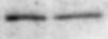
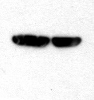
Western Blot Shows Human CD47 Specificity by Using Knockout Cell Line.
Western blot shows lysates of HEK293T human embryonic kidney parental cell line and CD47 knockout HEK293T cell line (KO). PVDF membrane was probed with 1 µg/mL of Sheep Anti-Human CD47 Antigen Affinity-purified Polyclonal Antibody (Catalog # AF4670) followed by HRP-conjugated Anti-Sheep IgG Secondary Antibody (Catalog # HAF016). Specific bands were detected for CD47 at approximately 50 kDa (as indicated) in the parental HEK293T cell line, but is not detectable in knockout HEK293T cell line. GAPDH (Catalog # AF5718) is shown as a loading control. This experiment was conducted under reducing conditions and using Immunoblot Buffer Group 1.
Detection of Human CD47 by Western Blot
Increased CD47 expression in aging and replicative senescence. (A) Quantification of relative CD47 mRNA levels by qPCR. Expression levels of senescent cells were normalized to the respective proliferating control (white bar). Data are representative of three independent experiments. All values are means ± SEM. **P < 0.005, ***P < 0.0005, ****P < 0.0001. Statistically significant differences were determined by unpaired Student’s t test. (B) Representative immunofluorescence images from indicated cell types stained for CD47 (green) and Hoechst (blue). Scale bar: 20 µm, except for 3T3 cells: 50 µm. (C) Whole-cell lysates from indicated cell types were isolated and analyzed by Western blotting for the indicated proteins. Senescence was induced by chemical treatment (palbociclib, except for A549: etopsoside). Shown are representative blots of three independent experiments. GAPDH images are derived from the same blot as Fig. S3 (for Panc1 cells the same GAPDH loading control is additionally used in Fig. 8 G). (D) Representative immunofluorescence images of proliferating or senescent small epithelial cells (SAEC) stained for CD47 (green) and Hoechst (blue). scale bars indicate 20 µm. n = 2–3. (E) Western blot analysis of CD47 expression in SAEC cells. Senescence was induced by chemical treatment (palbociclib). GAPDH was used as loading control. n = 2. GAPDH images are derived from the same blot as Fig. S3. (F) Representative immunofluorescence images of primary lung fibroblast (proliferative or replicative senescent by serially passaging; normal healthy [NHLF] or IPF-derived [IPF]) showing CD47 (green) and Hoechst (blue) staining. scale bars indicate 20 µm. n = 2. (G) Whole-cell lysates from primary NHLF or IPF lung fibroblasts were isolated and analyzed by Western blotting for the indicated proteins. Senescence was induced by replicative passing. Shown are representative blots of three to four independent experiments. Source data are available for this figure: SourceData
Detection of Human CD47 by Immunocytochemistry/Immunofluorescence
Adhesion of EpCAMpos/neg cells on multi-marker arrays.2x105 EpCAMpos (cell pool of MCF7, SKBR3, HCC1500, ZR-75-1, TMX2-28) (A) and EpCAMlow/neg cells (MDA-MB-231) (B) were either stained with 1 μM MitoTracker Green FM (A) or MitoTracker Orange CM (B) and incubated for cell adhesion experiments on NEXTERION slides AL, coated with different antibodies and ECM molecules, alone and in combination (0.1 mg/ml each). The labeling above the single spots indicates respective capture molecules (Iso = isotype control, ms = mouse, Lam = laminin, Col = collagen, HA = hyaluronic acid, rt = rat, T = Trop2, EpC = EpCAM, 49f = CD49f, rb = rabbit). (C) Overlay image of (A) and (B); scale bars (white) = 500 μm, 20x magnification. (D) Array layout (5x5 mm) with 36 spots (spot diameter = 500 μm; pitch = 800 μm) printed on NEXTERION slides AL. Image collected and cropped by CiteAb from the following publication (https://pubmed.ncbi.nlm.nih.gov/26695635), licensed under a CC-BY license. Not internally tested by R&D Systems.
Detection of Human CD47 by Western Blot
Impairment of efferocytosis by senescent cells is mediated by CD47 and CD24 expression. (G) Whole-cell lysates derived from Panc1 cells (WT and CD47 KO) were analyzed by Western blotting for the indicated proteins. GAPDH was used as loading control. Senescence was induced by chemical treatment (Palbociclib). Blots are representative of three independent experiments. GAPDH image is derived from the same blot as Fig. 7 and Fig. S3. CD24 image is derived from the same blot as in Fig. S3. Image collected and cropped by CiteAb from the following open publication (https://pubmed.ncbi.nlm.nih.gov/36459066), licensed under a CC-BY license. Not internally tested by R&D Systems.Applications for Human CD47 Antibody
Application
Recommended Usage
CyTOF-ready
Ready to be labeled using established conjugation methods. No BSA or other carrier proteins that could interfere with conjugation.
Flow Cytometry
2.5 µg/106 cells
Sample: Human whole blood lymphocytes
Sample: Human whole blood lymphocytes
Immunohistochemistry
5-15 µg/mL
Sample: Immersion fixed paraffin-embedded sections of human placenta
Sample: Immersion fixed paraffin-embedded sections of human placenta
Knockout Validated
CD47
is specifically detected in HEK293T human embryonic kidney parental cell line but is not detectable in
CD47 knockout HEK293T cell line.
Western Blot
1 µg/mL
Sample: U937 human histiocytic lymphoma cell line and human placenta tissue, not heated to minimize aggregation
Sample: U937 human histiocytic lymphoma cell line and human placenta tissue, not heated to minimize aggregation
Neutralization
Measured by its ability to neutralize SIRP alpha /CD172a-mediated adhesion of human erythrocytes. The adhesion of human erythrocytes to immobilized Recombinant Human SIRP alpha /CD172a/Fc Chimera (2 µg/mL, 100 µL/well) was maximally inhibited (70-100%) by 2 µg/mL of the antibody.
Reviewed Applications
Read 7 reviews rated 4.6 using AF4670 in the following applications:
Flow Cytometry Panel Builder
Bio-Techne Knows Flow Cytometry
Save time and reduce costly mistakes by quickly finding compatible reagents using the Panel Builder Tool.
Advanced Features
- Spectra Viewer - Custom analysis of spectra from multiple fluorochromes
- Spillover Popups - Visualize the spectra of individual fluorochromes
- Antigen Density Selector - Match fluorochrome brightness with antigen density

Formulation, Preparation, and Storage
Purification
Antigen Affinity-purified
Reconstitution
Reconstitute at 0.2 mg/mL in sterile PBS. For liquid material, refer to CoA for concentration.
Loading...
Formulation
Lyophilized from a 0.2 μm filtered solution in PBS with Trehalose. *Small pack size (SP) is supplied either lyophilized or as a 0.2 µm filtered solution in PBS.
Shipping
Lyophilized product is shipped at ambient temperature. Liquid small pack size (-SP) is shipped with polar packs. Upon receipt, store immediately at the temperature recommended below.
Stability & Storage
Use a manual defrost freezer and avoid repeated freeze-thaw cycles.
- 12 months from date of receipt, -20 to -70 °C as supplied.
- 1 month, 2 to 8 °C under sterile conditions after reconstitution.
- 6 months, -20 to -70 °C under sterile conditions after reconstitution.
Calculators
Background: CD47
Alternate Names
CD47, IAP, MER6, OA3
Entrez Gene IDs
Gene Symbol
CD47
UniProt
Additional CD47 Products
Product Documents for Human CD47 Antibody
Certificate of Analysis
To download a Certificate of Analysis, please enter a lot or batch number in the search box below.
Note: Certificate of Analysis not available for kit components.
Product Specific Notices for Human CD47 Antibody
For research use only
Citations for Human CD47 Antibody
 Powered by Bioz
Customer Reviews for Human CD47 Antibody (7)
4.6 out of 5
7 Customer Ratings
Have you used Human CD47 Antibody?
Submit a review and receive an Amazon gift card!
$25/€18/£15/$25CAN/¥2500 Yen for a review with an image
$10/€7/£6/$10CAN/¥1110 Yen for a review without an image
Submit a review
Customer Images

Showing
1
-
5 of
7 reviews
Showing All
Filter By:
-
Application: Western BlotSample Tested: BT549 and MDA-231 cell lysateSpecies: HumanVerified Customer | Posted 04/03/2020

-
Application: Western BlotSample Tested: BT-20 human breast cancer cell lineSpecies: HumanVerified Customer | Posted 12/27/2019
-
Application: Western BlotSample Tested: A549 human lung carcinoma cell lineSpecies: HumanVerified Customer | Posted 05/20/2019
-
Application: Immunofluorescence - paraffinSample Tested: Chronic lymphocytic leukemia/small lymphocytic lymphoma (CLL/SLL)Species: HumanVerified Customer | Posted 03/09/2019Human CLL/SLL stained with anti-CD47 1:50pH9 heat-induced antigen retrieval
-
Application: Western BlotSample Tested: BT549 and BT-20 human breast cancer cell lineSpecies: HumanVerified Customer | Posted 10/17/2018
-
Application: Western BlotSample Tested: Breast cancer cellsSpecies: HumanVerified Customer | Posted 10/17/2018
-
Application: Western BlotSample Tested: HepG2 human hepatocellular carcinoma cell lineSpecies: HumanVerified Customer | Posted 07/15/2018
There are no reviews that match your criteria.
Protocols
Find general support by application which include: protocols, troubleshooting, illustrated assays, videos and webinars.
- 7-Amino Actinomycin D (7-AAD) Cell Viability Flow Cytometry Protocol
- Antigen Retrieval Protocol (PIER)
- Antigen Retrieval for Frozen Sections Protocol
- Appropriate Fixation of IHC/ICC Samples
- Cellular Response to Hypoxia Protocols
- Chromogenic IHC Staining of Formalin-Fixed Paraffin-Embedded (FFPE) Tissue Protocol
- Chromogenic Immunohistochemistry Staining of Frozen Tissue
- ClariTSA™ Fluorophore Kits
- Detection & Visualization of Antibody Binding
- Extracellular Membrane Flow Cytometry Protocol
- Flow Cytometry Protocol for Cell Surface Markers
- Flow Cytometry Protocol for Staining Membrane Associated Proteins
- Flow Cytometry Staining Protocols
- Flow Cytometry Troubleshooting Guide
- Fluorescent IHC Staining of Frozen Tissue Protocol
- Graphic Protocol for Heat-induced Epitope Retrieval
- Graphic Protocol for the Preparation and Fluorescent IHC Staining of Frozen Tissue Sections
- Graphic Protocol for the Preparation and Fluorescent IHC Staining of Paraffin-embedded Tissue Sections
- Graphic Protocol for the Preparation of Gelatin-coated Slides for Histological Tissue Sections
- IHC Sample Preparation (Frozen sections vs Paraffin)
- Immunofluorescent IHC Staining of Formalin-Fixed Paraffin-Embedded (FFPE) Tissue Protocol
- Immunohistochemistry (IHC) and Immunocytochemistry (ICC) Protocols
- Immunohistochemistry Frozen Troubleshooting
- Immunohistochemistry Paraffin Troubleshooting
- Intracellular Flow Cytometry Protocol Using Alcohol (Methanol)
- Intracellular Flow Cytometry Protocol Using Detergents
- Intracellular Nuclear Staining Flow Cytometry Protocol Using Detergents
- Intracellular Staining Flow Cytometry Protocol Using Alcohol Permeabilization
- Intracellular Staining Flow Cytometry Protocol Using Detergents to Permeabilize Cells
- Preparing Samples for IHC/ICC Experiments
- Preventing Non-Specific Staining (Non-Specific Binding)
- Primary Antibody Selection & Optimization
- Propidium Iodide Cell Viability Flow Cytometry Protocol
- Protocol for Heat-Induced Epitope Retrieval (HIER)
- Protocol for Liperfluo
- Protocol for Making a 4% Formaldehyde Solution in PBS
- Protocol for VisUCyte™ HRP Polymer Detection Reagent
- Protocol for the Characterization of Human Th22 Cells
- Protocol for the Characterization of Human Th9 Cells
- Protocol for the Preparation & Fixation of Cells on Coverslips
- Protocol for the Preparation and Chromogenic IHC Staining of Frozen Tissue Sections
- Protocol for the Preparation and Chromogenic IHC Staining of Frozen Tissue Sections - Graphic
- Protocol for the Preparation and Chromogenic IHC Staining of Paraffin-embedded Tissue Sections
- Protocol for the Preparation and Chromogenic IHC Staining of Paraffin-embedded Tissue Sections - Graphic
- Protocol for the Preparation and Fluorescent IHC Staining of Frozen Tissue Sections
- Protocol for the Preparation and Fluorescent IHC Staining of Paraffin-embedded Tissue Sections
- Protocol for the Preparation of Gelatin-coated Slides for Histological Tissue Sections
- Protocol: Annexin V and PI Staining by Flow Cytometry
- Protocol: Annexin V and PI Staining for Apoptosis by Flow Cytometry
- R&D Systems Quality Control Western Blot Protocol
- TUNEL and Active Caspase-3 Detection by IHC/ICC Protocol
- The Importance of IHC/ICC Controls
- Troubleshooting Guide: Fluorokine Flow Cytometry Kits
- Troubleshooting Guide: Immunohistochemistry
- Troubleshooting Guide: Western Blot Figures
- Western Blot Conditions
- Western Blot Protocol
- Western Blot Protocol for Cell Lysates
- Western Blot Troubleshooting
- Western Blot Troubleshooting Guide
- View all Protocols, Troubleshooting, Illustrated assays and Webinars
Loading...
Associated Pathways
