
Human Complement Component C9 Antibody (887025)
R&D Systems | Catalog # MAB8126

Scientific images may be resized, cropped, or adjusted for brightness or contrast for presentation purposes. These updates are limited to presentation and do not affect the underlying scientific interpretation of the data. Where available, additional source data may be provided upon request.
Key Product Details
Species Reactivity
Applications
Label
Antibody Source
Product Specifications
Immunogen
Met1-Lys559
Accession # P02748
Specificity
Clonality
Host
Isotype
Scientific Data Images for Human Complement Component C9 Antibody (887025)
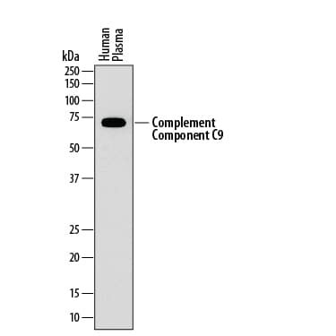
Detection of Human Complement Component C9 by Western Blot.
Western blot shows human plasma. PVDF membrane was probed with 1 µg/mL of Mouse Anti-Human Complement Component C9 Monoclonal Antibody (Catalog # MAB8126) followed by HRP-conjugated Anti-Mouse IgG Secondary Antibody (Catalog # HAF018). A specific band was detected for Complement Component C9 at approximately 70 kDa (as indicated). This experiment was conducted under reducing conditions and using Immunoblot Buffer Group 1.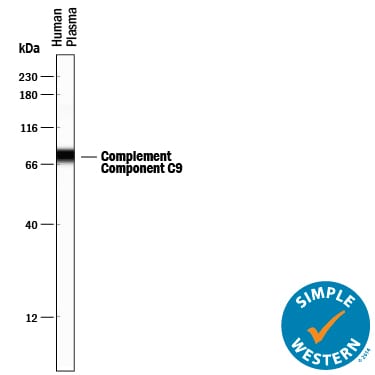
Detection of Human Complement Component C9 by Simple WesternTM.
Simple Western lane view shows human plasma, loaded at 0.5 mg/mL. A specific band was detected for Complement Component C9 at approximately 78 kDa (as indicated) using 10 µg/mL of Mouse Anti-Human Complement Component C9 Monoclonal Antibody (Catalog # MAB8126). This experiment was conducted under reducing conditions and using the 12-230 kDa separation system.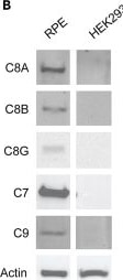
Detection of bjizl0bbottag9zqg7rj.jpg by Western Blot
Pathogenicity and effect of variants on protein function(A) Representation of the structure of MAC and C8 complex. The schematic representation of MAC shows the location of the identified variants in protein complex. 3D visualization of C8 complex showing the three subunits (C8 alpha – green, C8 beta – teal, C8 gamma – magenta). Variants are indicated by red residues, and WT residues are in yellow. Potential hydrogen bonds formed by residues are indicated by dashed yellow lines. Hydrogen bonds gained by introduction of variants are marked by white arrows, and hydrogen bonds lost are marked by a dashed white ellipsoid.(B) Immunoblots showing expression of C8 and its direct partners within the membrane attack complex (MAC) in human retinal pigment epithelium (RPE) and lack of expression in HEK293 cells.(C) Co-IP analyses on the interaction between C8 alpha, beta, and gamma subunits in protein lysates from HEK293 cells.(D and E) Quantification of IP signal in lysates from HEK293 cells. Relative amounts of C8 subunits are plotted on a Jitter plot. Individual measurements are marked by a dot, and the quadrant range is indicated by a box. Transfection (repeated in triplicates) conditions are indicated by a “+” under each panel. Quantification of C8 alpha, beta, and gamma subunits is indicated in green, teal and magenta, respectively. Wilcoxon test was done to assess statistical significance, and comparison approaching significance are indicated. Image collected and cropped by CiteAb from the following open publication (https://pubmed.ncbi.nlm.nih.gov/37153444), licensed under a CC-BY license. Not internally tested by R&D Systems.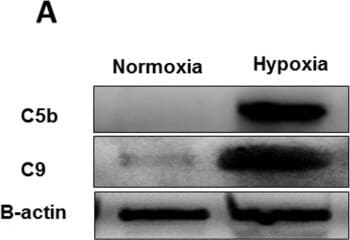
Detection of jw6qbcnnnxxxzw7bdjde.jpg by Western Blot
Molecular mechanisms of MAC assembly in severe hypoxia-exposed MSCs. MSCs were exposed to hypoxia for 24 h. A C5b and C9 levels were analyzed using Western blot. beta actin was used for normalization of protein loading. The levels of both B C5b and C C9 increased significantly in hypoxic MSCs compared to MSCs under normoxia. (*p < 0.05 vs. control, n = 3) Image collected and cropped by CiteAb from the following open publication (https://pubmed.ncbi.nlm.nih.gov/39485618), licensed under a CC-BY license. Not internally tested by R&D Systems.Applications for Human Complement Component C9 Antibody (887025)
Simple Western
Sample: Human plasma
Western Blot
Sample: Human plasma
Reviewed Applications
Read 1 review rated 2 using MAB8126 in the following applications:
Formulation, Preparation, and Storage
Purification
Reconstitution
Reconstitute at 0.5 mg/mL in sterile PBS. For liquid material, refer to CoA for concentration.
Formulation
Shipping
Stability & Storage
- 12 months from date of receipt, -20 to -70 °C as supplied.
- 1 month, 2 to 8 °C under sterile conditions after reconstitution.
- 6 months, -20 to -70 °C under sterile conditions after reconstitution.
Calculators
Background: Complement Component C9
Alternate Names
Gene Symbol
UniProt
Additional Complement Component C9 Products
Product Documents for Human Complement Component C9 Antibody (887025)
Certificate of Analysis
To download a Certificate of Analysis, please enter a lot or batch number in the search box below.
Note: Certificate of Analysis not available for kit components.
Product Specific Notices for Human Complement Component C9 Antibody (887025)
For research use only
Related Research Areas
Citations for Human Complement Component C9 Antibody (887025)
 Powered by Bioz
Customer Reviews for Human Complement Component C9 Antibody (887025) (1)
Have you used Human Complement Component C9 Antibody (887025)?
Submit a review and receive an Amazon gift card!
$25/€18/£15/$25CAN/¥2500 for a review with an image
$10/€7/£6/$10CAN/¥1110 for a review without an image
Submit a review
Customer Images
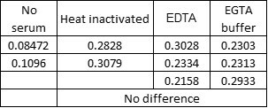
-
Application: complement membrane attack complex (C5b-9) detectionSample Tested: Human serumSpecies: human serumVerified Customer | Posted 01/18/2023unable to detect MAC formation performed by Zymosan activation in human serum
 Bio-Techne ResponseThank you for reviewing our product. We are sorry to hear that this product did not perform as expected. We have been in touch with the customer to resolve this issue according to our Product Guarantee and to the customer’s satisfaction. The customer worked with our Technical Support team and had success detecting the complexed form of Complement Component C9 in the complement membrane attack complex (C5b-9) while using a polyclonal antibody for Human Complement Component C9 (AF8126). We have not tested complement membrane attack complex (C5b-9) detection with MAB8126 in-house.
There are no reviews that match your criteria.
Protocols
Find general support by application which include: protocols, troubleshooting, illustrated assays, videos and webinars.
- Cellular Response to Hypoxia Protocols
- R&D Systems Quality Control Western Blot Protocol
- Troubleshooting Guide: Western Blot Figures
- Western Blot Conditions
- Western Blot Protocol
- Western Blot Protocol for Cell Lysates
- Western Blot Troubleshooting
- Western Blot Troubleshooting Guide
- View all Protocols, Troubleshooting, Illustrated assays and Webinars