Goat anti-Mouse IgG Secondary Antibody

R&D Systems | Catalog # AF007

R&D Systems

Key Product Details

Species Reactivity

Mouse

Applications

Western Blot

Label

Unconjugated

Antibody Source

Polyclonal Goat IgG
Loading...

Product Specifications

Immunogen

Mouse IgG

Specificity

Detects mouse IgG

Clonality

Polyclonal

Host

Goat

Isotype

IgG

Scientific Data Images for Goat anti-Mouse IgG Secondary Antibody

Detection of IgG by Western Blot

Detection of IgG by Western Blot

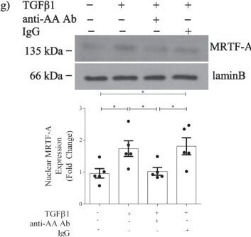
Activin A regulation of MRTF-A contributes to alpha SMA induction by TGF beta 1 in MC. a ActA (20 ng/ml) and TGF beta 1 show an additive effect on alpha SMA promoter transcriptional activation at 24 h (n = 6). Activin inhibition with b follistatin (n = 7) and c an actA neutralizing antibody decrease TGF beta 1-induced alpha SMA promoter transcriptional activity (n = 14–23). d actB neutralization does not decrease TGF beta 1-induced alpha SMA promoter transcriptional activity (n = 6–9). e actA increases MRTF-A nuclear localization at 18 h (n = 5). TGF beta 1-induced nuclear MRTF-A translocation is attenuated with f follistatin (n = 9) and g actA neutralization (n = 5). h ActA-induced alpha SMA promoter transcriptional activity is inhibited by MRTF-A siRNA (n = 9) and i the Smad3 inhibitor SIS3 (n = 3). *, **, ***, ****P < 0.05, 0.01, 0.001, 0.0001; one-way ANOVA with Tukey’s multiple comparisons post hoc test Image collected and cropped by CiteAb from the following open publication (https://pubmed.ncbi.nlm.nih.gov/36717814), licensed under a CC-BY license. Not internally tested by R&D Systems.
Detection of IgG by Western Blot

Detection of IgG by Western Blot

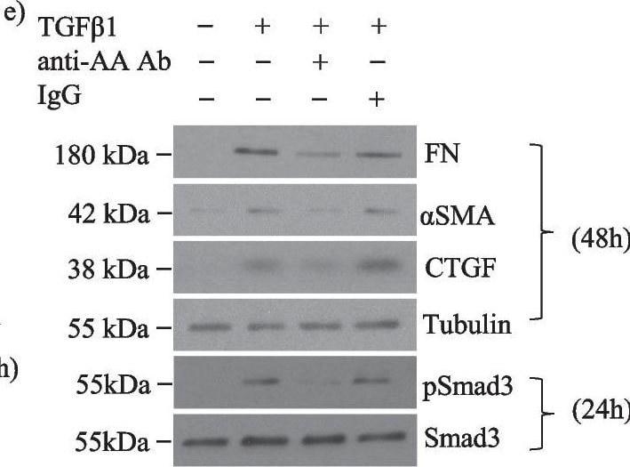
Specific activin A inhibition attenuates TGF beta 1-induced Smad3 activation and profibrotic responses in MC. a ELISA demonstrates that TGF beta 1 (24 h) increases actA and actB secretion (n = 3) to 19.5 ng/ml and 2.5 ng/ml, representing an 8.9- and 1.06-fold induction respectively. b TGF beta 1 increases actA in whole cell lysate by 1.8-fold (n = 3). c ActA (20 ng/ml) upregulates FN (n = 3–4), CTGF (n = 4) and alpha SMA (n = 6) at 48 h. d TGF beta 1 and actA both increase Smad3 transcriptional activity; no synergistic effect is seen (n = 6–12). e An actA neutralizing antibody attenuates TGF beta 1-induced FN (n = 5–6), alpha SMA (n = 5), CTGF (n = 5–6), and Smad3 activation (n = 10–12). f ActA neutralization decreases TGF beta 1-induced Smad3 transcriptional activity at 24 h (n = 9–15), but this is not decreased by actB neutralization (n = 6) (g). h MC were stimulated with TGF beta 1 or actA for 1 h, then treated with their type I receptor inhibitor SB431542 (50 µM). Restimulation with the same ligand shows that cells become refractory to TGF beta 1, but not actA (n = 4). *, **, ***, ****P < 0.05, 0.01, 0.001, 0.0001; one-way ANOVA with Tukey’s multiple comparisons post hoc test Image collected and cropped by CiteAb from the following open publication (https://pubmed.ncbi.nlm.nih.gov/36717814), licensed under a CC-BY license. Not internally tested by R&D Systems.
Detection of IgG by Western Blot

Detection of IgG by Western Blot

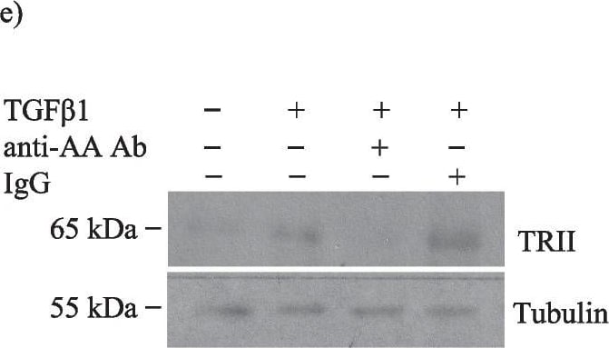
Activins facilitate TGF beta 1 receptor type II upregulation in response to TGF beta 1 in MC. a Follistatin (FST) inhibits the TGF beta 1-induced increase in TRII expression at 24 h in whole cell lysate (n = 10–11). b The TGF beta 1-induced increase in cell surface TRII was diminished by follistatin at 24 h (n = 3). c No change in response to either TGF beta 1 or follistatin were seen with TRI (n = 6). d The TGF beta 1-induced increase in TRII transcript was also diminished by follistatin at 24 h (n = 13–15). e TGF beta 1 induction of TRII expression is reduced with an actA neutralizing antibody (n = 6). *, **, ***, ****P < 0.05, 0.01, 0.001, 0.0001; one-way ANOVA with Tukey’s multiple comparisons post hoc test Image collected and cropped by CiteAb from the following open publication (https://pubmed.ncbi.nlm.nih.gov/36717814), licensed under a CC-BY license. Not internally tested by R&D Systems.
Detection of IgG by Western Blot

Detection of IgG by Western Blot

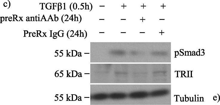
Activin A facilitates TGF beta 1-induced type II receptor expression without altering TGF beta 1 receptor sensitivity in MC. a TGF beta 1 for 30 min increased TRII expression and Smad3 activation in whole cell lysate, and both were prevented by 24 h pretreatment with follistatin (n = 6). b There was no change in TRII transcript after 24 h of follistatin with or without TGF beta 1 for 30 min. c Pretreatment with an actA neutralizing antibody for 24 h inhibited acute (30 min) TGF beta 1-induced Smad3 activation and TRII upregulation (n = 6–7). These were similarly inhibited by downregulation of the actA type I receptor ALK4 with siRNA (d) (n = 3). e Pretreatment with actA (20 ng/ml) for 24 h did not affect acute TGF beta 1-induced Smad3 activation (n = 6). *, **, ****P < 0.05, 0.01, 0.0001; one-way ANOVA with Tukey’s multiple comparisons post hoc test Image collected and cropped by CiteAb from the following open publication (https://pubmed.ncbi.nlm.nih.gov/36717814), licensed under a CC-BY license. Not internally tested by R&D Systems.

Applications for Goat anti-Mouse IgG Secondary Antibody

Application
Recommended Usage

Western Blot

Use at a 1:5000 dilution in conjunction with an appropriate tertiary detection antibody.

Formulation, Preparation, and Storage

Purification

Antigen Affinity-purified

Reconstitution

Reconstitute at 0.5 mg/mL in sterile PBS.

Formulation

Lyophilized from a 0.2 μm filtered solution in PBS with Trehalose.

Shipping

The product is shipped at ambient temperature. Upon receipt, store it immediately at the temperature recommended below.

Stability & Storage

Use a manual defrost freezer and avoid repeated freeze-thaw cycles.
  • 12 months from date of receipt, -20 to -70 °C as supplied.
  • 1 month, 2 to 8 °C under sterile conditions after reconstitution.
  • 6 months, -20 to -70 °C under sterile conditions after reconstitution.

Calculators

The reconstitution calculator allows you to quickly calculate the volume of a reagent to reconstitute your vial. Simply enter the mass of reagent and the target concentration and the calculator will determine the rest.

=
÷

Background: IgG

R&D Systems offers a range of secondary antibodies and controls for flow cytometry, immunohistochemistry, and Western blotting. We provide species-specific secondary antibodies that are available with a variety of conjugated labels.

Our NorthernLights fluorescent secondary antibodies are bright and resistant to photobleaching. We are currently offering secondary antibodies recognizing mouse, rat, goat, sheep, and rabbit IgG as well as chicken IgY. These reagents are available with three distinct excitation and emission maxima, making them ideal for multi-color fluorescence microscopy.

Long Name

Immunoglobulin G

Alternate Names

Immunoglobulin G, ImmunoglobulinG

Additional IgG Products

Product Documents for Goat anti-Mouse IgG Secondary Antibody

Certificate of Analysis

To download a Certificate of Analysis, please enter a lot or batch number in the search box below.

Note: Certificate of Analysis not available for kit components.

Product Specific Notices for Goat anti-Mouse IgG Secondary Antibody

For research use only

Citations for Goat anti-Mouse IgG Secondary Antibody

Customer Reviews for Goat anti-Mouse IgG Secondary Antibody

There are currently no reviews for this product. Be the first to review Goat anti-Mouse IgG Secondary Antibody and earn rewards!

Have you used Goat anti-Mouse IgG Secondary Antibody?

Submit a review and receive an Amazon gift card!

$25/€18/£15/$25CAN/¥2500 for a review with an image

$10/€7/£6/$10CAN/¥1110 for a review without an image

Submit a review
Amazon Gift Card

FAQs

No product specific FAQs exist for this product.

View all FAQs for Secondary Antibodies
Loading...
