
Loading...
Key Product Details
Species Reactivity
Validated:
Human, Mouse, Rat
Cited:
Human, Mouse, Rat, Canine, Xenograft
Applications
Validated:
Western Blot, Simple Western, Immunoprecipitation
Cited:
Western Blot, Immunoprecipitation, Western Blot Control
Label
Unconjugated
Antibody Source
Polyclonal Rabbit IgG
Loading...
Product Specifications
Immunogen
Synthetic peptide corresponding to a portion of the human GAPDH/G3PDH sequence
Specificity
Detects human, mouse, and rat GAPDH/G3PDH in Western blots.
Clonality
Polyclonal
Host
Rabbit
Isotype
IgG
Scientific Data Images for GAPDH Antibody
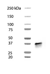
Detection of Human, Mouse, and Rat GAPDH/G3PDH by Western Blot.
Western blot shows lysates of U2OS human osteosarcoma cell line, A20 mouse B cell lymphoma cell line, and L6 rat myoblast cell line. PVDF membrane was probed with 0.1 µg/mL Rabbit Anti-Human/Mouse/Rat GAPDH Polyclonal Antibody (Catalog # 2275-PC) followed by HRP-conjugated Anti-Rabbit IgG Secondary Antibody (Catalog # HAF008). A specific band was detected for GAPDH at approximately 36 kDa (as indicated). This experiment was conducted under reducing conditions and using Immunoblot Buffer Group 1.
Detection of Human GAPDH by Simple WesternTM.
Simple Western shows lysates of Exosome Standards (LNCaP) (NBP3-11687), Exosome Standards (HT‑29) (NBP3-11685) and Jurkat human acute T cell leukemia cell line, loaded at 0.5 mg/ml. A specific band was detected for GAPDH at approximately 41 kDa (as indicated) using 5 µg/mL of Rabbit Anti-Human/Mouse/Rat GAPDH Antigen Affinity-purified Polyclonal Antibody (Catalog # 2275-PC-100). This experiment was conducted under reducing conditions and using the 12-230kDa separation system.
Detection of Human, Mouse and Rat GAPDH by Simple WesternTM.
Simple Western lane view shows lysates of Jurkat human acute T cell leukemia cell line, C2C12 mouse myoblast cell line, and C6 rat glioma cell line, loaded at 0.2 mg/mL. A specific band was detected for GAPDH at approximately 43 kDa (as indicated) using 1 µg/mL of Rabbit Anti-Human/Mouse/Rat GAPDH Antigen Affinity-purified Polyclonal Antibody (Catalog # 2275-PC-100). This experiment was conducted under reducing conditions and using the 12-230 kDa separation system.
Detection of Mouse Human/Mouse/Rat GAPDH Antibody by Western Blot
Higher levels of CD4 are found in macrophages expressing LCK.A) Schematic diagram of self-inactivating lentiviral construct used to express wild-type Lck and puroR (LCKWT P). A similar construct was used to express a kinase inactive form of Lck and puroR (LCKINACTIVE P) or to express puroR only (‘control P’). B) Detection of protein expression of LCK and CD4. Control and transgenic PSC-macrophages lysates were analysed by western blotting using anti-LCK and anti-CD4 antibodies. The loading control GAPDH was detected using anti-GAPDH antibody. C) Protein levels were measured with Odyssey software (Li-COR) and CD4 expression was normalised to GAPDH expression. Symbols represent normalised CD4 expression, relative to the PSC-macrophages control group, of two independent experiments. D) Detection of surface CD4 and total LCK expression. Representative two-colour immunofluorescence (dot plot) analysis is shown. Gates were determined by using the two relevant isotype control antibodies. Quantification of total LCK expression (E) and surface CD4 expression (F), expressed as the ratio of the geometric mean fluorescence intensity (MFI) over the isotype control ±SEM of independent experiments (n = 7). G) Detergent resistance of CD4, expressed as mean Flow Cytometric Detergent Resistance (FCDR) index of CD4 (n = 4) in PSC-macrophages, calculated as described in materials and methods. Image collected and cropped by CiteAb from the following publication (https://pubmed.ncbi.nlm.nih.gov/24465876), licensed under a CC-BY license. Not internally tested by R&D Systems.
Detection of Mouse Human/Mouse/Rat GAPDH Antibody by Western Blot
CD4 knock-down in genetically modified stem cell-derived macrophages. C) Detection of protein expression of CD4. Control and transgenic PSC-macrophages lysates were analysed by western blotting using anti-CD4 antibodies. GAPDH, a loading control, was detected using anti-GAPDH antibody. Representative blot is shown. Image collected and cropped by CiteAb from the following publication (https://pubmed.ncbi.nlm.nih.gov/24465876), licensed under a CC-BY license. Not internally tested by R&D Systems.
Detection of Mouse GAPDH by Western Blot
Higher levels of CD4 are found in macrophages expressing LCK.A) Schematic diagram of self-inactivating lentiviral construct used to express wild-type Lck and puroR (LCKWT P). A similar construct was used to express a kinase inactive form of Lck and puroR (LCKINACTIVE P) or to express puroR only (‘control P’). B) Detection of protein expression of LCK and CD4. Control and transgenic PSC-macrophages lysates were analysed by western blotting using anti-LCK and anti-CD4 antibodies. The loading control GAPDH was detected using anti-GAPDH antibody. C) Protein levels were measured with Odyssey software (Li-COR) and CD4 expression was normalised to GAPDH expression. Symbols represent normalised CD4 expression, relative to the PSC-macrophages control group, of two independent experiments. D) Detection of surface CD4 and total LCK expression. Representative two-colour immunofluorescence (dot plot) analysis is shown. Gates were determined by using the two relevant isotype control antibodies. Quantification of total LCK expression (E) and surface CD4 expression (F), expressed as the ratio of the geometric mean fluorescence intensity (MFI) over the isotype control ±SEM of independent experiments (n = 7). G) Detergent resistance of CD4, expressed as mean Flow Cytometric Detergent Resistance (FCDR) index of CD4 (n = 4) in PSC-macrophages, calculated as described in materials and methods. Image collected and cropped by CiteAb from the following open publication (https://pubmed.ncbi.nlm.nih.gov/24465876), licensed under a CC-BY license. Not internally tested by R&D Systems.
Detection of Mouse GAPDH by Western Blot
CD4 knock-down in genetically modified stem cell-derived macrophages.C) Detection of protein expression of CD4. Control and transgenic PSC-macrophages lysates were analysed by western blotting using anti-CD4 antibodies. GAPDH, a loading control, was detected using anti-GAPDH antibody. Representative blot is shown. Image collected and cropped by CiteAb from the following open publication (https://pubmed.ncbi.nlm.nih.gov/24465876), licensed under a CC-BY license. Not internally tested by R&D Systems.
Detection of Mouse GAPDH by Western Blot
Higher levels of CD4 are found in macrophages expressing LCK.A) Schematic diagram of self-inactivating lentiviral construct used to express wild-type Lck and puroR (LCKWT P). A similar construct was used to express a kinase inactive form of Lck and puroR (LCKINACTIVE P) or to express puroR only (‘control P’). B) Detection of protein expression of LCK and CD4. Control and transgenic PSC-macrophages lysates were analysed by western blotting using anti-LCK and anti-CD4 antibodies. The loading control GAPDH was detected using anti-GAPDH antibody. C) Protein levels were measured with Odyssey software (Li-COR) and CD4 expression was normalised to GAPDH expression. Symbols represent normalised CD4 expression, relative to the PSC-macrophages control group, of two independent experiments. D) Detection of surface CD4 and total LCK expression. Representative two-colour immunofluorescence (dot plot) analysis is shown. Gates were determined by using the two relevant isotype control antibodies. Quantification of total LCK expression (E) and surface CD4 expression (F), expressed as the ratio of the geometric mean fluorescence intensity (MFI) over the isotype control ±SEM of independent experiments (n = 7). G) Detergent resistance of CD4, expressed as mean Flow Cytometric Detergent Resistance (FCDR) index of CD4 (n = 4) in PSC-macrophages, calculated as described in materials and methods. Image collected and cropped by CiteAb from the following open publication (https://pubmed.ncbi.nlm.nih.gov/24465876), licensed under a CC-BY license. Not internally tested by R&D Systems.Applications for GAPDH Antibody
Application
Recommended Usage
Immunoprecipitation
1:1000-1:5000 dilution
Sample: L292 human fibrosarcoma cell line, WEHI 7.1 mouse T cell lymphoma cell line, and INT 407 human intestinal epithelial cell line
Sample: L292 human fibrosarcoma cell line, WEHI 7.1 mouse T cell lymphoma cell line, and INT 407 human intestinal epithelial cell line
Simple Western
1-5 µg/mL
Sample: Exosome Standards (LNCap) (Catalog # NBP3-11687), Exosome Standards (HT-29) (Catalog # NBP3-11685), Jurkat human acute T cell leukemia cell line, C2C12 mouse myoblast cell line, and C6 rat glioma cell line
Sample: Exosome Standards (LNCap) (Catalog # NBP3-11687), Exosome Standards (HT-29) (Catalog # NBP3-11685), Jurkat human acute T cell leukemia cell line, C2C12 mouse myoblast cell line, and C6 rat glioma cell line
Western Blot
0.1 µg/mL
Sample: U2OS human osteosarcoma cell line, A20 mouse B cell lymphoma cell line, and L6 rat myoblast cell line
Sample: U2OS human osteosarcoma cell line, A20 mouse B cell lymphoma cell line, and L6 rat myoblast cell line
Reviewed Applications
Read 2 reviews rated 5 using 2275-PC-100 in the following applications:
Formulation, Preparation, and Storage
Purification
Antigen Affinity-purified
Formulation
Supplied as a solution containg PBS.
Shipping
The product is shipped with dry ice or equivalent. Upon receipt, store it immediately at the temperature recommended below.
Stability & Storage
Use a manual defrost freezer and avoid repeated freeze-thaw cycles.
- 12 months from date of receipt, -20 to -70 °C as supplied.
- 1 month, 2 to 8 °C under sterile conditions after opening.
- 6 months, -20 to -70 °C under sterile conditions after opening.
Background: GAPDH
Long Name
Glyceraldehyde-3-phosphate Dehydrogenase
Alternate Names
G3PDH
Gene Symbol
GAPDH
Additional GAPDH Products
Product Documents for GAPDH Antibody
Certificate of Analysis
To download a Certificate of Analysis, please enter a lot or batch number in the search box below.
Note: Certificate of Analysis not available for kit components.
Product Specific Notices for GAPDH Antibody
For research use only
Related Research Areas
Citations for GAPDH Antibody
 Powered by Bioz
Customer Reviews for GAPDH Antibody (2)
5 out of 5
2 Customer Ratings
Have you used GAPDH Antibody?
Submit a review and receive an Amazon gift card!
$25/€18/£15/$25CAN/¥2500 Yen for a review with an image
$10/€7/£6/$10CAN/¥1110 Yen for a review without an image
Submit a review
Customer Images

Showing
1
-
2 of
2 reviews
Showing All
Filter By:
-
Application: Western BlotSample Tested: HCT-116 human colorectal carcinoma cell lineSpecies: HumanVerified Customer | Posted 02/03/2026Band of GAPDH in HCT cell line1:1000 dilution, strong band

-
Application: Western BlotSample Tested: MDA-MB-231 human breast cancer cell lineVerified Customer | Posted 02/03/2026GAPDH expression in MDA-231Very clear band with 1:1000 dilution
There are no reviews that match your criteria.
Protocols
Find general support by application which include: protocols, troubleshooting, illustrated assays, videos and webinars.
- Cellular Response to Hypoxia Protocols
- Immunoprecipitation Protocol
- R&D Systems Quality Control Western Blot Protocol
- Troubleshooting Guide: Western Blot Figures
- Western Blot Conditions
- Western Blot Protocol
- Western Blot Protocol for Cell Lysates
- Western Blot Troubleshooting
- Western Blot Troubleshooting Guide
- View all Protocols, Troubleshooting, Illustrated assays and Webinars
Loading...