GLI-2 is a 166 kDa biopotential transcription regulator of the hedgehog signaling pathway. It contains 5 conserved tandem C2H2 zinc finger domains that are flanked by a repression domain at the N-terminal region and an activation domain in the C-terminal region. At least four isoforms that differ in their N- or C-terminal regions have been described. Transcriptional activity of GLI-2 is regulated by proteolytic processing and degradation. Within the region used as immunogen, human and mouse GLI-2 share 88% amino acid sequence homology.

Key Product Details
Validated by
Biological Validation
Species Reactivity
Validated:
Human
Cited:
Human, Mouse
Applications
Validated:
Immunohistochemistry, Western Blot, Chromatin Immunoprecipitation (ChIP)
Cited:
Immunohistochemistry, Chromatin Immunoprecipitation (ChIP), Chromatin Immunoprecipitation Sequencing, Bioassay, Proximity Ligation Assay
Label
Unconjugated
Antibody Source
Polyclonal Sheep IgG
Loading...
Product Specifications
Immunogen
E. coli-derived recombinant human GLI-2
Ala2-Glu91
Accession # BAA25668
Ala2-Glu91
Accession # BAA25668
Specificity
Detects human GLI-2 in direct ELISAs and Western blots. In direct ELISAs and Western blots, approximately 25% cross-reactivity with recombinant mouse GLI-2 and 5% cross-reactivity with recombinant human (rh) GLI-1 and rhGLI-3 is observed.
Clonality
Polyclonal
Host
Sheep
Isotype
IgG
Scientific Data Images for Human GLI‑2 Antibody
Detection of GLI‑2-regulated Genes by Chromatin Immunoprecipitation.
Jurkat human acute T cell leukemia cell line treated with 50 ng/mL PMA and 200 ng/mL calcium ionomycin for 30 minutes was fixed using formaldehyde, resuspended in lysis buffer, and sonicated to shear chromatin. GLI-2/DNA complexes were immunoprecipitated using 5 µg Sheep Anti-Human GLI-2 Antigen Affinity-purified Polyclonal Antibody (Catalog # AF3526) or control antibody (Catalog # 5-001-A) for 15 minutes in an ultrasonic bath, followed by Biotinylated Anti-Sheep IgG Secondary Antibody (Catalog # BAF016).Immunocomplexes were captured using 50 µL of MagCellect Streptavidin Ferrofluid (Catalog # MAG999) and DNA was purified using chelating resin solution. Thebcl-2promoter was detected by standard PCR.Applications for Human GLI‑2 Antibody
Application
Recommended Usage
Chromatin Immunoprecipitation (ChIP)
5 µg/5 x 106 cells
Sample: PMA and calcium ionomycin treated Jurkat human acute T cell leukemia cell line chromatin, bcl-2 promoter detected by standard PCR
Sample: PMA and calcium ionomycin treated Jurkat human acute T cell leukemia cell line chromatin, bcl-2 promoter detected by standard PCR
Immunohistochemistry
5-15 µg/mL
Sample: Immersion fixed paraffin-embedded sections of human skin, basal cell carcinoma, and prostate cancer tissue
Sample: Immersion fixed paraffin-embedded sections of human skin, basal cell carcinoma, and prostate cancer tissue
Western Blot
0.1 µg/mL
Sample: Recombinant Human GLI‑2
Sample: Recombinant Human GLI‑2
Reviewed Applications
Read 3 reviews rated 3.7 using AF3526 in the following applications:
Formulation, Preparation, and Storage
Purification
Antigen Affinity-purified
Reconstitution
Reconstitute at 0.2 mg/mL in sterile PBS. For liquid material, refer to CoA for concentration.
Loading...
Formulation
Lyophilized from a 0.2 μm filtered solution in PBS with Trehalose. *Small pack size (SP) is supplied either lyophilized or as a 0.2 µm filtered solution in PBS.
Shipping
Lyophilized product is shipped at ambient temperature. Liquid small pack size (-SP) is shipped with polar packs. Upon receipt, store immediately at the temperature recommended below.
Stability & Storage
Use a manual defrost freezer and avoid repeated freeze-thaw cycles.
- 12 months from date of receipt, -20 to -70 °C as supplied.
- 1 month, 2 to 8 °C under sterile conditions after reconstitution.
- 6 months, -20 to -70 °C under sterile conditions after reconstitution.
Calculators
Background: GLI-2
Long Name
GLI-Kruppel family member GLI2
Alternate Names
GLI2, THP2
Gene Symbol
GLI2
UniProt
Additional GLI-2 Products
Product Documents for Human GLI‑2 Antibody
Certificate of Analysis
To download a Certificate of Analysis, please enter a lot or batch number in the search box below.
Note: Certificate of Analysis not available for kit components.
Product Specific Notices for Human GLI‑2 Antibody
For research use only
Related Research Areas
Citations for Human GLI‑2 Antibody
 Powered by Bioz
Customer Reviews for Human GLI‑2 Antibody (3)
3.7 out of 5
3 Customer Ratings
Have you used Human GLI‑2 Antibody?
Submit a review and receive an Amazon gift card!
$25/€18/£15/$25CAN/¥2500 Yen for a review with an image
$10/€7/£6/$10CAN/¥1110 Yen for a review without an image
Submit a review
Customer Images
Showing
1
-
3 of
3 reviews
Showing All
Filter By:
-
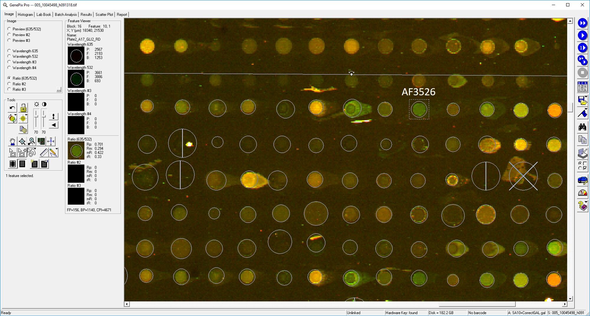
Application: MicroarraysSample Tested: EDTA PlasmaSpecies: HumanVerified Customer | Posted 03/11/2019

-
Application: ELISASample Tested: PlasmaSpecies: HumanVerified Customer | Posted 11/10/2018
-
Application: MicroarraySample Tested: EDTA PlasmaSpecies: HumanVerified Customer | Posted 11/02/2018
There are no reviews that match your criteria.
Protocols
Find general support by application which include: protocols, troubleshooting, illustrated assays, videos and webinars.
- Antigen Retrieval Protocol (PIER)
- Antigen Retrieval for Frozen Sections Protocol
- Appropriate Fixation of IHC/ICC Samples
- Cellular Response to Hypoxia Protocols
- ChIP Protocol Video
- Chromatin Immunoprecipitation (ChIP) Protocol
- Chromatin Immunoprecipitation Protocol
- Chromogenic IHC Staining of Formalin-Fixed Paraffin-Embedded (FFPE) Tissue Protocol
- Chromogenic Immunohistochemistry Staining of Frozen Tissue
- ClariTSA™ Fluorophore Kits
- Detection & Visualization of Antibody Binding
- Fluorescent IHC Staining of Frozen Tissue Protocol
- Graphic Protocol for Heat-induced Epitope Retrieval
- Graphic Protocol for the Preparation and Fluorescent IHC Staining of Frozen Tissue Sections
- Graphic Protocol for the Preparation and Fluorescent IHC Staining of Paraffin-embedded Tissue Sections
- Graphic Protocol for the Preparation of Gelatin-coated Slides for Histological Tissue Sections
- IHC Sample Preparation (Frozen sections vs Paraffin)
- Immunofluorescent IHC Staining of Formalin-Fixed Paraffin-Embedded (FFPE) Tissue Protocol
- Immunohistochemistry (IHC) and Immunocytochemistry (ICC) Protocols
- Immunohistochemistry Frozen Troubleshooting
- Immunohistochemistry Paraffin Troubleshooting
- Preparing Samples for IHC/ICC Experiments
- Preventing Non-Specific Staining (Non-Specific Binding)
- Primary Antibody Selection & Optimization
- Protocol for Heat-Induced Epitope Retrieval (HIER)
- Protocol for Making a 4% Formaldehyde Solution in PBS
- Protocol for VisUCyte™ HRP Polymer Detection Reagent
- Protocol for the Preparation & Fixation of Cells on Coverslips
- Protocol for the Preparation and Chromogenic IHC Staining of Frozen Tissue Sections
- Protocol for the Preparation and Chromogenic IHC Staining of Frozen Tissue Sections - Graphic
- Protocol for the Preparation and Chromogenic IHC Staining of Paraffin-embedded Tissue Sections
- Protocol for the Preparation and Chromogenic IHC Staining of Paraffin-embedded Tissue Sections - Graphic
- Protocol for the Preparation and Fluorescent IHC Staining of Frozen Tissue Sections
- Protocol for the Preparation and Fluorescent IHC Staining of Paraffin-embedded Tissue Sections
- Protocol for the Preparation of Gelatin-coated Slides for Histological Tissue Sections
- R&D Systems Quality Control Western Blot Protocol
- TUNEL and Active Caspase-3 Detection by IHC/ICC Protocol
- The Importance of IHC/ICC Controls
- Troubleshooting Guide: Immunohistochemistry
- Troubleshooting Guide: Western Blot Figures
- Western Blot Conditions
- Western Blot Protocol
- Western Blot Protocol for Cell Lysates
- Western Blot Troubleshooting
- Western Blot Troubleshooting Guide
- View all Protocols, Troubleshooting, Illustrated assays and Webinars
Loading...